knowt logo

Biology: Unit 2

Unit 2 Day 1: Studying Cells in a Laboratory Setting and Cell Types

Overview:

Cell Theory:

  • All organisms are made of cells

  • The cell is the smallest unit of life

  • All cells come from pre-existing cells

■ Remember….cell structure is correlated to cellular function

Studying Cells in a Lab

Microscopy

Why do scientists use microscopes? → To visualize cells too small to see with the naked eye and study their complexity

Types of Microscopes

Light Microscope (LM) - Visible light passes through a specimen and then through glass lesnses, which magnify the image

Image quality depends on…

Magnification: The ratio of an objects image size to its real size

Resolution: The measure of the clarity of the image

Contrast: Visible differences in parts of the sample

■ LMs can magnify effectively to about 1,000 times

What are the limitations of LMs? → Organelles are too small to be resolved (focused) by an LM

Electron Microscopes (EMs)

Types:

Scanning Electron Microscopes (SEMs) - Focus a beam of electrons onto the surface of a specimen, proving images that look 3D

Transmission Electron Microscopes (TEMs) - Used to study the internal structure of a cell

What are the limitations of EMs? → Cannot observe living cells

Cell Fractionation

What does cell fractionation do? → Takes cells apart and separates the major organelles from one another

What tool is used to seperate cells into their component parts? → Ultracentrifuge

Cell Types

■ There are two types of cells: prokaryotes and eukaryotes

Prokaryotic Cells

All Cells

Eukaryotic Cells

No nucleus

Are bound by a plasma membrane

DNA in a nucleus that is bound by a membranous nuclear envelope

DNA in an unbound region called the nucleoid

Contain Cytosol (Cytoplasm)

Membrane-bound organelles

No membrane-bound organelles

Contain chromosomes (carry genes)

Larger than Prokaryotic Cells

Typically small

Contain ribosomes (make proteins)

Multicellular

Unicellular

Ex: Bacteria

Ex: Animals, plants, fungi, and protists

What does compartmentalization do? → Allows for different reactions in different locations

What does it prevent? → Prevents interfering reactions from occurring in the same locations

- - -

The Endomembrane system

■ The Endomembrane System regulates protein traffic and performs metabolic functions

Components of the endomembrane system:

  • Nuclear envelope

  • Endoplasmic Reticulum (ER)

  • Golgi Apparatus

  • Lysosomes

  • Vacuoles

  • Plasma Membrane

How are these components connected? → They are connected continuously (already connected) or are connected via the transfer of vesicles

The eukaryotic cell’s genetic instructions are housed in the nucleus and carried out by the ribosomes

Nucleus

  • Contains chromosomes

  • Enclosed in a nuclear envelope

    Double Membrane

  • Has pore

    Regulates entry and exit of materials

  • Contains a nucleolus

    Nucleolus is a dense region of the nucleus that makes ribosomes

    Exits via nuclear pores

How are ribosomes formed? → rRNA and proteins make ribosomes

What do ribosomes do with eh mRNA message? → They translate message to the primary structure of proteins (Amino Acids)

Ribosomes

Function: Make proteins

Locations:

Cytosol: Free ribosomes are located in the Cytosol (Cytoplasm)

Bound on the outside of the endoplasmic reticulum or the nuclear envelope

Endoplasmic Reticulum

■ A network of membranous sacs and tubes

Functions:

  • Makes membranes

  • Compartmentalize (separates) the cell to keep proteins formed in the rough Er seperate from those of free ribosomes

  • Two Types: Rough Endoplasmic Reticulum and Smooth Endoplasmic Reticulum

Smooth ER

  • Lacks ribosomes

  • Makes lipids

  • Metabolizes carbohydrates (Breaks Carbs down)

  • Detoxifies poison

  • Stores calcium

Rough ER

  • Has bound ribosomes which secrete glycoproteins

    Glycoproteins are proteins bonded to carbohydrates

  • Distributes transport vesicles, proteins surrounded by membranes

Golgi Apparatus

■ Consists of flatted membranous sacs called Golgi Apparatus

  • Has directionality

Cis Face: Receives vesicles from the ER

- Faces ER

Trans Face: Sends vesicles back out into the cytosol to other locations or to the plasma membrane for secretion

- Faces plasma membrane

Functions:

  • Receives transport vesicles with materials from the ER

  • Modifies materials

  • Sorts materials

  • Adds molecular tag

  • Packages materials into new transport vehicles that exit the membrane via exocytosis

    (Exo - Exit, Cytosis - Cell)

Lysosomes

■ Membranous sacs of hydrolytic enzymes that digest macromolecules

Function:

  • Can hydrolyze proteins, fats, polysaccharides, and nucleic acid

Phagocytosis: Cellular eating (A cell engulfs another cell)

Autophagy: Cells eat itself for renewal (Breaks down own components)

Peroxisomes

  • Similar to lysosomes

  • Membrane bound metabolic component

  • Catalyze reactions that produce H²O² (Hydrogen Peroxide)

    Enzymes in peroxisomes then break down H²O² to water

Vacuoles

■ Large vesicles that stem from the ER and Golgi

Types:

  • Food Vacuole: Formed by phagocytosis and digested by lysosomes

  • Contractile Vacuole: Pump excess water out of cells

- Found in freshwater protists

  • Central Vacuole: Hold inorganic ions and water

- Found in many mature plant cells

- Important for turgor pressure (Holds water in plant cells)

- - -

Energy Organelles

Endosymboint Theory: The theory that explains the similarities mitochondria and chloroplasts have to a prokaryotes

■ Theory that states an early eukaryotic cell engulfed a prokaryotic cell

■ Prokaryotic cell became an endosyboint (cell that lives in another cell)

Evidence:

  • Double membrane

  • Ribosomes

  • Circular DNA

  • Capable of functioning on their own

Mitochondria
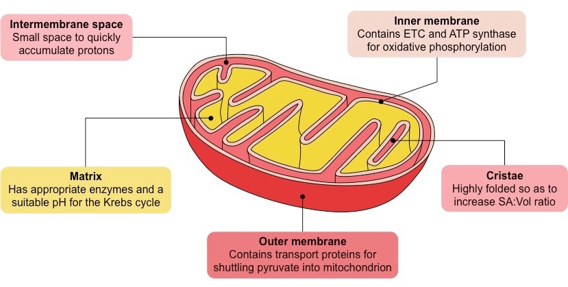
Function: Site of cellular respiration, a metabolic process that generates ATP energy

■ ATP Energy = Usable Energy

Structure of the double membrane: Has a smooth outer membrane and inner membrane is folded into Cristae (<------ The electron transport chain)

  1. Intermembrane: Space between inner and outer membrane

  2. Mitochondrial Matrix: Enclosed by the inner membrane

    Location for the Krebs Cycle

    Contains:

  • Enzymes that catalyze cellular respiration and ATP energy

  • Mitochondrial DNA

  • Ribosomes

■ The number of mitochondria in a cell correlated with metabolic activity

- Cells with high metabolic activity have more mitochondria

Ex: Cells that move/contract (Muscles in humans)

Chloroplasts
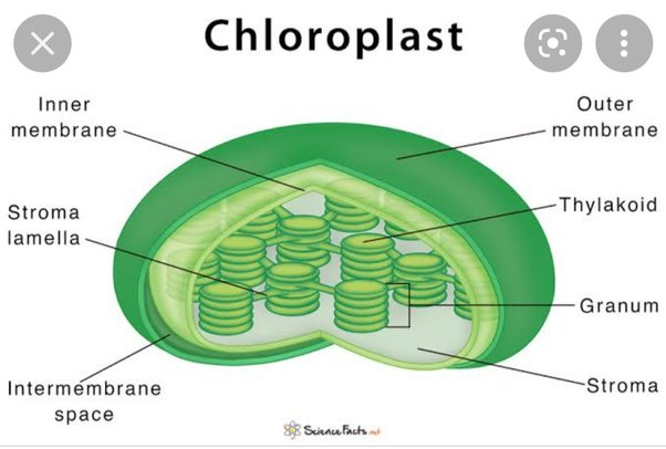
Found in the leaves of plants and algae, are the sites of photosynthesis

- Chlorophyll: Green pigment in plants (absorbs light energy)

Inside of its double membrane:

  • Thylakoids

- Membranous sacs that can organize into stacks called Granum

  • Stroma: The internal fluid

- Calvin Cycle occurs here

- - -

The Cytoskeleton

The Cytoskeleton

■ A network of fibers throughout the cytoplasm

Functions: Structural support (shape) and mechanical support (movement)

There are three types of fibers in the cytoskeleton:

  • Microtubules (Thickets of the three)

  • Microfilaments (Thin and skinny)

  • Intermediate Filaments (Smaller than Microtubules, but larger than Microfilaments)

Microtubules:

Functions:

  • Shapes cell (structural support)

  • Guides movements of organelles (Think: Tracks)

  • Separates choromosomes during cell division

  • Cell motility (i.s. Cilia and Flagellum)

Centrosomes and Centrioles

- The centriole is a “microtubule-organizing center”

- Separates chromosomes in animal cells

- Microtubules grow out from a centrosome

■ Only found in animal cells

Cilia and Flagella

- Microtubules control beating of Cilia and Flagella

- Cilia and Flagella have different beating patterns

Common Structure:

- Core of microtubules

- Anchored by a “basel body” (Thing attached to the cell membrane)

Dynein is a motor protein that drives the movements of cilium and flagellum

Microfilaments (Actin Filaments)

Functions:

- Maintains cell shape

  • Bear tension

  • Forms a 3D network called cortex (Supports shape)

- Assists in muscle contraction and cell motility

  • Actin works with myosin to cause contraction

- Division of animal cells

○ Pseudopodia: Cellular extension of amoeba

○ Cytoplasmic Streaming: Circular flow of cytoplasm within cells

Intermediate Filaments:

Functions:

- Maintains cell shape

- Anchor nucleus and organelles

- Form the nuclear Lamina

  • Lines the Nuclear Envelope

- - -

Cell Size

Cell Size

Cellular metabolism depends on cell size

Metabolism - How they break apart

■ Cell waste must leave

■ Dissipate thermal energy

■ Nutrients and other resources (chemical materials) must enter

At a certain size, it begins to be too difficult for a cell to regulate what comes in and what goes out of the plasma membrane

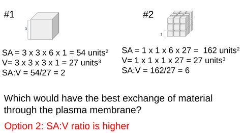
Surface Area to Volume

The size of a cell will dictate the function

Cells need a high surface area-to-volume ratio to optimize the exchange of material through the plasma membrane

Formulas
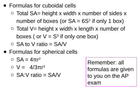
Example: Cubiodal Cell
Example: Spherical Cell
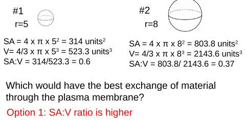
Eukaryotic cells can reach above 100nm (nanometer) in diameter, while prokaryotic cells, like bacteria, remain small, reaching only to about 10nm in diameter. How are eukaryotic cells able to become so much larger than prokaryotic cells while still efficiently exchanging material through the plasma membrane?

What does this mean for a cell?

Cells tend to be small

  • Small cells have a high SA:V ratio

- Optimizes exchange of materials at the plasma membrane

  • Larger cells have a lower SA:V ratio

- Lose efficiency exchanging materials

■ The cellular demand for resources increases

■ Rate of heat exchange decreases

- - -

Plasma Membrane and Membrane Permeability

Phospholipid Bilayer Structure

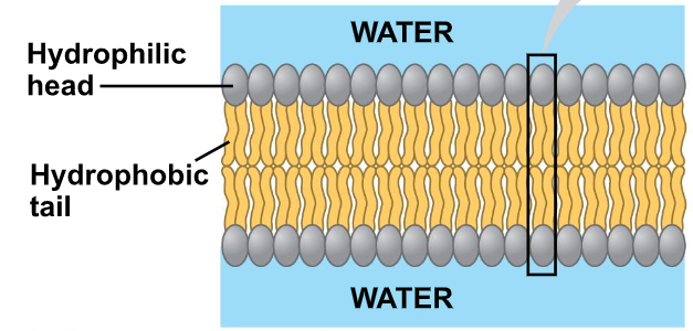
Phospholipid Bilayer: A double layer of phospholipids

■ Phospholipids are amphipathic molecules

Amphipathic: Has both a hydrophobic region and a hydrophilic region

Examine the phospholipid to the right and identify the structures:
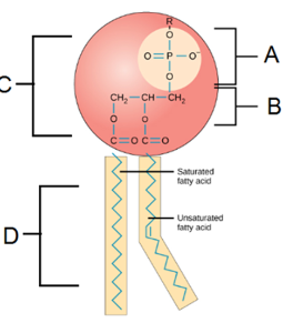
A: Phosphate Group

B: Glycerol

C: Hydrophilic Head

D: Hydrophobic Tails

■ A bilayer forms because they are amphipathic

■ Hydrophilic heads orient toward aqueous environments

■ Hydrophobic tails are facing inwards away from aqueous environments

Fluid Mosaic Model

A model to describe the structure of cell membranes

Fluidity: Phospholipids are capable of movement

Mosaic: Comprised of many macromolecules

  • Phospholipids

  • Cholesterol

  • Proteins

  • Carbohydrates

The fluidity of Membranes

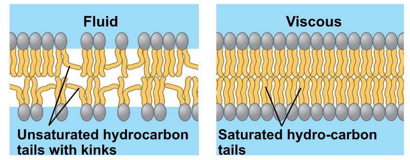
Fluid: Membrane is held together by weak hydrophobic interactions so phospholipids can move and shift

Which way do lipids drift? → Laterally/Rarely flip-flops transversely (Across the membrane)

Effects of Temperature on Fluidity

As temperatures cool, membranes switch from a fluid to a solid state

Which types of fatty acids are more fluid? Why? → Unsaturated fatty acids are more fluid than saturated fats due to kinks
Cholesterol has different effects on membrane fluidity at different temperatures

- High Temp: Reduces movements

- Low Temp: Maintains fluidity by preventing tight packing

Membrane Proteins and Their Functions

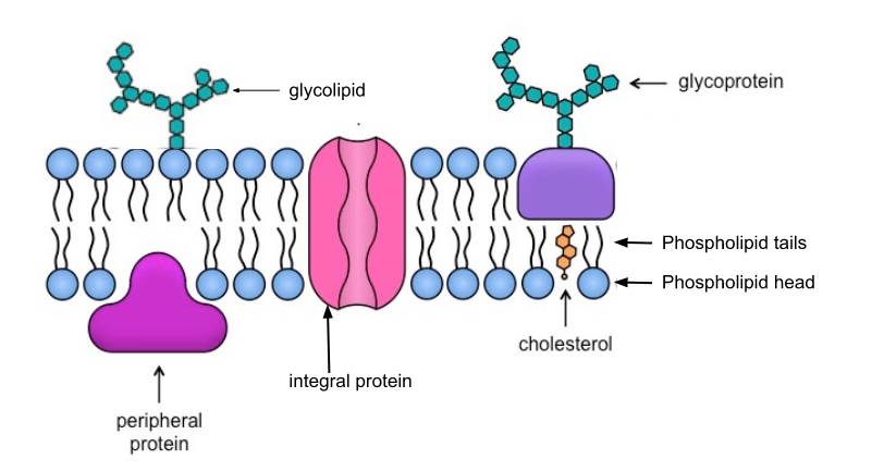
Proteins determine most of the membrane’s specific functions

Categories:

  1. Integral proteins

- Embedded in the lipid bilayer

- Transmembrane proteins (spans the membrane)

- Amphipathic

  1. Peripheral Proteins

- Bound to the surface of the membrane

Membrane Carbohydrates

Important for cell-to-cell recognition

  • Glycolipids

- Carbohydrates bonded to lipids

  • Glycoproteins

- Carbohydrates bonded to proteins

- Most abundant

Draw and Label the cell membrane:
Membrane Functions

  • Transport

  • Enzymatic Activity

  • Signal Transductions

  • Cell-cell Recognition

  • Intercellular Joining

  • Attachment to the cytoskeleton and extracellular matrix (ECM)

Transport

A cell must exchange materials with its surroundings, a process controlled by the plasma membrane

Selectively Permeable: Selectively letting some things pass through, but not others (like a screen)

Which types of molecules can pass through easily and rapidly? → Small hydrophobic (nonpolar) molecules

Which types of molecules cannot pass through easily and require assistance? → Large, hydrophilic (polar) molecules

Transport Proteins

Are transport proteins integral or peripheral? Integral

Channel Proteins: Has a hydrophilic channel that is used as a tunnel

Ex: Aquaporins

Carrier Proteins: Bind to molecules and change shape to shuttle them across the membrane

A transport protein is specific for the substance it moves

Plant Cell Barriers

Plants have a cell wall that covers their plasma membranes

Extracellular structure (not found in animal cells)

■ Made of cellulose

Provides:

  • Shape/Structure

  • Protection

  • Regulation of water intake

Communication

Plants communicate through plasmodesmata

Plasmodesmata: Hole-like structures in the cell wall filled with cytosol that connect adjacent cells

- - -

Passive Transport

Selective Permeability

Some substances can cross the membrane more easily than others

Easy passage across the membrane: small, hydrophobic (nonpolar) molecules

Difficult passage or protein assisted passage: Large hydrophilic (polar) molecules + ions

Transport Across a Membrane

There are two types of transport across a membrane: passive and active

Passive: Without the use of energy

Active: Uses energy

Passive Transport

Passive Transport:

■ Molecules move down their concentration gradient from high to low concentration

Diffusion:

Diffusion: Molecules spread out evenly in the available space

■ Substances move from a high to a low concentration

■ Move with the concentration gradient

Dynamic Equilibrium:

■ Molecules diffuse directly across the membrane

■ Different rates of diffusion for different molecules

What factors impact the rate of diffusion? → Concentration gradient, distance, area, and the presence of transport proteins

Osmosis:

Osmosis: The diffusion of water across a selectively permeable membrane

■ Osmosis can happen without a protein

■ Water diffused from a region of lower solute (high water) concentration to a region of higher solute (low water) concentration

Facilitated Diffusion:

Facilitated Diffusion: To help

■ Speeds up the rate of diffusion of: small ions, water, and carbohydrates

■ Two categories of transport proteins: channel and carrier

■ Each transport protein is specific for substances it can facilitate movement for

Channel Proteins:

Channel Proteins: Provides channels to allow specific molecules or ions to cross the membrane

■ Channel is hydrophilic

Which type of molecules pass through channel proteins? → Small polar molecules and ions

Aquaporins: Proteins used for the facilitated diffusion of water

Gated channels: Only open and close in response to stimulus

Carrier Proteins:

Carrier Proteins: Change shape that translocates the solute-binding site across the membrane

Which type of molecules pass through carrier proteins? → Lipid insoluble molecules

- - -

KR

Biology: Unit 2

Unit 2 Day 1: Studying Cells in a Laboratory Setting and Cell Types

Overview:

Cell Theory:

  • All organisms are made of cells

  • The cell is the smallest unit of life

  • All cells come from pre-existing cells

■ Remember….cell structure is correlated to cellular function

Studying Cells in a Lab

Microscopy

Why do scientists use microscopes? → To visualize cells too small to see with the naked eye and study their complexity

Types of Microscopes

Light Microscope (LM) - Visible light passes through a specimen and then through glass lesnses, which magnify the image

Image quality depends on…

Magnification: The ratio of an objects image size to its real size

Resolution: The measure of the clarity of the image

Contrast: Visible differences in parts of the sample

■ LMs can magnify effectively to about 1,000 times

What are the limitations of LMs? → Organelles are too small to be resolved (focused) by an LM

Electron Microscopes (EMs)

Types:

Scanning Electron Microscopes (SEMs) - Focus a beam of electrons onto the surface of a specimen, proving images that look 3D

Transmission Electron Microscopes (TEMs) - Used to study the internal structure of a cell

What are the limitations of EMs? → Cannot observe living cells

Cell Fractionation

What does cell fractionation do? → Takes cells apart and separates the major organelles from one another

What tool is used to seperate cells into their component parts? → Ultracentrifuge

Cell Types

■ There are two types of cells: prokaryotes and eukaryotes

Prokaryotic Cells

All Cells

Eukaryotic Cells

No nucleus

Are bound by a plasma membrane

DNA in a nucleus that is bound by a membranous nuclear envelope

DNA in an unbound region called the nucleoid

Contain Cytosol (Cytoplasm)

Membrane-bound organelles

No membrane-bound organelles

Contain chromosomes (carry genes)

Larger than Prokaryotic Cells

Typically small

Contain ribosomes (make proteins)

Multicellular

Unicellular

Ex: Bacteria

Ex: Animals, plants, fungi, and protists

What does compartmentalization do? → Allows for different reactions in different locations

What does it prevent? → Prevents interfering reactions from occurring in the same locations

- - -

The Endomembrane system

■ The Endomembrane System regulates protein traffic and performs metabolic functions

Components of the endomembrane system:

  • Nuclear envelope

  • Endoplasmic Reticulum (ER)

  • Golgi Apparatus

  • Lysosomes

  • Vacuoles

  • Plasma Membrane

How are these components connected? → They are connected continuously (already connected) or are connected via the transfer of vesicles

The eukaryotic cell’s genetic instructions are housed in the nucleus and carried out by the ribosomes

Nucleus

  • Contains chromosomes

  • Enclosed in a nuclear envelope

    Double Membrane

  • Has pore

    Regulates entry and exit of materials

  • Contains a nucleolus

    Nucleolus is a dense region of the nucleus that makes ribosomes

    Exits via nuclear pores

How are ribosomes formed? → rRNA and proteins make ribosomes

What do ribosomes do with eh mRNA message? → They translate message to the primary structure of proteins (Amino Acids)

Ribosomes

Function: Make proteins

Locations:

Cytosol: Free ribosomes are located in the Cytosol (Cytoplasm)

Bound on the outside of the endoplasmic reticulum or the nuclear envelope

Endoplasmic Reticulum

■ A network of membranous sacs and tubes

Functions:

  • Makes membranes

  • Compartmentalize (separates) the cell to keep proteins formed in the rough Er seperate from those of free ribosomes

  • Two Types: Rough Endoplasmic Reticulum and Smooth Endoplasmic Reticulum

Smooth ER

  • Lacks ribosomes

  • Makes lipids

  • Metabolizes carbohydrates (Breaks Carbs down)

  • Detoxifies poison

  • Stores calcium

Rough ER

  • Has bound ribosomes which secrete glycoproteins

    Glycoproteins are proteins bonded to carbohydrates

  • Distributes transport vesicles, proteins surrounded by membranes

Golgi Apparatus

■ Consists of flatted membranous sacs called Golgi Apparatus

  • Has directionality

Cis Face: Receives vesicles from the ER

- Faces ER

Trans Face: Sends vesicles back out into the cytosol to other locations or to the plasma membrane for secretion

- Faces plasma membrane

Functions:

  • Receives transport vesicles with materials from the ER

  • Modifies materials

  • Sorts materials

  • Adds molecular tag

  • Packages materials into new transport vehicles that exit the membrane via exocytosis

    (Exo - Exit, Cytosis - Cell)

Lysosomes

■ Membranous sacs of hydrolytic enzymes that digest macromolecules

Function:

  • Can hydrolyze proteins, fats, polysaccharides, and nucleic acid

Phagocytosis: Cellular eating (A cell engulfs another cell)

Autophagy: Cells eat itself for renewal (Breaks down own components)

Peroxisomes

  • Similar to lysosomes

  • Membrane bound metabolic component

  • Catalyze reactions that produce H²O² (Hydrogen Peroxide)

    Enzymes in peroxisomes then break down H²O² to water

Vacuoles

■ Large vesicles that stem from the ER and Golgi

Types:

  • Food Vacuole: Formed by phagocytosis and digested by lysosomes

  • Contractile Vacuole: Pump excess water out of cells

- Found in freshwater protists

  • Central Vacuole: Hold inorganic ions and water

- Found in many mature plant cells

- Important for turgor pressure (Holds water in plant cells)

- - -

Energy Organelles

Endosymboint Theory: The theory that explains the similarities mitochondria and chloroplasts have to a prokaryotes

■ Theory that states an early eukaryotic cell engulfed a prokaryotic cell

■ Prokaryotic cell became an endosyboint (cell that lives in another cell)

Evidence:

  • Double membrane

  • Ribosomes

  • Circular DNA

  • Capable of functioning on their own

Mitochondria

Function: Site of cellular respiration, a metabolic process that generates ATP energy

■ ATP Energy = Usable Energy

Structure of the double membrane: Has a smooth outer membrane and inner membrane is folded into Cristae (<------ The electron transport chain)

  1. Intermembrane: Space between inner and outer membrane

  2. Mitochondrial Matrix: Enclosed by the inner membrane

    Location for the Krebs Cycle

    Contains:

  • Enzymes that catalyze cellular respiration and ATP energy

  • Mitochondrial DNA

  • Ribosomes

■ The number of mitochondria in a cell correlated with metabolic activity

- Cells with high metabolic activity have more mitochondria

Ex: Cells that move/contract (Muscles in humans)

Chloroplasts

Found in the leaves of plants and algae, are the sites of photosynthesis

- Chlorophyll: Green pigment in plants (absorbs light energy)

Inside of its double membrane:

  • Thylakoids

- Membranous sacs that can organize into stacks called Granum

  • Stroma: The internal fluid

- Calvin Cycle occurs here

- - -

The Cytoskeleton

The Cytoskeleton

■ A network of fibers throughout the cytoplasm

Functions: Structural support (shape) and mechanical support (movement)

There are three types of fibers in the cytoskeleton:

  • Microtubules (Thickets of the three)

  • Microfilaments (Thin and skinny)

  • Intermediate Filaments (Smaller than Microtubules, but larger than Microfilaments)

Microtubules:

Functions:

  • Shapes cell (structural support)

  • Guides movements of organelles (Think: Tracks)

  • Separates choromosomes during cell division

  • Cell motility (i.s. Cilia and Flagellum)

Centrosomes and Centrioles

- The centriole is a “microtubule-organizing center”

- Separates chromosomes in animal cells

- Microtubules grow out from a centrosome

■ Only found in animal cells

Cilia and Flagella

- Microtubules control beating of Cilia and Flagella

- Cilia and Flagella have different beating patterns

Common Structure:

- Core of microtubules

- Anchored by a “basel body” (Thing attached to the cell membrane)

Dynein is a motor protein that drives the movements of cilium and flagellum

Microfilaments (Actin Filaments)

Functions:

- Maintains cell shape

  • Bear tension

  • Forms a 3D network called cortex (Supports shape)

- Assists in muscle contraction and cell motility

  • Actin works with myosin to cause contraction

- Division of animal cells

○ Pseudopodia: Cellular extension of amoeba

○ Cytoplasmic Streaming: Circular flow of cytoplasm within cells

Intermediate Filaments:

Functions:

- Maintains cell shape

- Anchor nucleus and organelles

- Form the nuclear Lamina

  • Lines the Nuclear Envelope

- - -

Cell Size

Cell Size

Cellular metabolism depends on cell size

Metabolism - How they break apart

■ Cell waste must leave

■ Dissipate thermal energy

■ Nutrients and other resources (chemical materials) must enter

At a certain size, it begins to be too difficult for a cell to regulate what comes in and what goes out of the plasma membrane

Surface Area to Volume

The size of a cell will dictate the function

Cells need a high surface area-to-volume ratio to optimize the exchange of material through the plasma membrane

Formulas

Example: Cubiodal Cell

Example: Spherical Cell

Eukaryotic cells can reach above 100nm (nanometer) in diameter, while prokaryotic cells, like bacteria, remain small, reaching only to about 10nm in diameter. How are eukaryotic cells able to become so much larger than prokaryotic cells while still efficiently exchanging material through the plasma membrane?

What does this mean for a cell?

Cells tend to be small

  • Small cells have a high SA:V ratio

- Optimizes exchange of materials at the plasma membrane

  • Larger cells have a lower SA:V ratio

- Lose efficiency exchanging materials

■ The cellular demand for resources increases

■ Rate of heat exchange decreases

- - -

Plasma Membrane and Membrane Permeability

Phospholipid Bilayer Structure

Phospholipid Bilayer: A double layer of phospholipids

■ Phospholipids are amphipathic molecules

Amphipathic: Has both a hydrophobic region and a hydrophilic region

Examine the phospholipid to the right and identify the structures:

A: Phosphate Group

B: Glycerol

C: Hydrophilic Head

D: Hydrophobic Tails

■ A bilayer forms because they are amphipathic

■ Hydrophilic heads orient toward aqueous environments

■ Hydrophobic tails are facing inwards away from aqueous environments

Fluid Mosaic Model

A model to describe the structure of cell membranes

Fluidity: Phospholipids are capable of movement

Mosaic: Comprised of many macromolecules

  • Phospholipids

  • Cholesterol

  • Proteins

  • Carbohydrates

The fluidity of Membranes

Fluid: Membrane is held together by weak hydrophobic interactions so phospholipids can move and shift

Which way do lipids drift? → Laterally/Rarely flip-flops transversely (Across the membrane)

Effects of Temperature on Fluidity

As temperatures cool, membranes switch from a fluid to a solid state

Which types of fatty acids are more fluid? Why? → Unsaturated fatty acids are more fluid than saturated fats due to kinks

Cholesterol has different effects on membrane fluidity at different temperatures

- High Temp: Reduces movements

- Low Temp: Maintains fluidity by preventing tight packing

Membrane Proteins and Their Functions

Proteins determine most of the membrane’s specific functions

Categories:

  1. Integral proteins

- Embedded in the lipid bilayer

- Transmembrane proteins (spans the membrane)

- Amphipathic

  1. Peripheral Proteins

- Bound to the surface of the membrane

Membrane Carbohydrates

Important for cell-to-cell recognition

  • Glycolipids

- Carbohydrates bonded to lipids

  • Glycoproteins

- Carbohydrates bonded to proteins

- Most abundant

Draw and Label the cell membrane:

Membrane Functions

  • Transport

  • Enzymatic Activity

  • Signal Transductions

  • Cell-cell Recognition

  • Intercellular Joining

  • Attachment to the cytoskeleton and extracellular matrix (ECM)

Transport

A cell must exchange materials with its surroundings, a process controlled by the plasma membrane

Selectively Permeable: Selectively letting some things pass through, but not others (like a screen)

Which types of molecules can pass through easily and rapidly? → Small hydrophobic (nonpolar) molecules

Which types of molecules cannot pass through easily and require assistance? → Large, hydrophilic (polar) molecules

Transport Proteins

Are transport proteins integral or peripheral? Integral

Channel Proteins: Has a hydrophilic channel that is used as a tunnel

Ex: Aquaporins

Carrier Proteins: Bind to molecules and change shape to shuttle them across the membrane

A transport protein is specific for the substance it moves

Plant Cell Barriers

Plants have a cell wall that covers their plasma membranes

Extracellular structure (not found in animal cells)

■ Made of cellulose

Provides:

  • Shape/Structure

  • Protection

  • Regulation of water intake

Communication

Plants communicate through plasmodesmata

Plasmodesmata: Hole-like structures in the cell wall filled with cytosol that connect adjacent cells

- - -

Passive Transport

Selective Permeability

Some substances can cross the membrane more easily than others

Easy passage across the membrane: small, hydrophobic (nonpolar) molecules

Difficult passage or protein assisted passage: Large hydrophilic (polar) molecules + ions

Transport Across a Membrane

There are two types of transport across a membrane: passive and active

Passive: Without the use of energy

Active: Uses energy

Passive Transport

Passive Transport:

■ Molecules move down their concentration gradient from high to low concentration

Diffusion:

Diffusion: Molecules spread out evenly in the available space

■ Substances move from a high to a low concentration

■ Move with the concentration gradient

Dynamic Equilibrium:

■ Molecules diffuse directly across the membrane

■ Different rates of diffusion for different molecules

What factors impact the rate of diffusion? → Concentration gradient, distance, area, and the presence of transport proteins

Osmosis:

Osmosis: The diffusion of water across a selectively permeable membrane

■ Osmosis can happen without a protein

■ Water diffused from a region of lower solute (high water) concentration to a region of higher solute (low water) concentration

Facilitated Diffusion:

Facilitated Diffusion: To help

■ Speeds up the rate of diffusion of: small ions, water, and carbohydrates

■ Two categories of transport proteins: channel and carrier

■ Each transport protein is specific for substances it can facilitate movement for

Channel Proteins:

Channel Proteins: Provides channels to allow specific molecules or ions to cross the membrane

■ Channel is hydrophilic

Which type of molecules pass through channel proteins? → Small polar molecules and ions

Aquaporins: Proteins used for the facilitated diffusion of water

Gated channels: Only open and close in response to stimulus

Carrier Proteins:

Carrier Proteins: Change shape that translocates the solute-binding site across the membrane

Which type of molecules pass through carrier proteins? → Lipid insoluble molecules

- - -