Body Systems (Respiratory)
The respiratory system is responsible for supplying oxygen to the bloodstream and at the same time removing the waste product of carbon dioxide, which has been produced during cellular metabolism.
 Components of the respiratory system
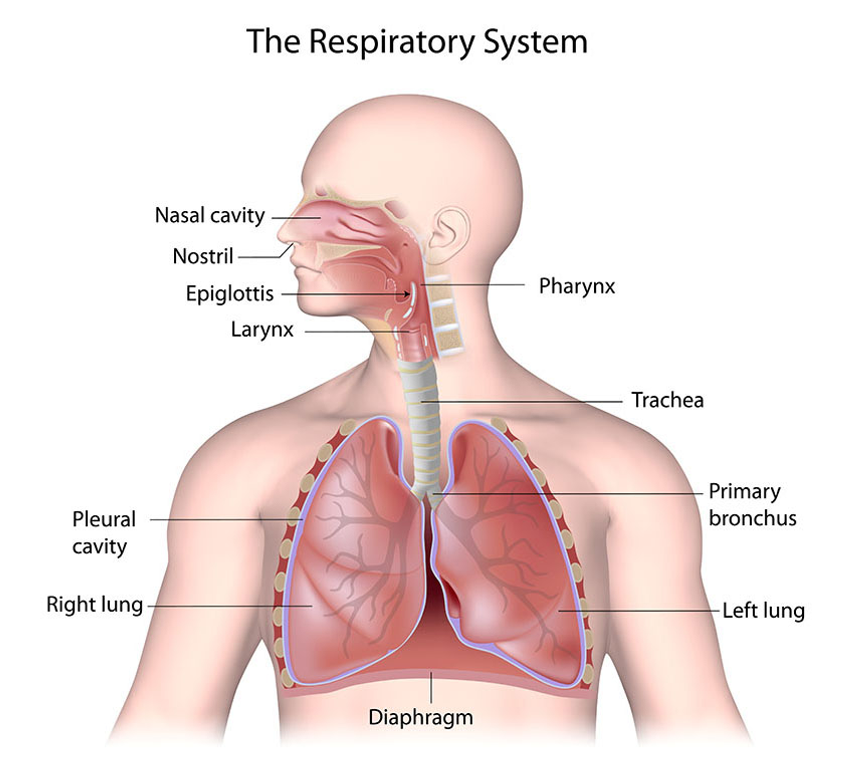
The major organs and components of the respiratory system are as follows:
nose and nasal cavity
sinuses, tonsils and adenoids
pharynx
larynx
trachea
bronchi and bronchioles
lungs and pleura
alveoli
diaphragm and intercostal muscles.
Medical terminology relating to the respiratory system
Root word | Meaning (origin) |
|---|---|
Bronch(o) | Airway (Greek) |
Laryng(o) | Voice box [Larynx] (Greek) |
Nas(o) | Nose (Latin) |
Pnea | Breathing, breath (Greek) |
Pneum(o) | Air, breathe [Lung] (Greek) |
Pulm(o) | Lung (Latin) |
Respir(o) | Breathing (Latin) |
Rhin(o) | Nose (Greek) |
Thorac(o) | Chest (Greek) |
Trache(o) | Rough [Trachea] (Greek) |
Medical terminology | Meaning |
|---|---|
Cilia | Small hair-like structures that line the airways |
Cyanosis | Bluish discolouration around the lips, mucous membranes or fingernail beds |
Diaphragm | The large muscle across the chest below the lungs. This muscle is vital for the work of breathing. |
Dyspnoea | Difficulty breathing |
Hypoxia | A lack of oxygen |
Thoracic cavity | Thorax is the word for chest. Thoracic cavity is the chest cavity in which the lungs sit. |
Wheezing | A whistling sound in the airways heard when the airways narrow |
Major Components
Nose and Nasal cavity
The nose and the nasal cavity are composed of:
nostrils (external nares)
nasal cavity (internal nares) and these are lined with ciliated mucous membranes
the palate.
The nose has several functions such as:
being an airway for respiration
warming, moistening and filtering inspired air
serving as a resonating chamber for speech
containing receptors for smell.
Sinuses, tonsils and adenoids
The sinuses or paranasal sinuses and nasolacrimal ducts drain into the nasal cavity.
The frontal, sphenoid, ethmoid and maxillary sinuses are hollow chambers for speech sounds. The eustachian tubes drain the middle ear into the pharynx and the tonsils and adenoids are found in the pharynx and nasopharynx. These have a part to play in protecting the body from infection.
Pharynx (throat) — consists of a muscular tube, lined with mucous membrane.
A common passageway for both food and air.
Larynx (voice box) — made of cartilage and it contains the vocal cords, together with connecting the pharynx to trachea.
Laryngeal opening (glottis) is hooded by the epiglottis and prevents food/ liquid from entering trachea.
The larynx also helps to produce sound as air passes the vocal cords.
Trachea — the air passage from the larynx to the bronchus.
It is a smooth muscular tube lined with ciliated mucosa.
It has C-shaped cartilage to keep the trachea open and divides to form right and left bronchi.
Bronchi — The right and left primary bronchi result from subdivision of the trachea and enter hilus of lung on each side.
The right bronchus is less angled which means that foreign bodies are more likely to enter the right side.
Lungs and the pleura
Right and left lung occupy entire thoracic cavity except where heart and great vessels lie in the mediastinum (central part of thoracic cavity).
Right lung (divided into three lobes - superior, middle and inferior)
Left lung (divided into two lobes - superior and inferior)
The pleura cover the surface of each lung and contain a small amount of serous fluid.
Diaphragm and intercostal muscles — The role of these muscles is to contract which allows for expansion of the lungs and the intake of air (inspiration), and then to relax and air is expired.
Alveoli
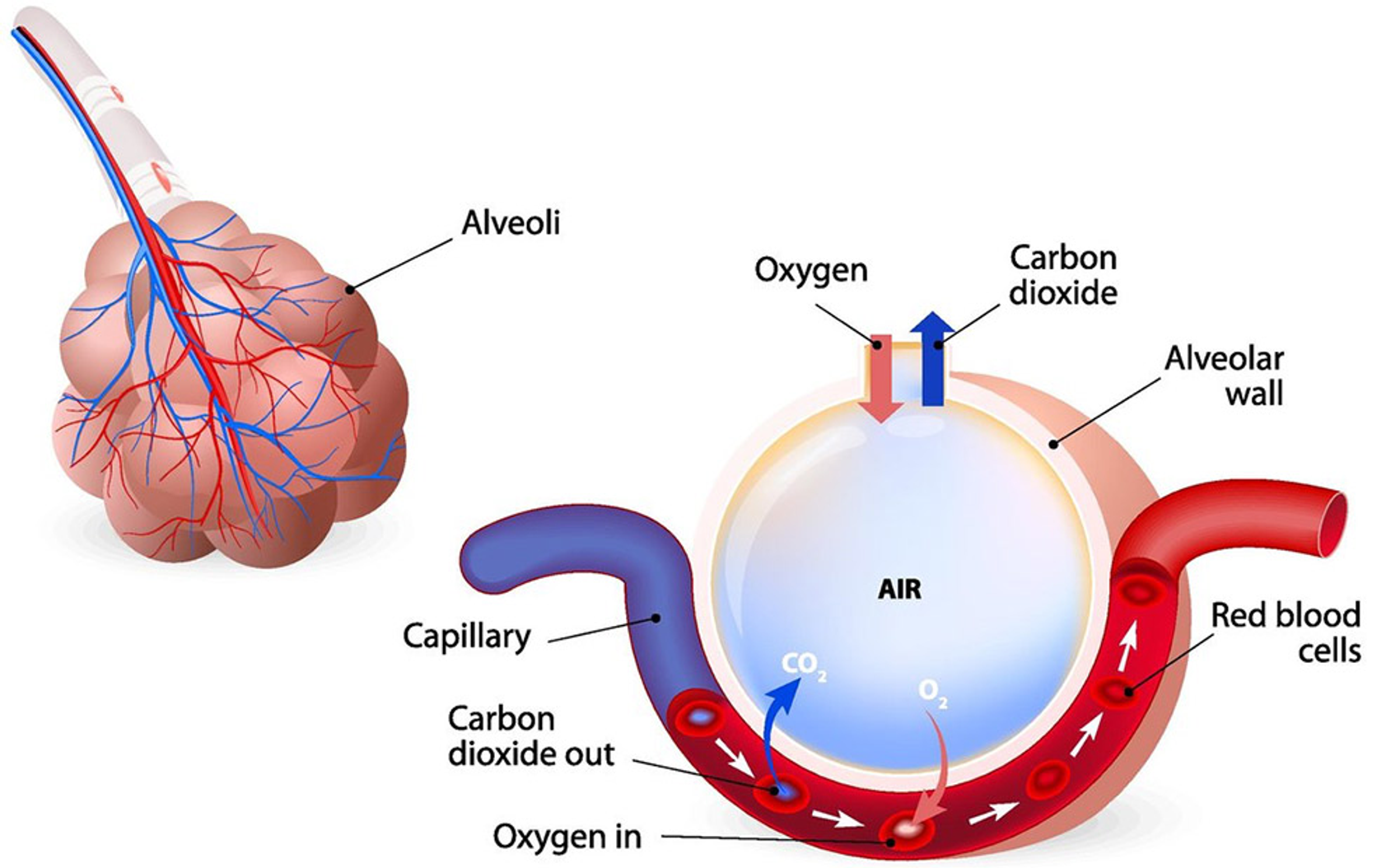
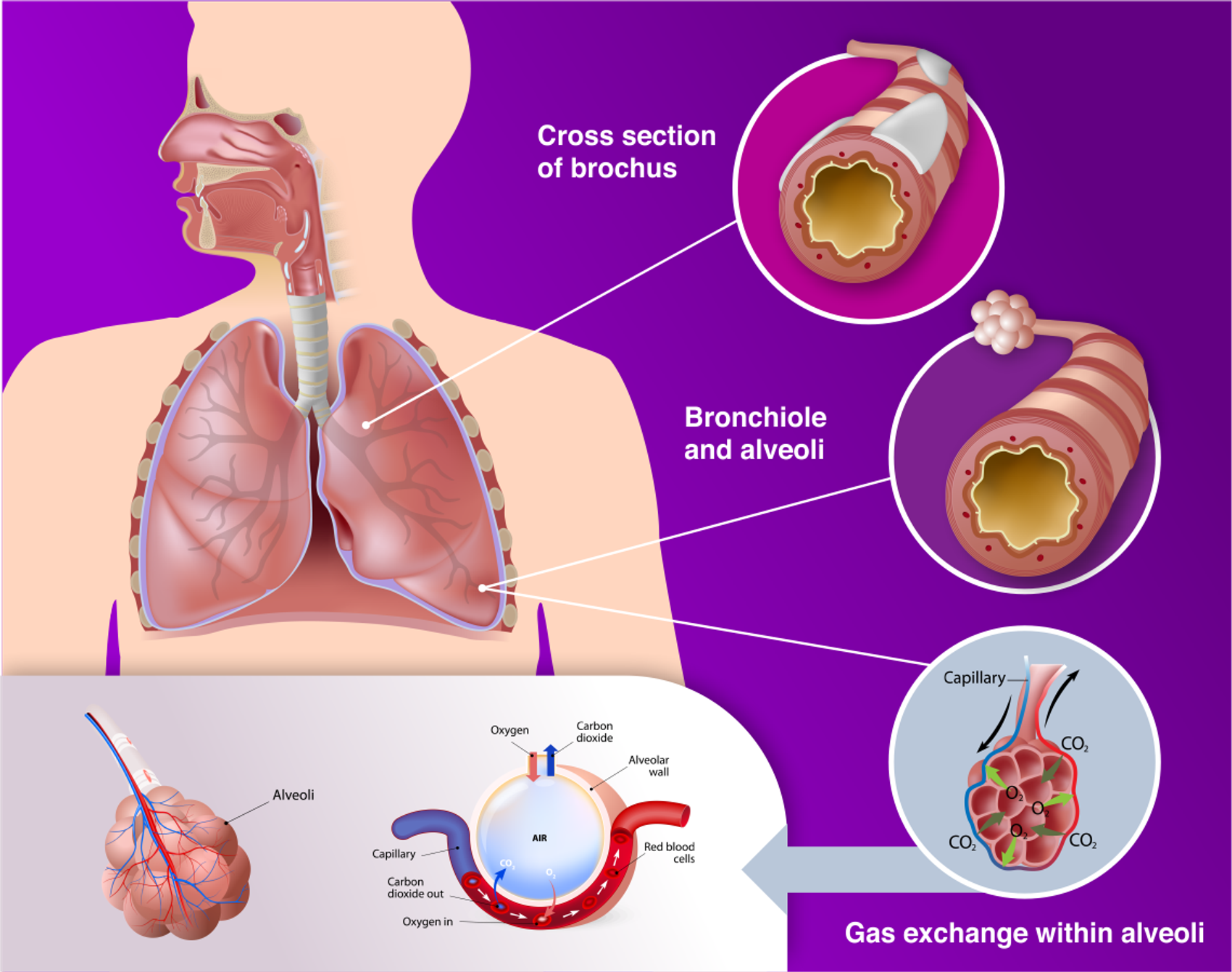
The smaller bronchioles terminate in the alveoli which are small air sacs. There are millions of grape-like clusters of alveoli in the lungs and they are where gas exchange occurs.
Alveolus gas exchange
The exchange of gases in the alveoli is referred to as external respiration. At the cellular level, the exchange of gases from the tissue cells of the body to the blood and vice versa is referred to as internal or cell respiration.

Respiratory changes
The signs that there are changes happening in the respiratory system relate to the rate of breathing, the depth of the breath, the sounds of breathing, effort made to breathe, presence of a cough and colour of the skin or mucous membranes of the person.
Rate of Breathing
Breathing is normally involuntarily and rhythmical.
One breath is considered to be a breath taken in and then let out. For an adult at rest this normally occurs between 12 – 20 times every minute.
When a person exercises, the breathing rate will go up to accommodate the oxygen needs of the body, but if the respiratory rate is getting faster when a person is at rest this may indicate a problem.
Depth of Breathing
Normally this pattern is also even with an occasional deeper breath. If the breathing is shallow and the person is only taking small breaths this may indicate pain or some other issue.
Sounds of Breathing
Breathing is normally quiet.
Noisy breathing is indicative of changes in the respiratory system.
If the airways are narrowed, as in asthma, a whistling sound is produced (wheezing).
Rales are sounds in the bronchial tubes that are caused by mucous in the bronchioles or by thickening of the walls.
Effort made to breathe
When someone is struggling to breathe, the nostrils of the nose will widen, the skin may suck in around the ribs and also at the top of the sternum.
The person will want to sit up and lean slightly forward to maximise the amount of air that they get into their lungs.
Presence of a cough
A persistent cough, either dry or moist cough are changes that may indicate some sort of infection or problem in the respiratory system.
Colour of skin and membranes
When a person is having trouble breathing, there is not enough oxygen being taken into the body, and they may have a bluish colour around their lips and in their mucous membranes or fingernail beds (this bluish discolouration is referred to as cyanosis).
Changes related to ageing and disability respiratory
Changes related to ageing or disability | Impact on the person's well-being |
|---|---|
Decrease in lung capacity with fewer alveoli leading to a decreased ability to take in oxygen | Increased shortness of breath when mobile |
Chest wall becomes more rigid | Restricts full expansion of the lungs leading to a decrease in air intake |
Changes in cardiovascular system which results in a decreased blood flow to the lungs | Reduces the ability of gas exchange in the lungs |
Cough reflex is reduced and there is a decrease in the number of cilia in the airways | |
— (Croft & Croft, 2017; Vafeas & Slatyer, 2021). | More difficult to cough up mucous which leads to increased risk of chest infections and respiratory complications |
People who have dysphagia or who need support for drinking, eating or swallowing | Risk of aspiration which can lead to respiratory infections |
People who have epilepsy or some physical disability that restricts movement | |
— (NDIS, 2022). | Increased risk of aspiration which can lead to respiratory infections |
<aside> 💡 The 2019 NDIS Quality and Safeguards Commission scoping review into the causes and contributors to the deaths of people with a disability, found that respiratory infections and disease were the major underlying causes of death for people with disability.
</aside>
Respiratory disorders and diseases
There are many diseases and disorders related to the respiratory system, and the common ones that will be considered are:
asthma
chronic obstructive pulmonary disease
covid-19
lung cancer
pneumonia.
Asthma
A common disease of the airways.
During an asthma attack, the airways narrow, reducing the flow of air in and out of the lungs. This may lead to wheezing and coughing. Pollen, cigarette smoke, colds and flu can trigger an asthma attack.
About one in ten Australians has asthma. A range of programs and services are available to support people with asthma.
Chronic obstructive pulmonary disease
It is an overarching term for chronic conditions such as emphysema and chronic bronchitis which get progressively worse and cause obstruction to the airways.
Chronic bronchitis — the mucous pools in the lower airways and consequently gas exchange and ventilation are impaired.
Ephysema — here the air trapped in the alveoli as the airways collapse during expiration and people expend a huge amount of energy to try and breathe out.
The conditions are usually caused by exposure to particles and irritants of the respiratory system, the most common of which is cigarette smoke.

COVID-19
This respiratory condition is caused by the coronavirus SARS-CoV-2 virus. The symptoms can range from mild to severe and may include:
fever
coughing
sore throat
shortness of breath
fatigue
aches and pains
headaches
sneezing
runny or stuffy nose
<aside> 💡 Covid-19 is spread mainly by respiratory droplets or by indirect contact with a contaminated surface. When providing support for a person with Covid-19 the current infection control policies of the workplace must be followed.
</aside>
Lung Cancer
Lung cancer is when abnormal cells in one or both lungs grow in an uncontrolled way.
As lung cancer is quite aggressive, a diagnosis is often not made until the cancer has metastasised (spread to other parts of the body). Most lung cancers are caused by cigarette smoking. Normally the lungs are protected by sticky mucus, cilia and nasal hairs, but the irritants from the cigarette smoke reduce the effect of these cleansing agents. The result is that the carcinogens in the cigarette smoke often lead to lung cancer.
Pneumonia
This condition occurs when there is inflammation of the bronchioles and the alveoli, as a result of an infection from a bacteria, virus or other pathogen. If there is inflammation of the lower airways without infection being present, this is called pneumonitis. Pneumonitis usually occurs when the person is hypersensitive to chemicals or dust.
When the pathogen invades the alveoli, there is inflammation and an excessive production of fluid that fills the alveoli, and gas exchange is reduced leading to hypoxia (a lack of oxygen). The symptoms of pneumonia include difficulty breathing, weakness, fever, chills, chest pain and coughing.
Maintain a healthy respiratory system
Stop smoking
The single most important change that can be made to improve respiratory health is to stop smoking if people are smokers.
Cigarette smoke contains tar and thousands of chemicals that damage lungs and cause inflammation. There are many tools and resources to help with quitting which include medication and hypnotherapy and nicotine replacement therapy.
Exercise
Regular exercise is another method of improving respiratory health and lung function. When you exercise, the increased need for oxygen in the muscles causes the brain to stimulate the respiratory system to increase ventilation or breathing.
Breathing exercises such as yoga and pilates are good for the respiratory system.
Eat a healthy diet
Eating a healthy balanced diet is also important to maintaining healthy lungs. The body requires vitamins and other nutrients to effectively build and maintain all tissues, including those that make up the respiratory system.
Practice good hygiene
Viral or bacterial infection and illnesses, such as influenza and pneumonia, can cause short- and longer-term damage to the respiratory system, seriously decreasing the lung's capacity and ability to absorb oxygen. Maintaining proper hygiene is the best way to minimise the chances of contracting microbial illnesses. One of the main ways this is achieved is by regular handwashing.