Anatomy & Physiology
Circulatory System: Red Blood Cells
Red Blood Cells - carry oxygen and nutrients throughout the body; structure allows it to grab onto iron and oxygen for transport.
Functions of the spleen:
filters the blood
removes old blood cells
recycles iron
makes antibodies
Arteries that supply blood to the spleen:
aorta
aeliac
spenic
Phlebotomist - person trained to draw blood from a patient
Cooley’s anemia - inherited disorder that interferes with the blood’s ability to carry oxygen; typical for the cells to appear faded (hypochromia)
Blood is a type of CONNECTIVE TISSUE
cells (rbc, wbc, platelets) = 45%
plasma (water, proteins, amino acids) = 55%
Hematocrit - the percentage of blood & plasma
blood cells = 45%
fluid (plasma) = 55%
Three Types of Blood Cells
erythrocytes = red blood cells
leukocytes = white blood cells
thrombocytes = platelets
Red Blood Cells - made from a protein called hemoglobin (Hb) which contains iron used to transport oxygen.
Shape = biconcave discs
5 million per cubic millimeter (drop)
contain no nuclei
Person with a low amount of iron in their diet has iron-deficient anemia.
Hematopoiesis - formation of blood cells
occurs in the bone marrow
an error in the genetic code can cause the protein to be abnormally shaped
Erythropoietin (EPO) - a hormone that increases the production of red blood cells
Oxyhemoglobin - plenty of oxygen; “bright red”
Deoxyhemoglobin - low in oxygen; “dark red”
Blood delivers oxygen to tissues and then returns to the heart through veins. The heart sends blood to lungs where it is oxygenated and sent to tissue via arteries. Veins and arteries meet at tiny vessels called capillaries which deliver to tissues.
Sickle Cell Disease
Caused by:
genetic mutation in the DNA code
incorrect formation of the hemoglobin protein
cells are abnormally shaped
cannot carry oxygen efficiently
Symptoms:
splenomegaly
fatigue/weakness
pain crisis
strokes
shortness of breath
The Nervous System MS (A) - Nerves
The nervous system includes
the brain
spinal cord
the nerves
The nervous system - coordinate the body’s systems by receiving and sending information; maintaining homeostasis
Sensory - receives information
Integrative - determines where information is sent
Motor - responds to signals
Central Nervous System (CNS)
brain
spinal cord
Peripheral Nervous System (PNS)
Nerves throughout the body
31 pairs of spinal nerves
12 pairs of cranial nerves
Somatic Nervous System
skeletal
voluntary
Autonomic Nervous System
smooth muscles
glands
involuntary
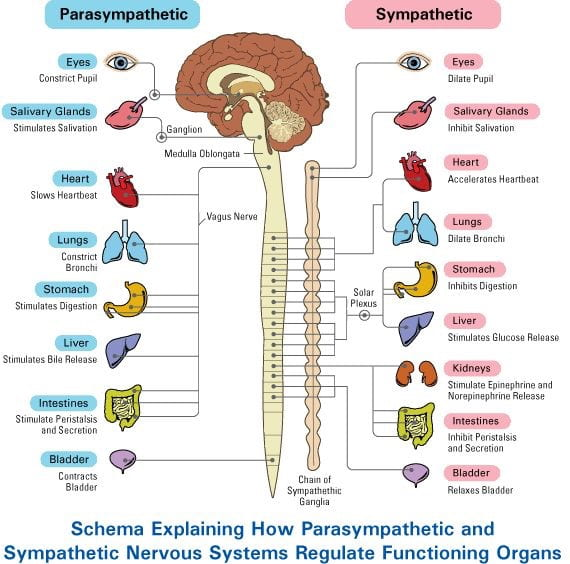
Parasympathetic (rest and digest)
Sympathetic (fight or flight)
Neurons - masses of nerve cells that transmit information (functional unit of the nervous system)
Cell Body - contains the nucleus and other cell organelles
Dendrites - shorter, more numerous, receive information
Axons - single long fibers, conducts information away from the cell
Chromatophilic substance (rough ER) - transport system
Myelin - insulation surrounding axons
Nodes of Ranvier - gaps in the insulation
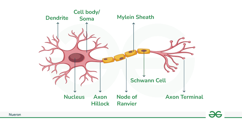
Neuroglial Cells - support for neurons
Microglial Cells - immune function; digest debris, kills bacteria
Oligodendrocytes - make myelin sheath that provides insulation around the axons
Astrocytes - connect blood vessels to neurons
Ependymal Cells - forms membranes around tissue
Schwann cells - form the insulating myelin sheath around the neurons in PNS (same function as oligodendrocytes)
Myelin Sheaths - insulate axons
Schwann cells supply the myelin for peripheral neurons while oligodendrocytes myelinate the axons of the central nervous system
Gaps in the sheath are called NODES OF RANVIER
As the neurons lose their myelin, the nerves are unable to send or receive signals.
Myelinated (white matter) - myelinated axons
Unmyelinated (grey matter) - unmyelinated
Lesions are evidence of nerve cell damage in your brain or spinal cord
lesions on the spinal cord may cause motor problems
lesions on the back of the brain may cause balance problems
Facts about neurons
Longevity - can live and function for a lifetime
Do not divide - fetal neurons lose their ability to undergo mitosisl neural stem cells are an exception
High metabolic rate - require abundant oxygen and glucose
Nerve impulses - wake electric current, like a wave
neuron membrane maintains resting potential
threshold stimulus is received
sodium channels open
sodium ions diffuse inward, depolarizing the membrane
potassium channels open
potassium ions diffuse outward, repolarizing the membrane
the resulting action potential causes a local bioelectric current that stimulates the membrane
Wave of action potentials travel the length of the axon as a nerve impulse
Depolarization - loss of the difference in charge between the inside and outside of the plasma membrane of a muscle or nerve cell due to a change in permeability and migration of sodium ions to the interior
Nerve impulse - speed is proportional to the size of the axon
greater diameter = faster impulse
Myelinated axons conduct impulses faster than unmyelinated one
Communication between neurons
Synapse - function between two communicating neurons
Nerve pathway - nerve impulse travels from neuron to neuron
Dendrite → cell body → along axon → synapse (gap) → dendrite
To complete the signals, a neurotransmitter is released at the gap to signal the next neuron; receptors on the dendrite receive the chemical message
Types of neurotransmitters
Excitatory - increase membrane permeability, increases chance for threshold to be achieved
Inhibitory - decrease membrane permeability, decrease chance for threshold to be achieved
Examples of neurotransmitters
Acetylcholine - stimulates muscle contraction
Dopamine - mood, happiness
Serotonin - sleepiness and mood
Endorphins - pain reduction, mood
Agonist - molecule that has the same effect on the postsynaptic neuron as the neurotransmitter itself does
Antagonist - molecule that blocks the effect that neurotransmitter normally has on the postsynaptic neuron
Antidepressants
example: selective serotonin reuptake inhibitors
Cocaine - it attaches to the dopamine transporter and blocks the normal recycling process, resulting in a buildup of dopamine in the synapse, which contributes to the pleasurable effects of cocaine
Ecstasy - takes the upkeep transporters and reveres their roles; this causes a massive flood of serotonin from the brain cells into the synapse
Heroin - activates opiate receptors; blocks release of GABA, and thus more dopamine is released
Amphetamines - mimics dopamine; binding to receptors; dopamine does not re-enter the cell, depleting the cell’s supply
Disorders Related to Neuron
Amyotrophic Lateral Sclerosis (ALS) - progressive degeneration of nerve cells in the spinal cord and brain
Neurodegenerative - symptoms will get worse over time
Epilepsy - caused by excessive electrical activity within networks of neurons in the brain; a fine balance between excitation and inhibition must be maintained in order for the brain to function normally; if there is too much glutamate, neurons can become hyperexcitable and a seizure may result; too little GABA (inhibitory) can also result in a seizure.
Myasthenia Gravis - autoimmune disorder in which antibodies destroy neuromuscular connections
Nervous System (B) - Brain
The meninges - membranes located between bone and soft tissues
Meningitis - can be bacterial of viral
fever
stiff neck
photosensitive
vomiting
rash
There is a vaccine!
Cerebrospinal fluid (CSF) - a clear liquid that bathes the brain and spinal cord; cushions the bran and serves as a shock absorber
Subdural hematoma - associated with traumatic brain injury; bleeding within the brain; puts pressure on the brain and can be life threatening
Spinal cord passes down the vertebral canal, has 31 segments (each with a pair of spinal nerves)
Locations:
C1 - C8
T1 - T12
L1 - L5
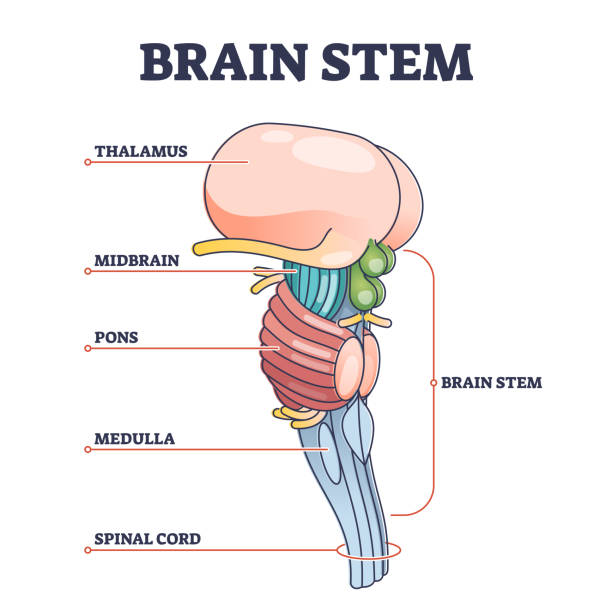
3 major parts of The Brain
cerebrum
brain stem
cerebellum
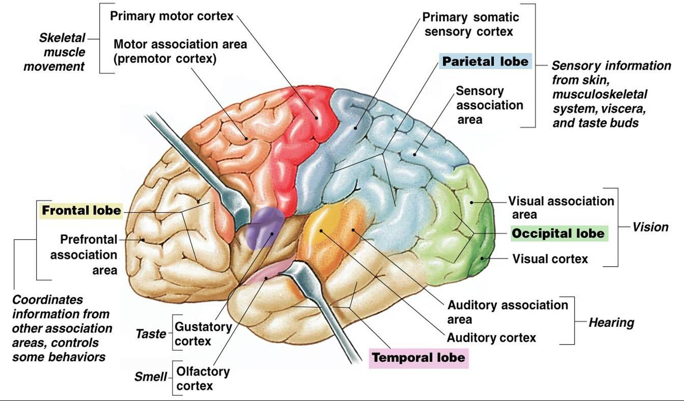
Cerebrum - wrinkly large part of the brain (cerebral cortex); higher mental function, solving problems
Cerebellum - balance and coordination; white matter within the cerebellum give it a tree-like appearance, this is called the arbor vitae (“tree of life”)
Brain stem - regulates visceral functions (autonomic system)
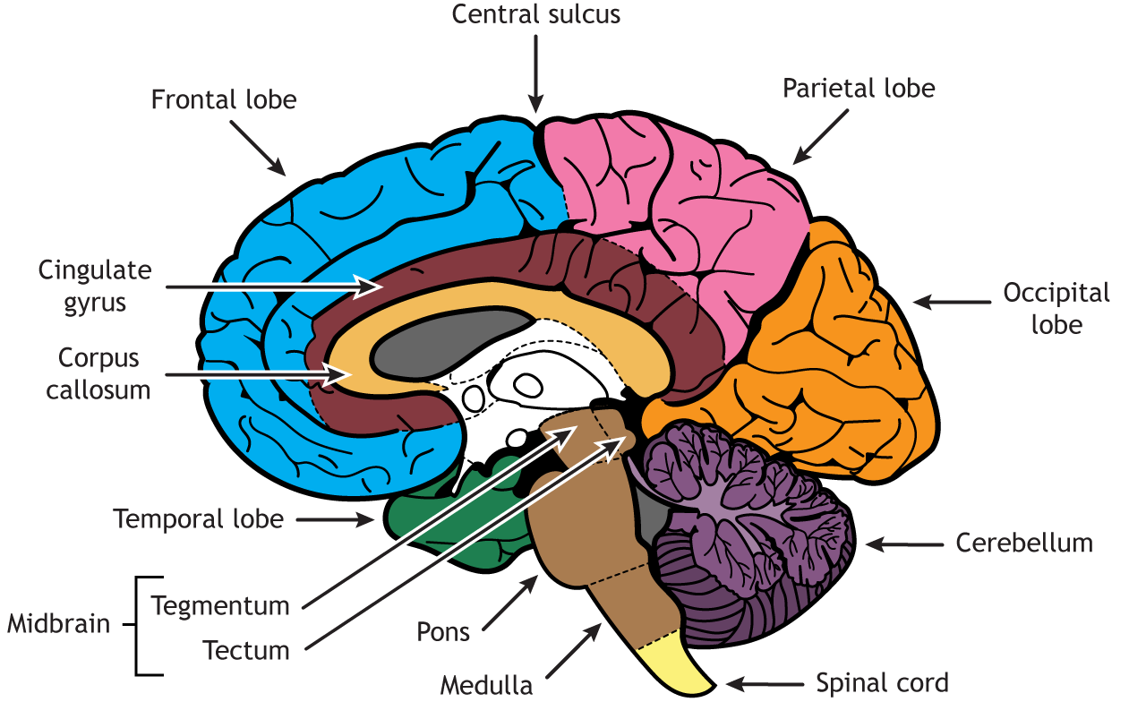
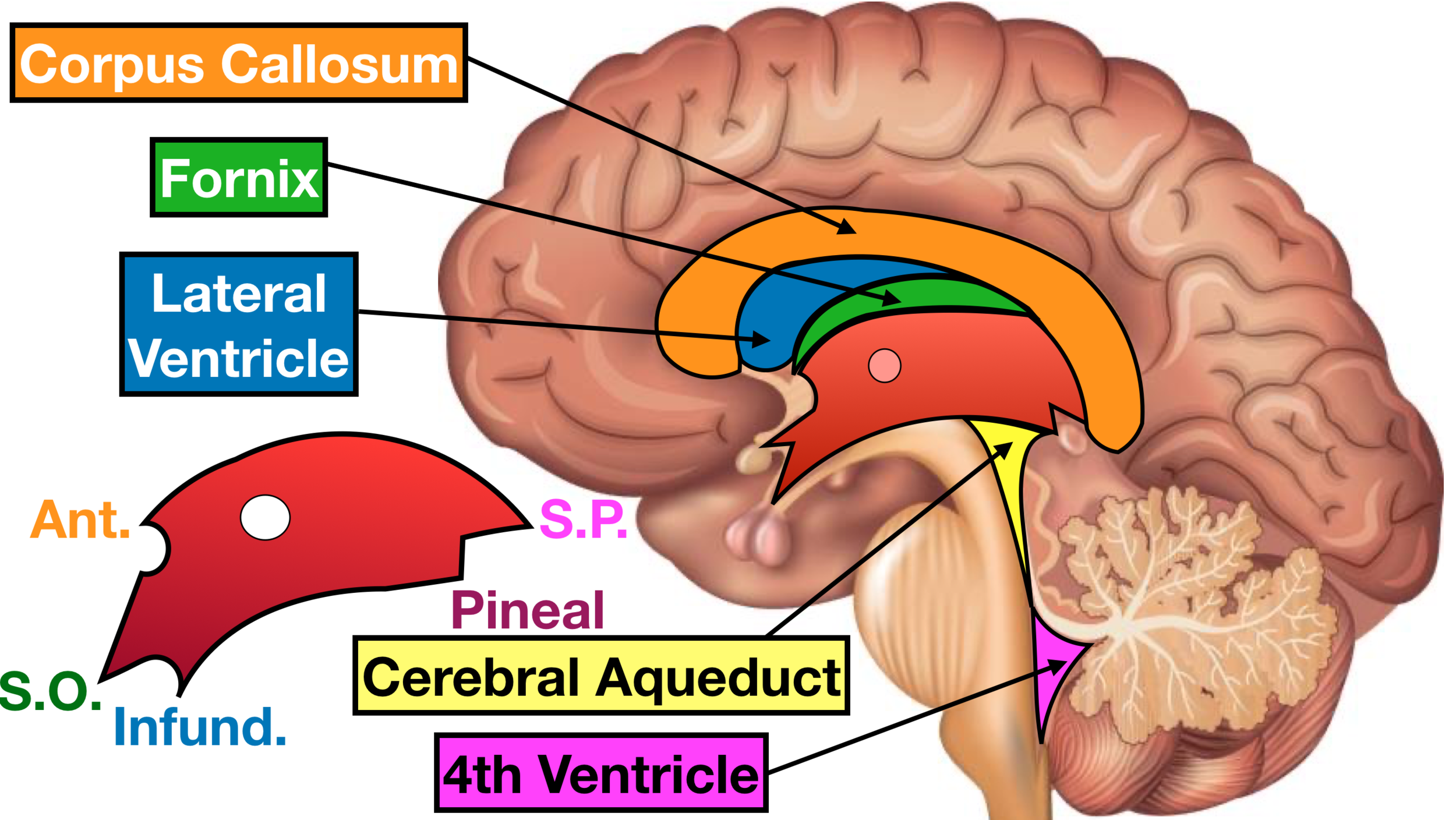
Cerebral hemispheres - left and right side separated by the corpus callosum - connects the two hemispeheres
Brain convolutions - the wrinkles and grooves of the cerebrum
Fissues - deep groove
Sulcus - shallow grooce
Gyrus - bump
Frontal lobe - executive functions
Parietal lobe - perception, sense-making, math
Occipital lobe - vision
Temporal lobe - memory, language
Longitudinal fissue - separate right and left sides
Transverse fissue - separates cerebrum and cerebellum
Ventricles of the brain - fluid filled cavities, contain CSF
Association areas - interpreting and analyzing information
Diencephalon has 2 parts…
Hypothalamus - hormones, heart rate, blood pressure, body temperature, hunger, attaches to pituitary
Thalamus - relay station
Optic tract / chiasma - optic nerves cross over each other
THE LEFT SIDE OF THE BRAIN CONTROLS THE RIGHT SIDE OF THE BODY
Midbrain - visual reflexes, eye movements
Pons - relay sensory information
Medulla - heart respiration, blood pressure
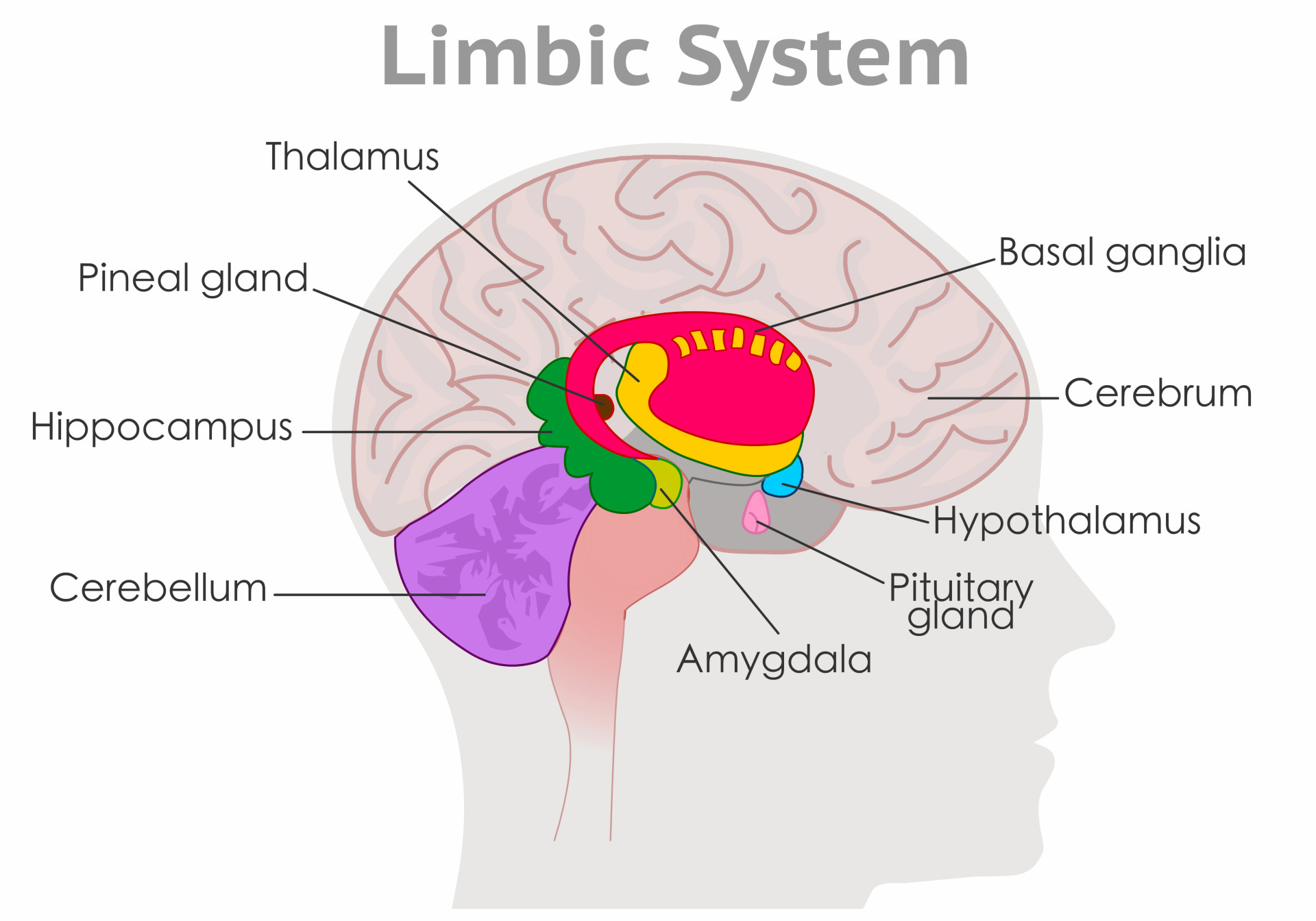
Pituitary gland - the “master gland” of the endocrine system; controls hormones
Hippocampus - (“sea horse”) storage and retrieval of memories
Amygdala - storage of memories associated with emotion
also associated with fear response and aggression
The limbic system
hypothalamus
hippocampus
amygdala
has a role in emotions
Peripheral Nervous System and Cranial Nerves
Peripheral nervous system - consists of the nerves that branch out from the CNS and connect it to other body parts, also includes the cranial nerves
Somatic nervous system - (conscious activities) skin, skeletal system
Autonomic nervous system - (unconscious activities) heart, viscera, glands
Peripheral nervous system - the spinal nerves comes out of the spine, and the cranial nerves come out of the brain directly
there are 12 pairs of cranial nerves
they are numbered with roman numerals
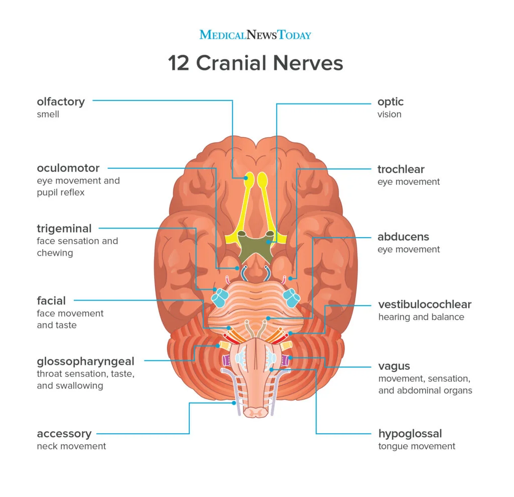
I. Olfactory - sense of SMELL; outside of the CNS, they are called olfactory nerves, and inside of the CNS, they are called the olfactory tract
II. Optic nerve - transmits information from the eye’s retina; VISION
III. Oculomotor nerve - controls most of the muscles of the eye that MOVES THE EYEBALL
IV. Trochlear nerve - innervates an extrinsic EYE MUSCLE, EYELID
V. Trigeminal nerve - the main SENSORY NERVE OF THE FACE; it has a large branch that passes through the foramen of the skull; it has 3
VI. Abducens - CONTROLS ONE OF THE EYE MUSCLES (lateral rectus)
VII. Facial nerve - innervates the muscle of FACIAL EXPRESSION
a person who cannot blink or smile may have damage to this nerve
Bell’s palsy is damage of the facial nerve causing paralysis on one side
VIII. Vestibulocochlear - hearing and balance (also called Auditory nerve)
IX. Glossopharyngeal - pharynx, tongue; SWALLOWING, speech, saliva
X. Vagus nerve - (vagrant = “wanders”) the only cranial nerve that travels into the abdomen
this is the most important cranial nerve because it innervates all of the organs in the thoracic and abdominal
XI. Accessory nerve - supplies the SHOULDER muscles
enters the skull through the foramen magnum
XII. Hypoglossal nerve - supplies the TONGUE
damage causes impairment of speech
Nerve | Function |
|---|---|
1. olfactory | sense of smell |
2. optic | sight |
3. occulomotor | moves the eyeballs |
4. trochlear | moves the eyelids |
5. trigeminal | face, jaw, chewing |
6. abducens | eyes |
7. facial | facial expressions |
8. vestibulocochlear (auditory) | sense of equilibrium, hearing |
9. glossopharyngeal | pharynx, tongue |
10. vagus | major organs, viscera |
11. accessory | shoulders |
12. hypoglossal | tongue |
On Old Olympus Towering Top A Fin And German Viewed A Hop
Spinal Nerves - 31 total
8 pairs of cervical nerves (C1 - C8)
12 pairs of thoracic nerves (T1 - T12)
5 pairs of lumbar nerves (L1 - L5)
5 pairs of sacral nerves (S1 -S5)
1 pair of coccygeal nerves (Co)
*Spinal cords ends at the level between the 1st and 2nd lumbar vertebrae
*The lumbar, sacral, coccygeal nerves descend from the end of the cord - CAUDA EQUINA (horse’s tail)
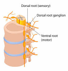
Each nerve emerges from the spinal cord at points called ROOTS
Dorsal root ganglion
Ventral root ganglion
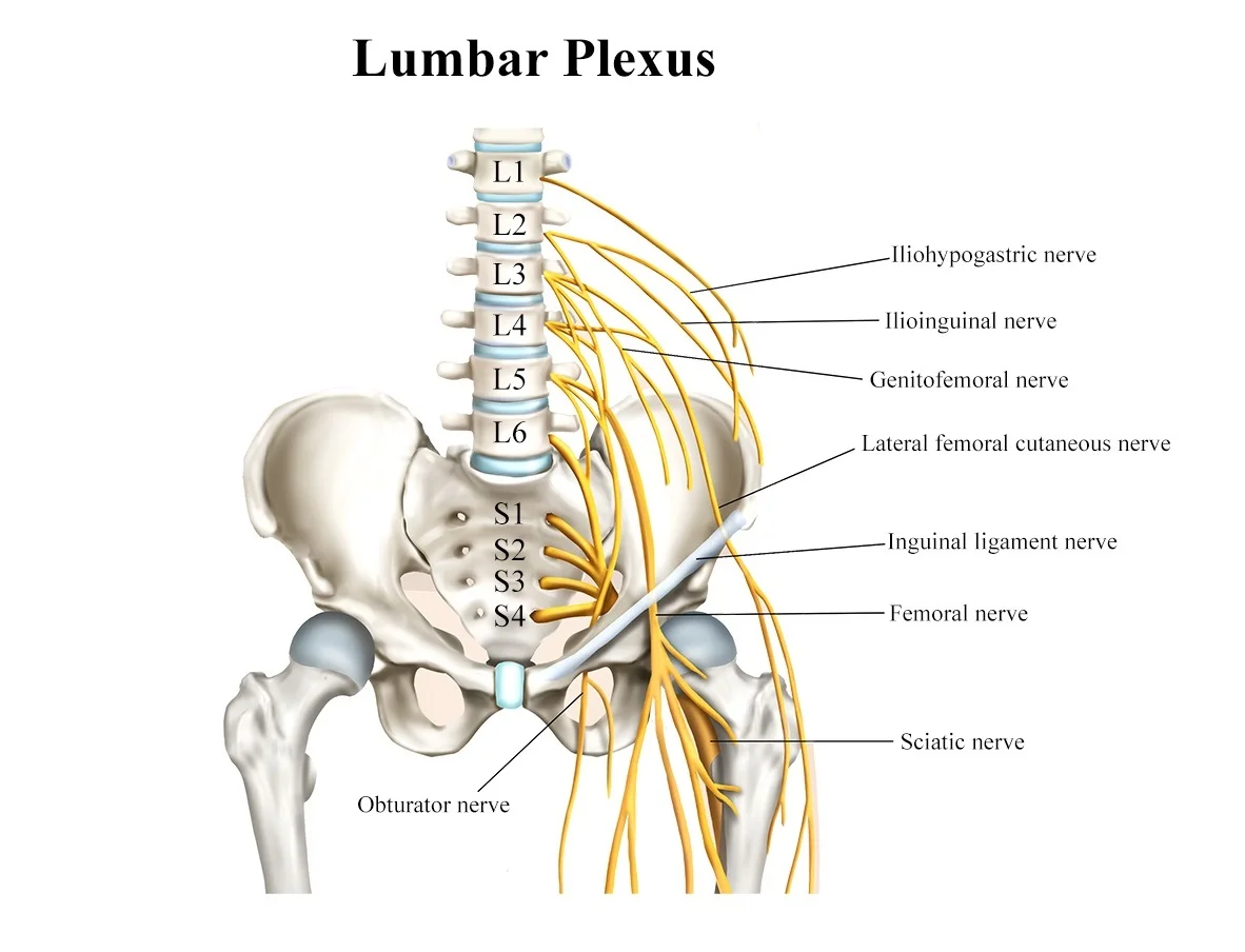
Main portions of the spinal nerves combine to form complex networks called PLEXUSES
Cervical Plexus - neck
Brachial Plexus - shoulders, arms, hands
Lumbosacral Plexus
Obturator nerve - obturator
Sciatic - lower spine to buttocks (largest nerve)
Femoral - femur, knee
Autonomic nervous system
Sympathetic - “fight or flight;” energy, high stress, emergency
Parasympathetic - “resting;” resting, normal
Divisions act antagonistically - one is excitatory, other inhibits
The Senses
Synesthesia is the perceptual phenomenon in which stimulation of one sensory or cognitive pathway leads to automatic, involuntary experiences in a second sensory or cognitive pathway
General senses - receptors found throughout the body, including joints and organs
Special senses - specialized receptors found in the head (eyes, ears, mouth)
Sensations & Receptors
Sensation - feeling that occurs when a brain interprets and sensory nerve impulse
Projection - the brain causes a feeling to stem from a source
Sensory adaption - sensory receptors stop sending signals when they are repeatedly stimulated
Sensory deprivation - a technique initially used by neuropsychiatrists designed to deliberately reduce or completely remove stimuli from one or all of the senses
Receptor cells in the PNS are activated by stimuli in the environment
Receptor cells can be classified into types on the basis of three different criteria
cell type
position
function
Structure receptor types
Free nerve endings - dendrites embedded in tissue as receivers; respond to pain and temperature
Encapsulated - embedded in connective tissue to increase sensitivity (pressure and touch)
Specialized - receptors in the retina of the eye
Location-based receptor types
Exteroreceptor - located near external environment (skin)
Interoreceptor - interprets stimuli from internal organs
Proprioreceptor - located near moving body parts, interprets position
Functional receptor types
Chemoreceptor - chemicals, mostly found in nose/mouth
Osmoreceptor - respond to solutes in body fluids
Thermoreceptor - temperature
Mechanoreceptor - pressure, vibration, body position
Nocireceptor - pain (chemicals released when tissue is damaged
Sense of pain
Visceral pain - occurs in visceral tissues such as heart, lungs, intestine
Referred pain - feels as though it is coming from a different part (heart pain may be felt as pain in arm or shoulder)
Acute pain - originates from skin, usually stops when stimulus stops (needle prick)
Chronic pain - dull aching sensation
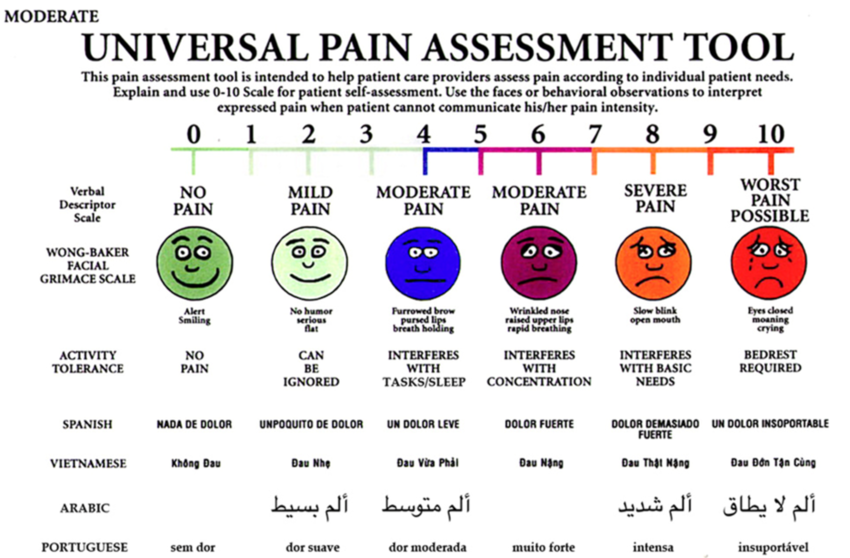
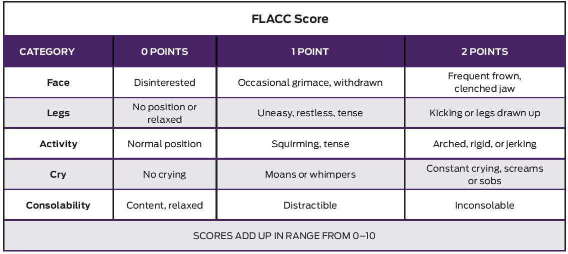
Measuring pain in babies and young children - face, legs, activity, cry, consolability scale (FLACC scale)
An overview of how opioids work to block pain and why they become less effective the longer you use them
nerve sends signal
opioids slow/block signal
limbic system (emotions) and reward pathway are stimulated
Special sense
olfactory - smell
gustatory - taste
hearing & equilibrium
sight
Sense of smell (olfactory)
odor → receptor cell → olfactory bulb → olfactory tract → limbic system
Sense of taste (gustatory)
Taste buds are found on papillae; some papillae do not have taste buds
Taste senstaions
sweet
sour
bitter
salty
savory (umami) - a savory taste, is one of the five basic tastes, together with sweet, sour, bitter, and salty; a loanword from the Japanese umami can be translated “pleasant savory taste"
Sense of hearing
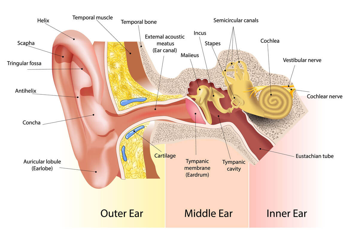
External ear
Auricle (pinna) - outer ear
Auditory canal (external auditory meatus) - opening to the eardrum
A group of muscles called the auriculares are responsible for this movement
Ear wigging is considered a vestigial trait - a trait that no longer function but is part of our evolutionary past
Middle ear (tympanic cavity)
Eardrum (tympanum)
Auditory ossicles - malleus, incus, stapes; transmit vibrations and amplify the signal
Auditory tube (eustachian tube) - connects the middle ear to the throat; helps maintain air pressue
Inner ear
Labyrinth - communicating chambers and tubes
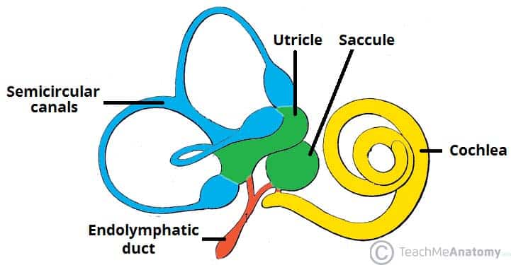
Semicircular canals - sense of equilibrium
Cochlea - sense or hearing
Organ of Corti - contains hearing receptors, hair cells detect vibrations
Why do we lose our hearing?
Inside the cochlea are special neurons called hair cells; loud noises damage these fibers
Steps in hearing
Sound waves enter external auditory canal
Eardrum vibrates
Auditory ossicles (malleus, incus, stapes) amplify vibrations
Stapes hits oval window and transmits vibrations to cochlea
Organs of corti contain receptor cells (hair cells) that deform from vibrations
Impulses sent to the vestibulocochlear nerve
Auditory cortex of the temporal lobe interprets sensory impulses
Round window dissipates vibrations within the cochlea
Cochlear implants - receives sound from the outside environment, processes it, and sends small electric currents near the auditory nerve; the brain learns to recognize this signal and the person experiences this as “hearing”
Sense of equilibrium
Static equilibrium - maintain stability and posture
Dynamic equilibrium - balance during sudden movement
Cerebellum - interprets impulses from the semicircular canals and maintains overall balance
Otolaryngology - physicians trained in the medical and surgical management and treatment of patients with diseases and disorders of the ear, nose, throat (ENT), and related structures of the head and neck; they are commonly referred to as ENT physicians
Disorders of sensory systems
Synethesia
Anosmia
Tinnitus
Anhidrosis (inability to sweat)
Congenital analgesia (CIP)
Mutations in genes prevents nerve impulsves from pain receptors (nociceptors) from sending signals to the brain.
Hereditary (Congenital) Deafness - occurs in 1 of every 1000 to 2000 newborns
syndromic → pendred syndrome → usher syndrome
Syndrome - a disease that has more than one feature or symptom; a person with usher syndrome also has vision problems
Deafness can also be caused by prenatal infections from “ToRCH” organisms
toxoplasmosis
rubella
cytomegalovirus (CMV)
herpes
Postnatal infections can also cause deafness
meningitis
streptococcus
listeria
influenza
Presbycusis - age related hearing loss
dimished hearing sensitivity
poor speech comprehension in noisy environments
slowed central processing of acoustic information
high frequencies are more difficult to hear
Careers related to hearing
audiologist
speech-language pathologist
sign language interpreter
teacher: deaf & hearing imparied
The Eyes
Vision tests
response to light
pupil response
ability to follow a target
ability to navigate obstacles
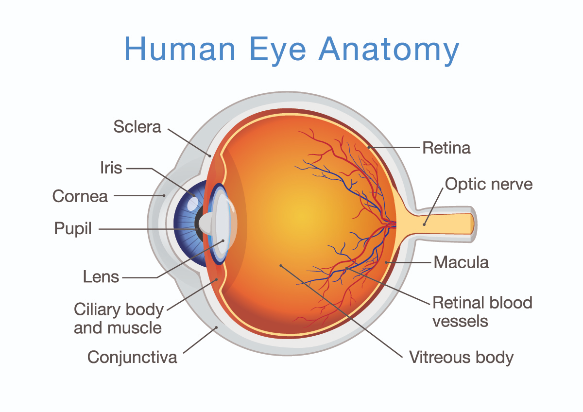
Structure of the eye & accessory organs
Eyelid - covers and protects the eye, thin skin
*Skill will not protect you from intense radiation, that’s why we use special goggles in tanning bed
Eyelashes - fine hairs that function to protect the eyes from dust and debris
Conjunctiva - covering around the eye and under the eyelids
Pink eye - also known as CONJUNCTIVITIS (from bacteria, very contagious)
Extrinsic eye muscle - move the eye
Lacrimal glands - produce tears, which drain into the nasal cavity via the LACRIMAL DUCT
Tears - moisten and lubricate the eye surface, and it has enzymes that kill bacteria
Outer tunic of the eye
Cornea - transparent dome that focuses light
Sclera - continuation of cornea, going toward the back of the eye (white of the eye)
Optic nerve - exits at optic disk and transmits information from the eye to occipital lobe of the brain
Middle tunic of the eye
Choroid - contains blood vessels
Ciliary body - holds the lens in place
Lens - focusing
Iris - colored portion of the eye
Pupil - opening for light to enter; dilate or constrict to adjust light entering eye
Aqueous humor - liquid surrounding the lens
Eye color
Melanin - a brownish pigment that adds color to your hair, eyes, and skin
Heterochromia - one eye is different color than the other
Inner tunic
Retina - visual receptor cells; has PHOTORECEPTORS, which are sensors for light
Fovea centralis - region of the sharpest vision, also called the macula
Optic disc - where nerve fibers leave the eye, creating the blind spot
Vitreous humor - supports internal parts, fluid within the eye
The region where the optic nerve and blood vessels goes in and out of the eye has no photoreceptors = BLIND SPOT
Rods - monochromatic (b&w)
Cones - color vision
ROYGBIV!
How does the eye work?
Accommodation - lens change shape to facilitate focusing
Images are then projected to the back of the eye (focal points), the lens reverse the image (objects are seen upside down)
Zonules of Zinn
connect ciliary body to lens
can change the shape of the lens
also called suspensory ligaments
Information from retina passes through the optic nerve, crosses at the chiasma, and is sent to the occipital lobe of the brain; the brain is responsible for interpreting those images
Problems with the eyes
Cataracts - clouding of the lens leads to a clinical condition (cataracts)
Treatment is to remove the lens and replace it with an artificial one called an intraocular lens
Problems with the iris and pupil - function is to constrict or dilate the pupil (opening) to allow light in; therefore, it regulates the amount of light passing to the visual receptors of the eye
Aniridia - a condition where a person is born without an iris
Onchocerciasis - (river blindness) a disease caused by infection by a parasitic worm; symptoms include severe itching, bumps under the skin, and blindness
Color blindness - a genetic trait that affects boys more than girls; the location of the gene is on the X chromosome
Floaters - occur when the vitreous substance clumps and casts shadows on the retina; floaters don’t actually move, the eye just tries to track them
Retinal detachment - occurs when the retina is pulled away from its normal position; blindness can occur if it is not treated right away; symptoms include flashing lights, new floaters, a shadow in the periphery of your field of vision, a gray curtain moving across your field of vision
Glaucoma - pressure from vitreous humor damages the optic nerve and can lead to blindness; can be managed with medication
Hyperopia - farsightedness; you can see distant objects fine, but close objects appear blurry
Presbyopia - the loss of ability to see close objects or small print, age-related
Myopia - nearsightedness; you can see near objects, fine, but distance objects appear blurry
Astigmatism - where the cornea has an irregular shape; part of field of view is out of focus
Most visions problems are treated with glasses or contact lenses; both change the focal point of the light entering the eye; surgery is also a option + gene therapy
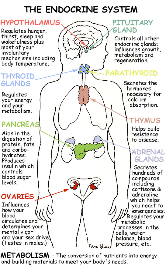
Endocrine System
made up of glands that produce and secrete hormones, chemical messengers
regulation of growth, metabolism, sexual development
responses to stress and injury
maintains homeostasis
BIG IDEA: HORMONES ARE CHEMICAL MESSENGERS THAT ACT ON TARGET CELLS
Major structures of the endocrine system
pineal hypothalamus pituitary | brain |
thyroid parathyroid | throat |
thymus | above heart |
adrenal | above kidneys |
pancreas | under stomach |
ovary / testes | lower abdomen / groin |
Endocrine - secretions inside the body
Exocrine - secretions outside the body (sweat)
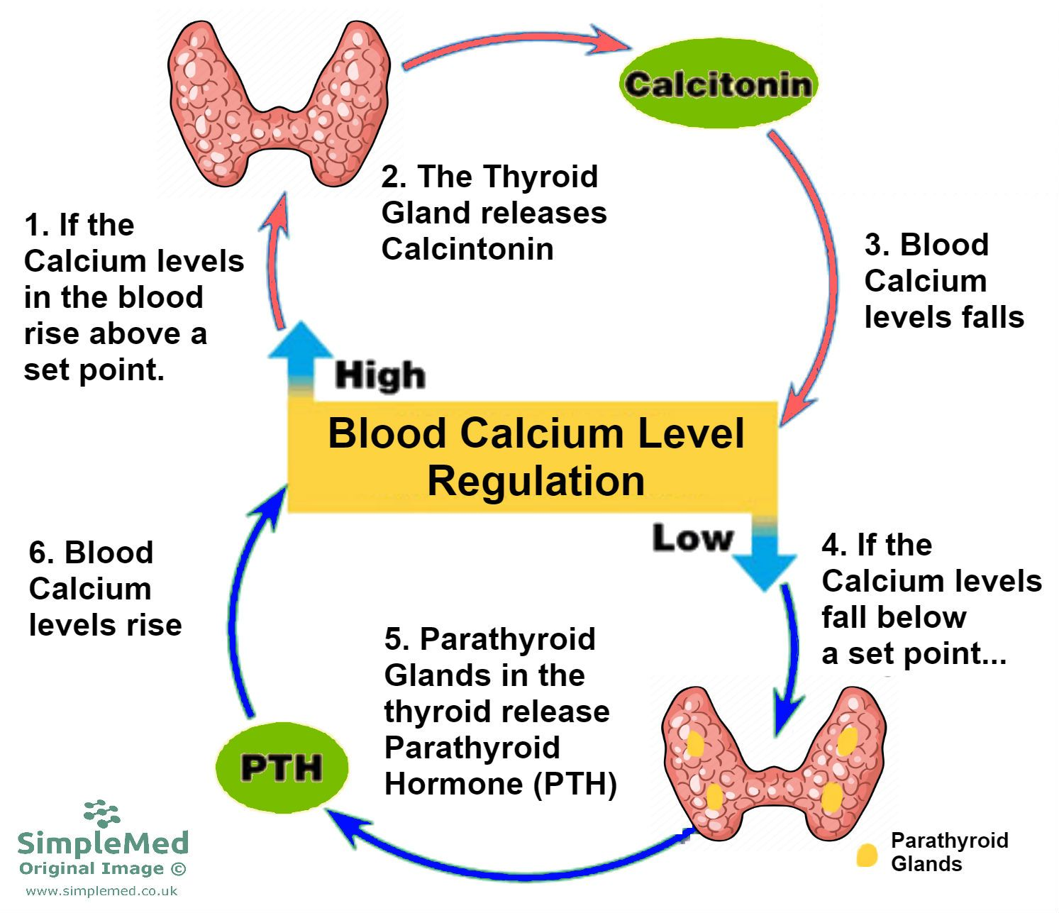
Control of hormones
Negative feedback system - when the levels go above or below a SET POINT, the endocrine system secretes hormones to lower or raise the level; think of it like a thermostat
Example of negative feedback:
Calcium regulation by the thyroid/parathyroid
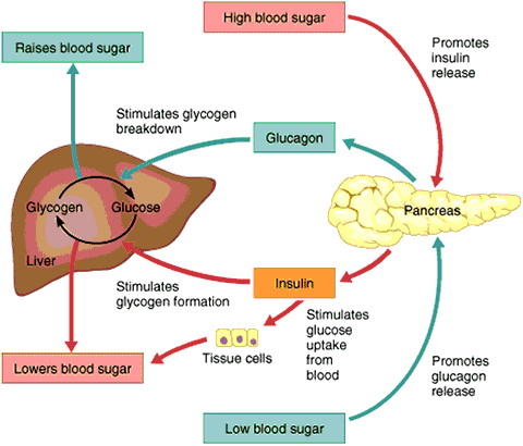
Glucose levels rise - insulin is produced to cause sugar to be taken up by the cells (and out of the blood)
Positive feedback system - ex. contractions during labor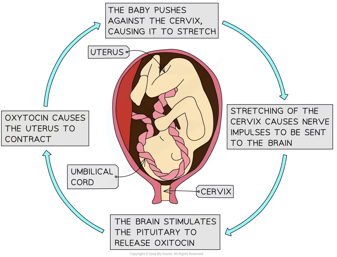
Pituitary gland - located below the brain
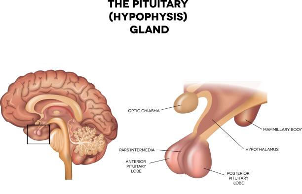
Hormone control
The pituitary is often called the “master gland” because it controls all of the other glands
Its actions are controlled by the hypothalamus in the brain
Anterior pituitary hormones
Prolactin or PRL- PRL stimulates milk production from a woman’s breasts after childbirth and can affect sex hormone levels from the ovaries in women and the testes in men
Growth hormone or GH - GH stimulates growth in childhood and is important for maintaining a healthy body composition; in adults, it is also important for maintaining muscle mass and bone mass; can affect fat distribution in the body
Myostatin - inhibits muscle growth
Myostatin inhibitor drugs are being developed with the intent of treating muscle-wasting diseases like muscular dystrophy in humans
Problems with the pituitary gland can result in dwarfism or a person can grow too much
Adrenocorticotropin or ACTH - ACTH stimulates production of cortisol by the adrenal glands
Cortisol, a so-called “stress hormone, is vital to surivial; it helps maintain blood pressure and glucose levels
Thyroid-stimulating hormone or TSH - TSH stimulates the thryoid gland to make thyroid hormones, which, in turn, control (regulate) the body’s metabolism, energy, growth and development, and nervous system activity
Luteinizing hormone or LH - regulates testosterone in men and estrogen in women
Follicle-stimulating hormone or FSH - FSH promotes sperm production in men and stimulates the ovaries to release eggs in women; LH and FSH work together to allow normal function of the ovaries or testes
Posterior pituitary hormone
made up of glands that produce and secrete hormones, chemical messengers
regulation of growth, metabolism, sexual development
responses to stress and injury
maintains homeostasis
BIG IDEA: HORMONES ARE CHEMICAL MESSENGERS THAT ACT ON TARGET CELLS
Major structures of the endocrine system
pineal hypothalamus pituitary | brain |
thyroid parathyroid | throat |
thymus | above heart |
adrenal | above kidneys |
pancreas | under stomach |
ovary / testes | lower abdomen / groin |
Endocrine - secretions inside the body
Exocrine - secretions outside the body (sweat)
Control of hormones
Negative feedback system - when the levels go above or below a SET POINT, the endocrine system secretes hormones to lower or raise the level; think of it like a thermostat
Example of negative feedback:
Calcium regulation by the thyroid/parathyroid
Glucose levels rise - insulin is produced to cause sugar to be taken up by the cells (and out of the blood)
Positive feedback system - ex. contractions during labor
Pituitary gland - located below the brain
Hormone control
The pituitary is often called the “master gland” because it controls all of the other glands
Its actions are controlled by the hypothalamus in the brain
Anterior pituitary hormones
Prolactin or PRL- PRL stimulates milk production from a woman’s breasts after childbirth and can affect sex hormone levels from the ovaries in women and the testes in men
Growth hormone or GH - GH stimulates growth in childhood and is important for maintaining a healthy body composition; in adults, it is also important for maintaining muscle mass and bone mass; can affect fat distribution in the body
Myostatin - inhibits muscle growth
Myostatin inhibitor drugs are being developed with the intent of treating muscle-wasting diseases like muscular dystrophy in humans
Problems with the pituitary gland can result in dwarfism or a person can grow too much
Adrenocorticotropin or ACTH - ACTH stimulates production of cortisol by the adrenal glands
Oxytocin - oxytocin causes milk letdown in nursing mothers and contractions during childbirth
Pitocin - synthetic form of oxytocin; generally used in two ways 1. to induce labor, and 2. to augment (speed up) labor
Antidiuretic hormone or ADH - ADH, also called, vasopressin, is stored in the back part of the pituitary gland and regulates water balance
Diuretics - increase urine production
Thyroid gland - controls your metabolism which is the body’s ability to break down food and store it as energy and release of energy
Thyroid hormones
Thyroxin (T4) & Tri-iodothyronine (T3) - both increase the rate at which cells release energy from carbohydrates
Calcitonin - regulates the blood concentration of calcium
Basal metabolic rate (BMR) - how many calories the body must consume to maintain life
Problems with the Thyroid
Iodine is essential for the formation of thyroxine
Lack of iodine causes a swelling of the thyroid → GOITER
Parathyroid glands - located behind the thyroid, four tiny glands
Parathyroid hormone (PTH) - takes calcium from the bones to make it available in the blood
Adrenal glands - located at the top of the kidneys; produce adrenaline
Adrenal cortex - outer area
Aldosterone - helps kidneys conserve sodium and excrete potassium, maintaining blood pressure
Cortisol - keeps blood glucose levels stable, response to stress
Adrenal sex hormones - androgens (male) and estrogens (female)
Adrenal medulla - inner area
Epinephrine & norepinephrine - increased heart rate, breathing rate, elevated blood pressure (fight or flight, response to stress)
Adrenal gland disorders
Cushing’s disease - hyperadrenocorticism; happens when the adrenal glands make too much cortisol
increased thirst and urination
increased hunger
increased panting
pot-bellied abdomen
obesity
loss of hair
Addison’s disease
hyposecretion of cortisol
low blood pressure results
increased pigmentation
Pancreas - a large gland behind the stomach that hel0ps the body to maintain healthy blood (glucose) levels; contains islands of cells called the islets of langerhans which secret glucagon and insulin
Glucagon - stimulates the liver to break down glycogen, raises blood sugar
Insulin - decreases blood sugar concentrations, affects the uptake of glucose by cells
Both hormones work together to maintain a balance in the blood sugar
Diabetes Mellitus - results from an insulin deficiency, blood sugar rises (hyperglycemia) and excess is excreted in the urine
Type I - insulin dependent diabetes mellitus or juvenile onset diabetes, often caused by inherited immune disorder that destroys pancreatic cells
Type II - mature onset diabetes (usually after the age of 40), often individuals are overweight, can be controlled with diet and exercise
Hypoglycemia - can occur if levels become too low, can be cured with direct injection of glucose or with eating something high in sugar; this is why diabetes often have candy
Diabetes insipidus - an uncommon condition that occurs when the kidneys are unable to conserve water as they perform their function of filtering blood
The amount of water conserved is controlled by antidiuretic hormone (ADH), also called vasopressin
ADH - a hormone produced in a region of the brain called the hypothalamus
Gestational diabetes - pregnancy hormones can blood insulin from doing its job; when this happens, glucose levels may increase in a pregnant woman’s blood
Diabetic neuropathies are a family of nerve disorders caused by diabetes; people with diabetes can develop nerve damage throughout the body; symptoms include pain, tingling, or numbness in the hands, arms, feet, and legs; this can result in wounds that are slow to heal
Other endocrine glands
Pineal gland - located between the cerebral hemispheres; secretes melatonin & maintains circadian rhythms
Thymus gland - large in young children, gradually shrinks with age, secretes thymosins, important to immune function
Reproductive glands - testes and ovaries, testosterone, progesterone, estrogen
Gonadotropins - include any hormone that affect the gonads