Lab Lecture #2

Nasal Cavity and Associated Structures
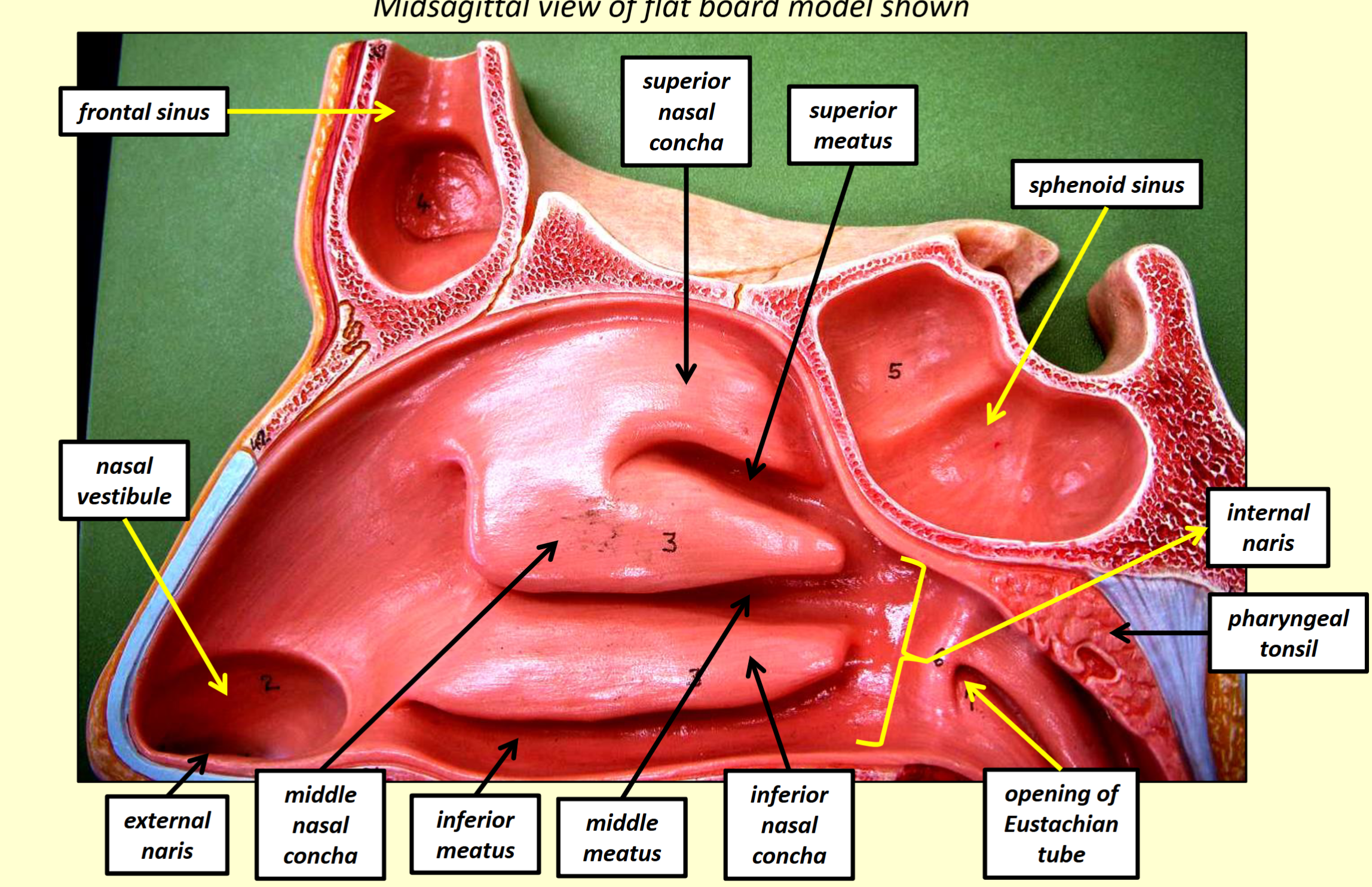
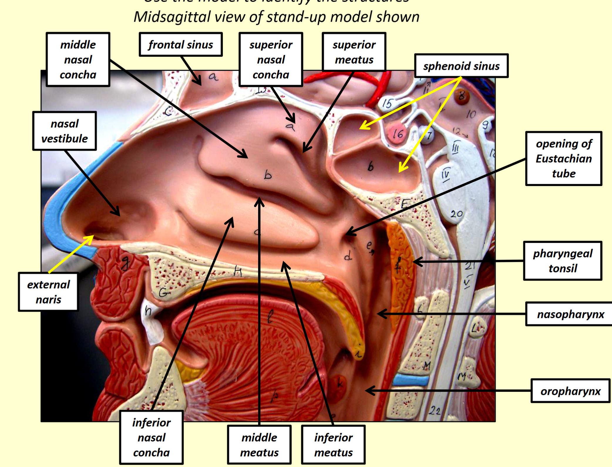
Nasal Cavity: Divided by the nasal septum.
Nasal Conchae: Increase mucosal surface area and enhance air turbulence.
Superior nasal concha
Middle nasal concha
Inferior nasal concha
Meatuses:
Superior meatus
Middle meatus
Inferior meatus
Internal Nares:
External Nares: Allow air to enter and exit the nasal cavity.
Nasal Vestibule: Composed of skin with vibrissae (hairs) to filter inspired air.
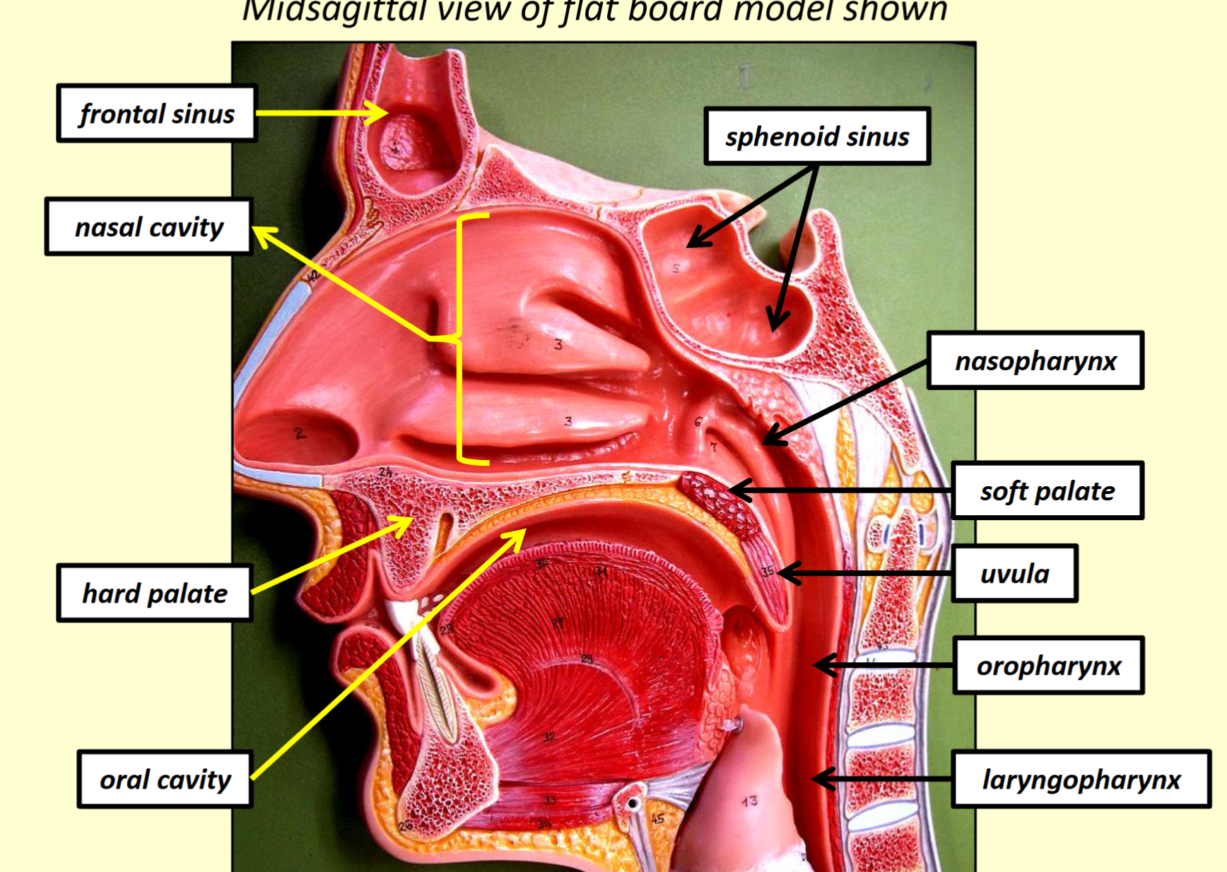
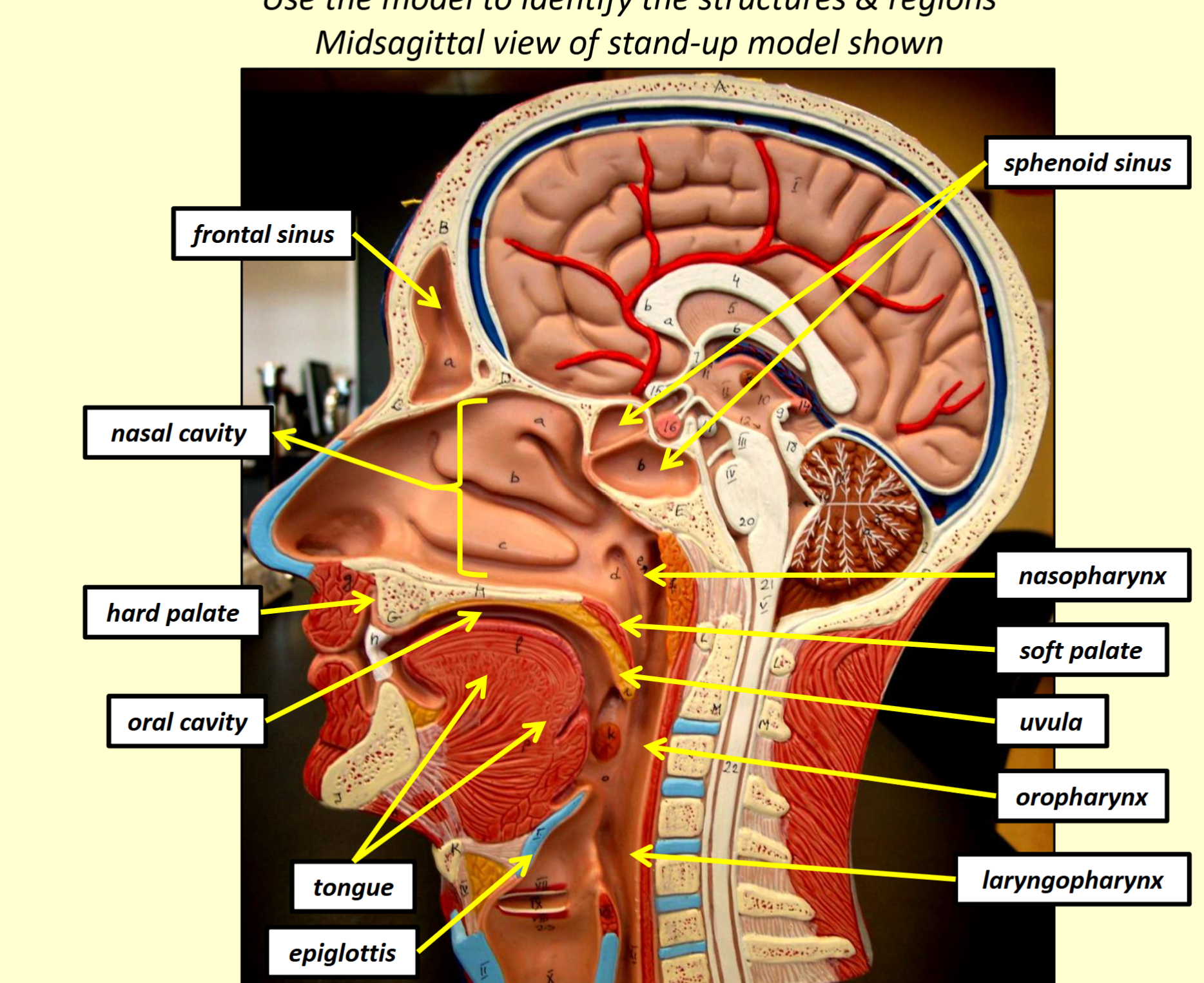
Paranasal Sinuses: Frontal and sphenoid sinuses lighten the skull and produce mucus to trap foreign matter.
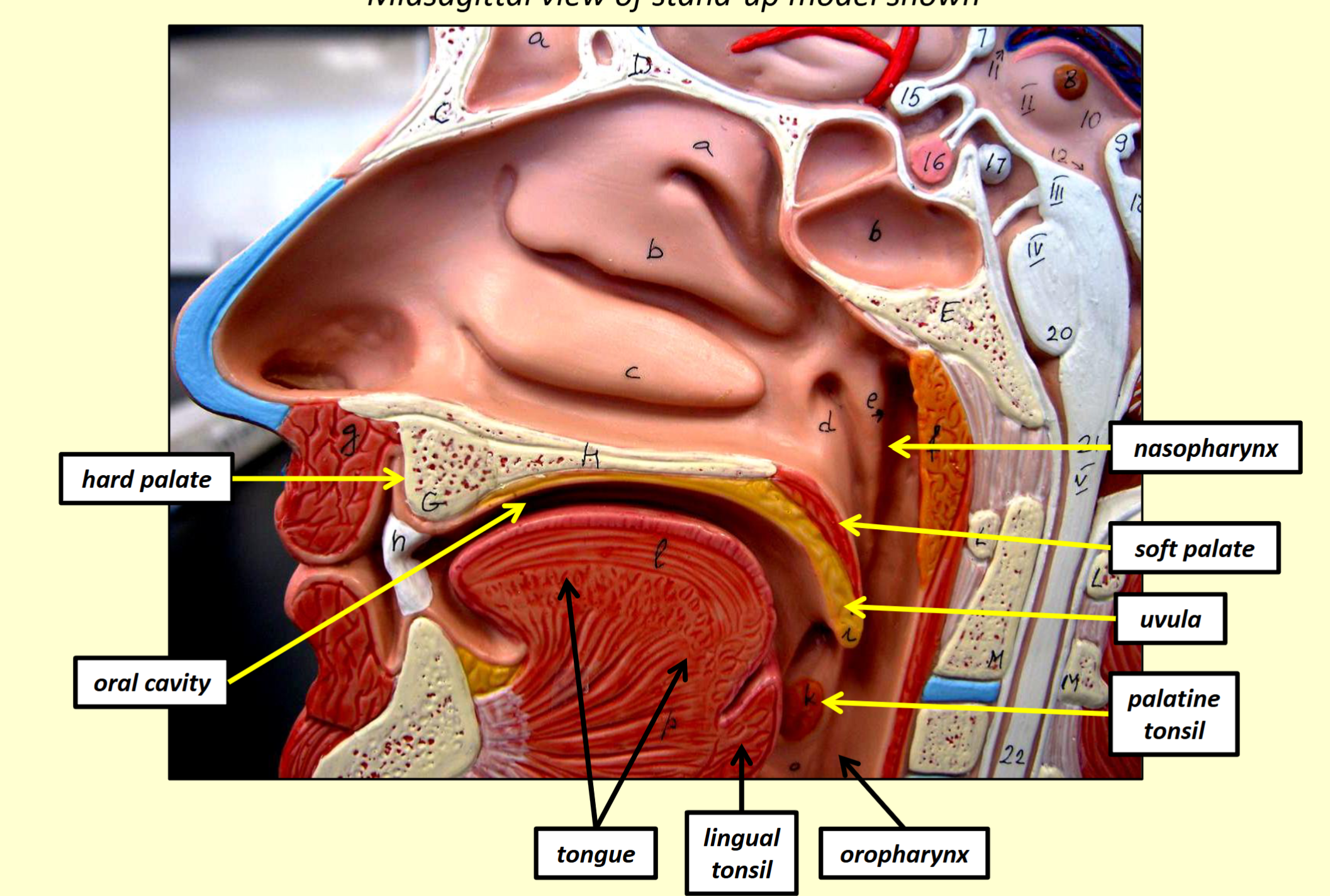
Hard Palate: Composed of maxillae and palatine bones.
Soft Palate: Composed of skeletal muscle and connective tissues.
Uvula: Closes off the nasopharynx during swallowing to prevent food/fluids from entering the nasal cavity.
Pharynx and Related Structures
Pharynx:
Nasopharynx: Passageway for air from nasal cavity through internal nares.
Oropharynx: Passageway for air, food, and fluids from oral cavity through fauces.
Laryngopharynx: Passageway for air to enter the larynx and food/fluids into the esophagus.
Tonsils:
Pharyngeal Tonsil: Traps pathogens in the nasopharynx.
Palatine Tonsil: Traps pathogens in the oropharynx.
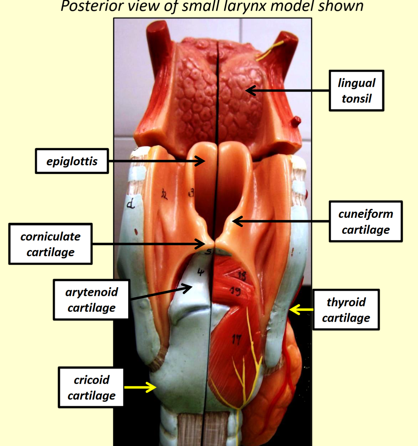
Lingual Tonsil: Traps pathogens in the oropharynx.
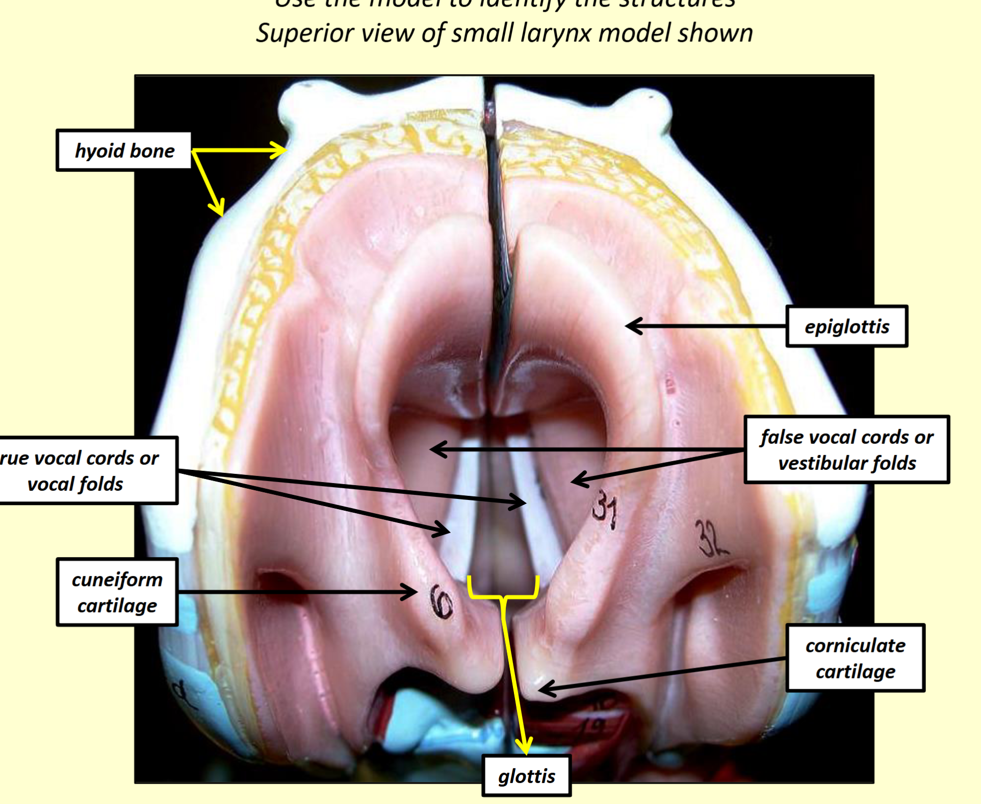
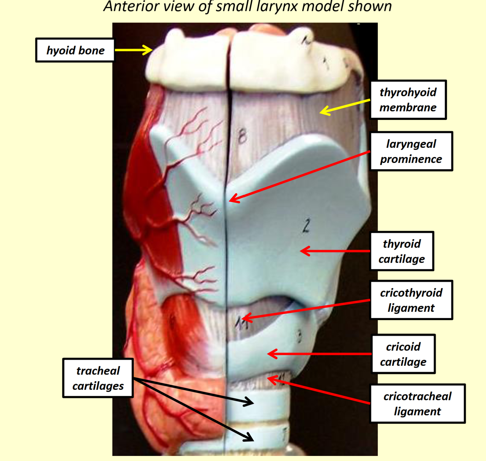
Larynx and Vocal Structures
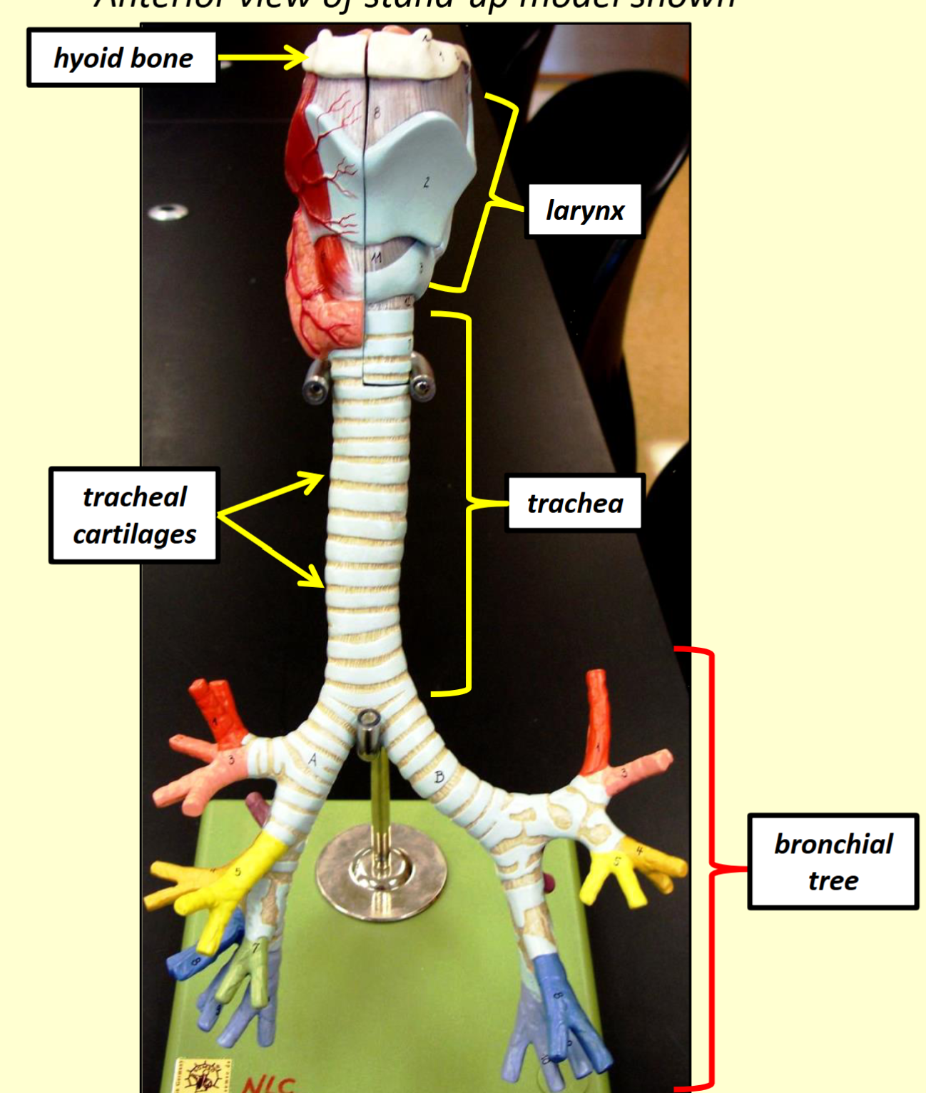
Larynx: Facilitates voice production and allows air to enter/exit the trachea.
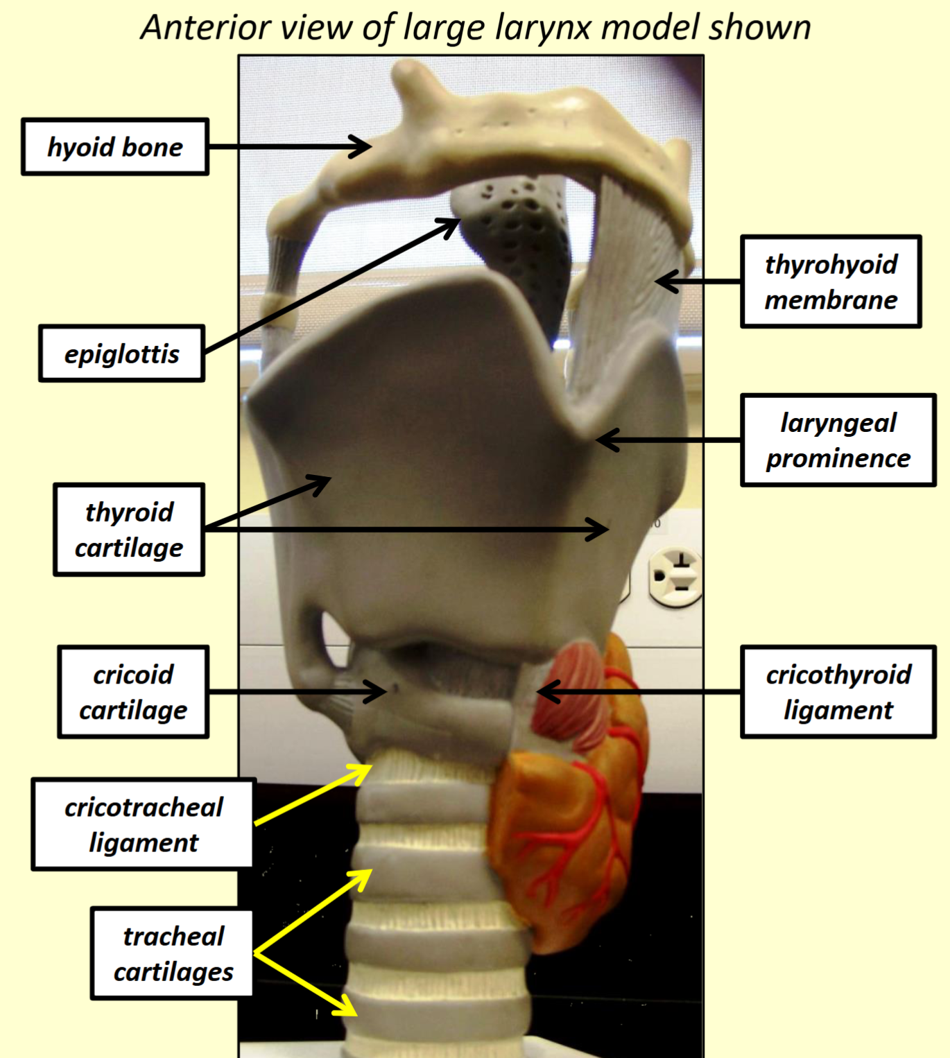
Hyoid Bone
Thyroid Cartilage: Forms Adam's apple (laryngeal prominence).
Cricoid Cartilage
Cuneiform Cartilages
Corniculate Cartilages
Arytenoid Cartilages: Change the position and tension of true vocal cords.
Epiglottis: Covers the glottis during swallowing.
Vocal Folds (True Vocal Cords): Produce sound when air passes.
Vestibular Folds (False Vocal Cords): Help close the glottis during swallowing.
Glottis: Space between true vocal cords.
Laryngeal Inlet: Opening to the larynx.
Thyrohyoid Membrane
Cricothyroid Ligament
Cricotracheal Ligament
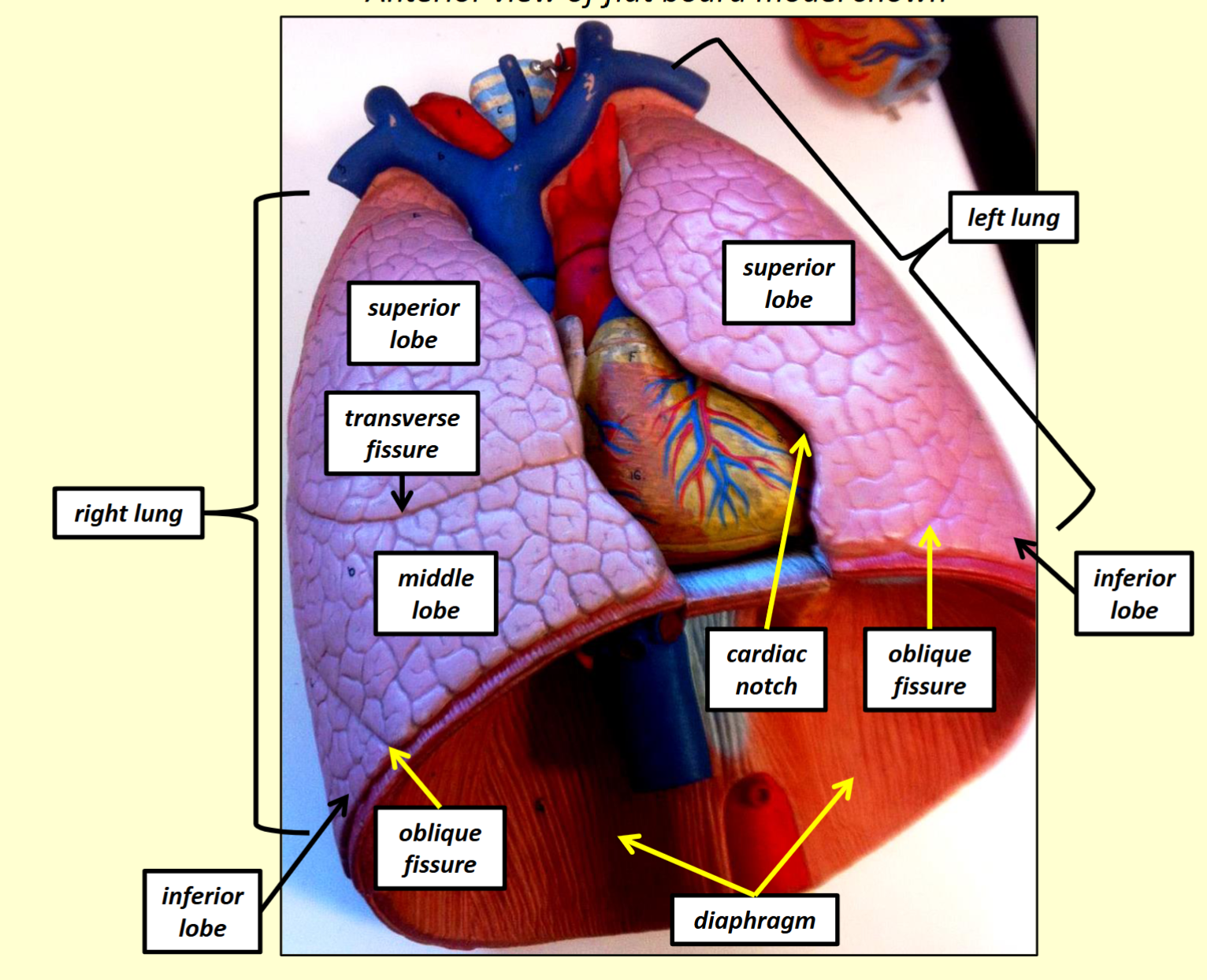
Lungs, Trachea, and Bronchial Tree
Right Lung
Superior Lobe
Middle Lobe
Inferior Lobe
Transverse (Horizontal) Fissure
Oblique Fissure
Left Lung
Superior Lobe
Inferior Lobe
Oblique Fissure
Cardiac Notch
Hilum
Opening that allows bronchi, blood vessels, lymphatic vessels, and nerves to enter/exit the lungs.
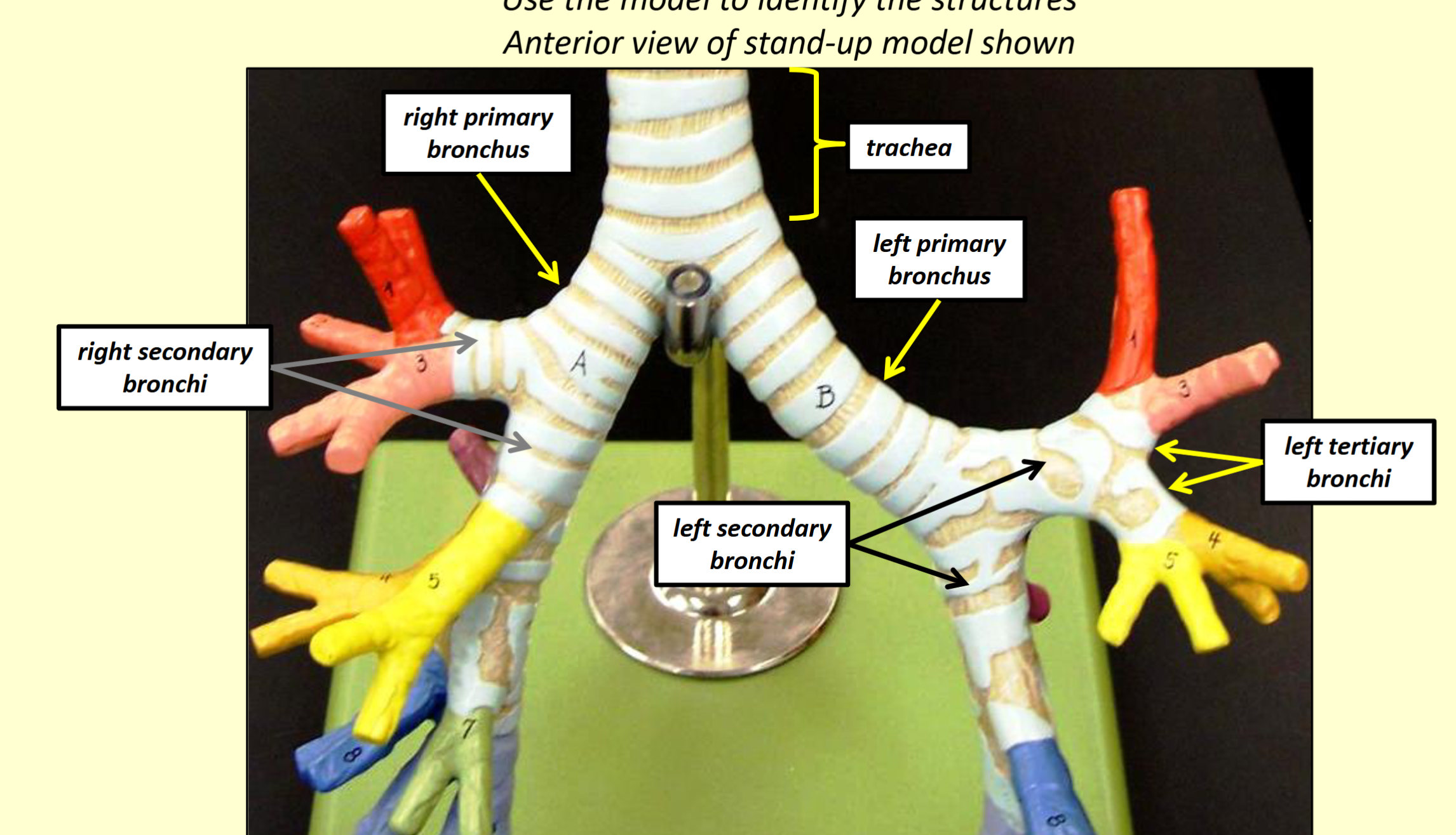
Trachea
Tracheal Cartilages: Tubular structure that functions as a passageway for air to enter/exit the lungs.
Primary Bronchi
Tubular structures that branch from the trachea, functioning as passageways for air to enter/exit lungs.
Right Primary Bronchus
Left Primary Bronchus
Secondary Bronchi
Tubular structures that branch from primary bronchi, functioning as passageways for air to enter/exit lungs.
Tertiary Bronchi
Tubular structures that branch from the secondary bronchi, functioning as passageways for air to enter/exit lungs, leading to bronchopulmonary segments.
Alveoli
Hollow, microscopic air sacs that function to exchange respiratory gases between the blood and air.
Terminal Bronchiole
Microscopic tubular structures that branch from bronchioles.
Respiratory Bronchioles
Microscopic tubular structures that branch from terminal bronchioles.
Alveolar Ducts
Microscopic tubular structures that branch from respiratory bronchioles and connect to alveoli.
Visceral Pleura
Respiratory Volumes and Capacities (Approximate Normal Values)
Tidal Volume: about 500 mL
Inspiratory Reserve Volume: about 3100 mL
Expiratory Reserve Volume: about 1200 mL
Residual Volume: about 1200 mL
Inspiratory Capacity: about 3600 mL
Functional Residual Capacity: about 2400 mL
Vital Capacity: about 4800 mL (measured via forced vital capacity)
Total Lung Capacity: about 6000 mL
Summary of Volumes
Inspiratory Capacity: 3600 mL
Vital Capacity: 4800 mL
Total Lung Capacity: 6000 mL
Functional Residual Capacity: 2400 mL