Session 3:Nervous System Notes
Divisions of the Nervous System
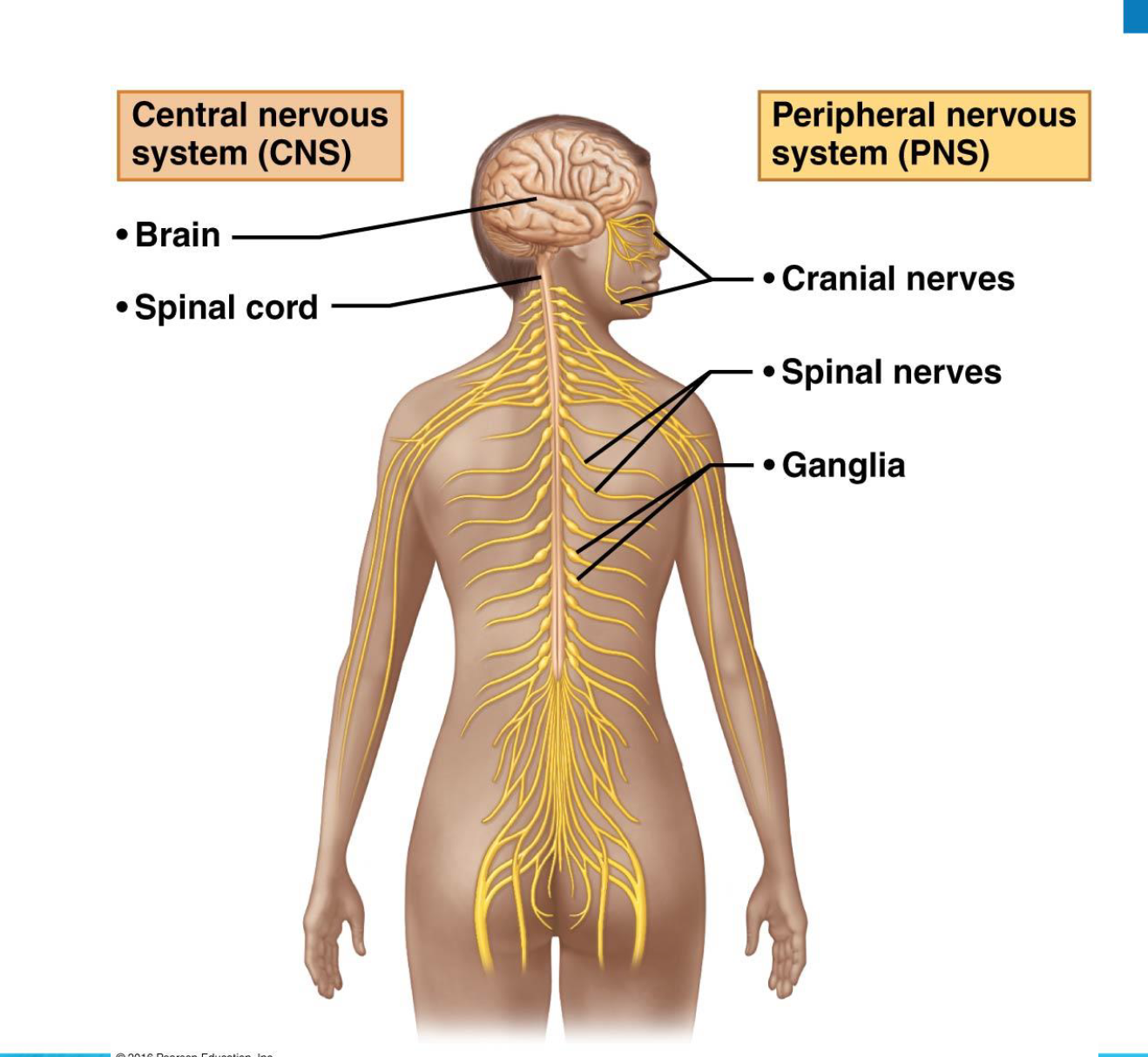
The nervous system (NS) is divided into two principal parts:
Central nervous system (CNS)
Brain and spinal cord of the dorsal body cavity
Integration and control center that interprets sensory input and dictates motor output
Peripheral nervous system (PNS)
The portion of the nervous system outside the CNS
Consists mainly of nerves that extend from the brain and spinal cord
Spinal nerves to and from the spinal cord
Cranial nerves to and from the brain
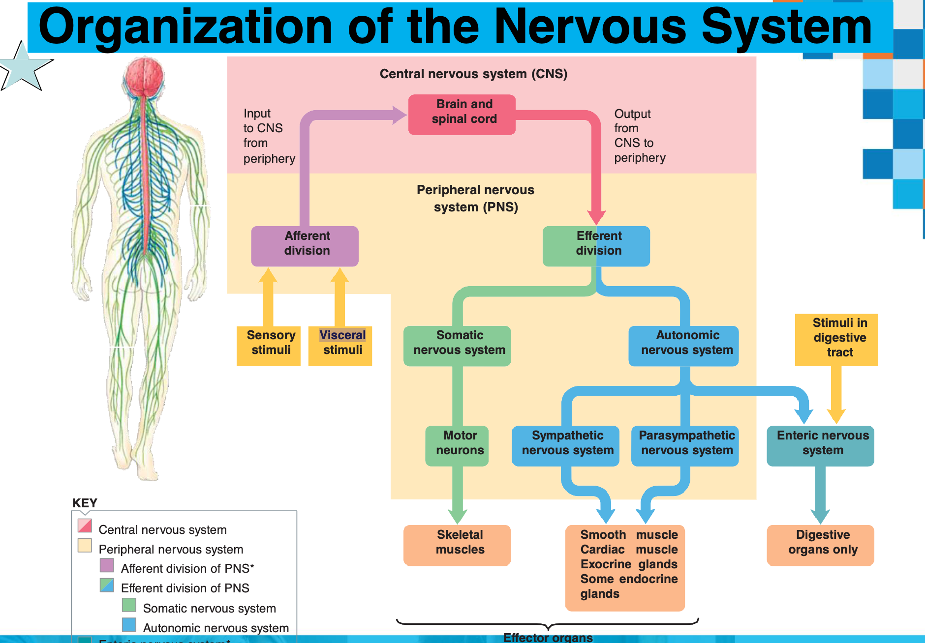
Organization of the Nervous System
Central Nervous System (CNS)
Brain
Spinal cord
Peripheral Nervous System (PNS)
Cranial nerves
Spinal nerves
Ganglia
Input and Output
Input to CNS from periphery (Afferent division)
Output from CNS to periphery (Efferent division)
Afferent Division
Sensory stimuli
Visceral stimuli (stimuli in the digestive tract)
Efferent Division
Somatic nervous system (motor neurons to skeletal muscles)
Autonomic nervous system (to digestive organs, smooth muscle, cardiac muscle, exocrine and some endocrine glands)
Sympathetic nervous system
Parasympathetic nervous system
Enteric nervous system
Functions of the Nervous System
The nervous system is the master controlling and communicating system of the body.
Cells communicate via electrical and chemical signals, leading to rapid and specific responses.
The function of the nervous system is to integrate sensory input and initiate appropriate motor output (action).
Functional Classification of Neurons
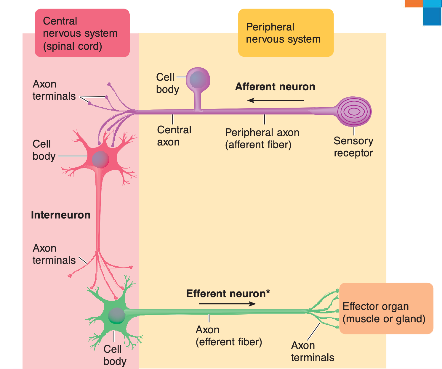
Sensory (afferent) neurons
detect changes in the environment called stimuli
transmit information to the brain or spinal cord
Interneurons (association neurons)
lie between sensory & motor pathways in the CNS
90% of our neurons are interneurons
Responsible for processing and interpretation of sensory input
Motor (efferent) neurons
send signals to muscle & gland cells (effectors) to carry out a response
Divisions of the PNS
Peripheral nervous system (PNS) has two functional divisions:
Sensory (afferent) division
Somatic sensory fibers: convey impulses from skin, skeletal muscles, and joints to the CNS
Visceral sensory fibers: convey impulses from visceral organs to the CNS
Motor (efferent) division
Transmits impulses from CNS to effector organs – muscles and glands
Two divisions:
Somatic nervous system
Autonomic nervous system
Nervous Tissue
Two basic cell types:
neurons
glial cells
Neurons (nerve cells)
functional unit (over 100 billion)
transmit & process information
Glial cells (neuroglia)
supporting cells
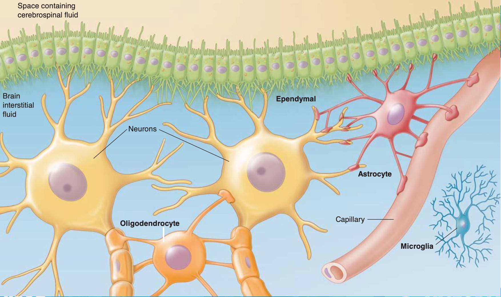
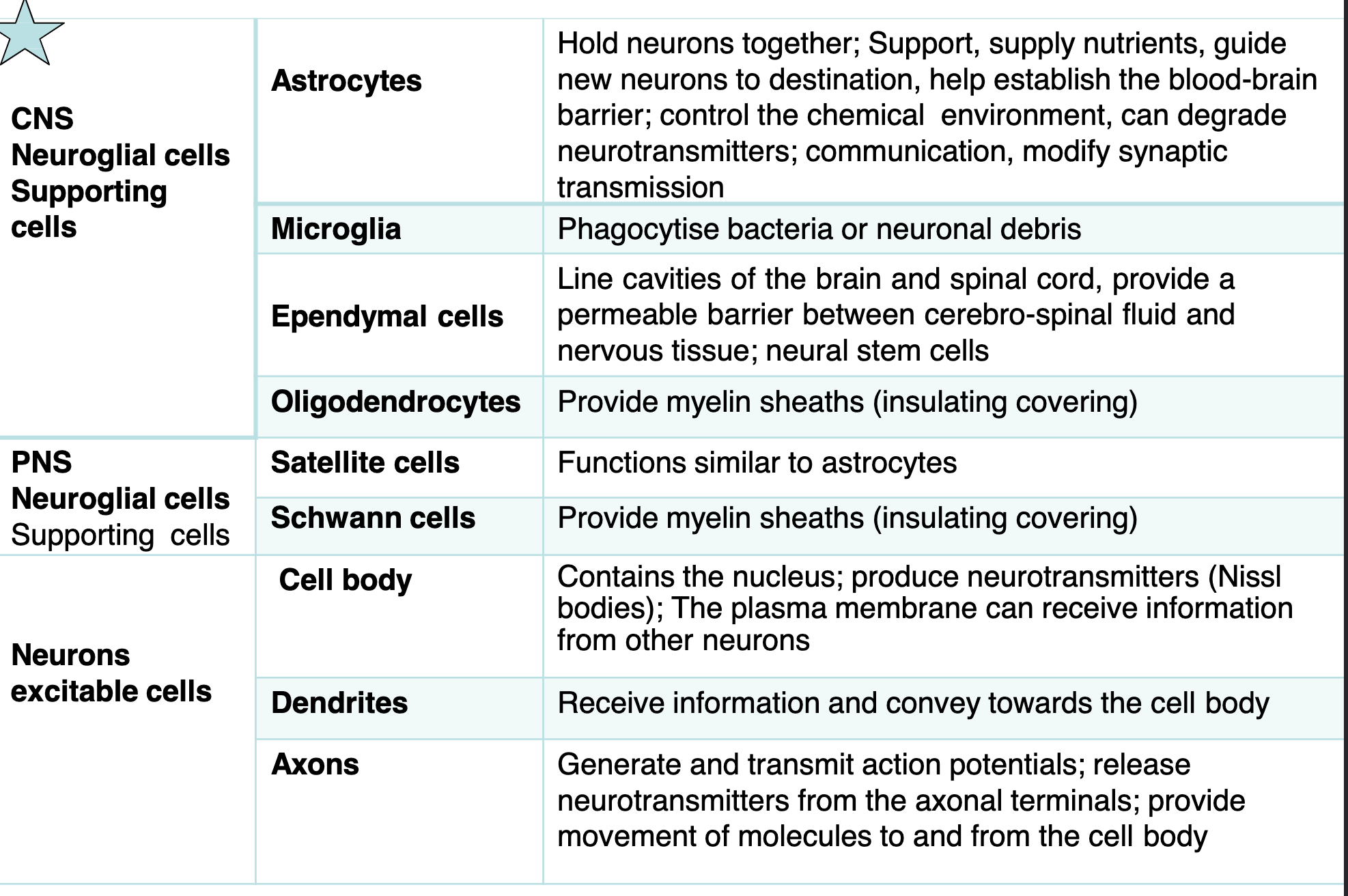
Neuroglial Cells
CNS Neuroglial cells (Supporting cells)
Astrocytes: Hold neurons together; Support, supply nutrients, guide new neurons to destination, help establish the blood-brain barrier; control the chemical environment, can degrade neurotransmitters; communication, modify synaptic transmission
Microglia: Phagocytize bacteria or neuronal debris
Ependymal cells: Line cavities of the brain and spinal cord, provide a permeable barrier between cerebro-spinal fluid and nervous tissue; neural stem cells
Oligodendrocytes: Provide myelin sheaths (insulating covering)
PNS Neuroglial cells (Supporting cells)
Satellite cells: Functions similar to astrocytes
Schwann cells: Provide myelin sheaths (insulating covering)
Neurons
Neurons = nerve cells
Cells specialized to transmit messages
Important properties:
Excitability – highly responsive to stimuli; generate electrical impulses (action potential)
Conductivity – ability to transmit an impulse
Major regions of neurons:
Cell body – nucleus and metabolic center of the cell
Processes – fibers that extend from the cell body
Dendrites – vast numbers; receive signals from receptors or other neurons
Axon – single (nerve fibre); generate and conduct electrical signal
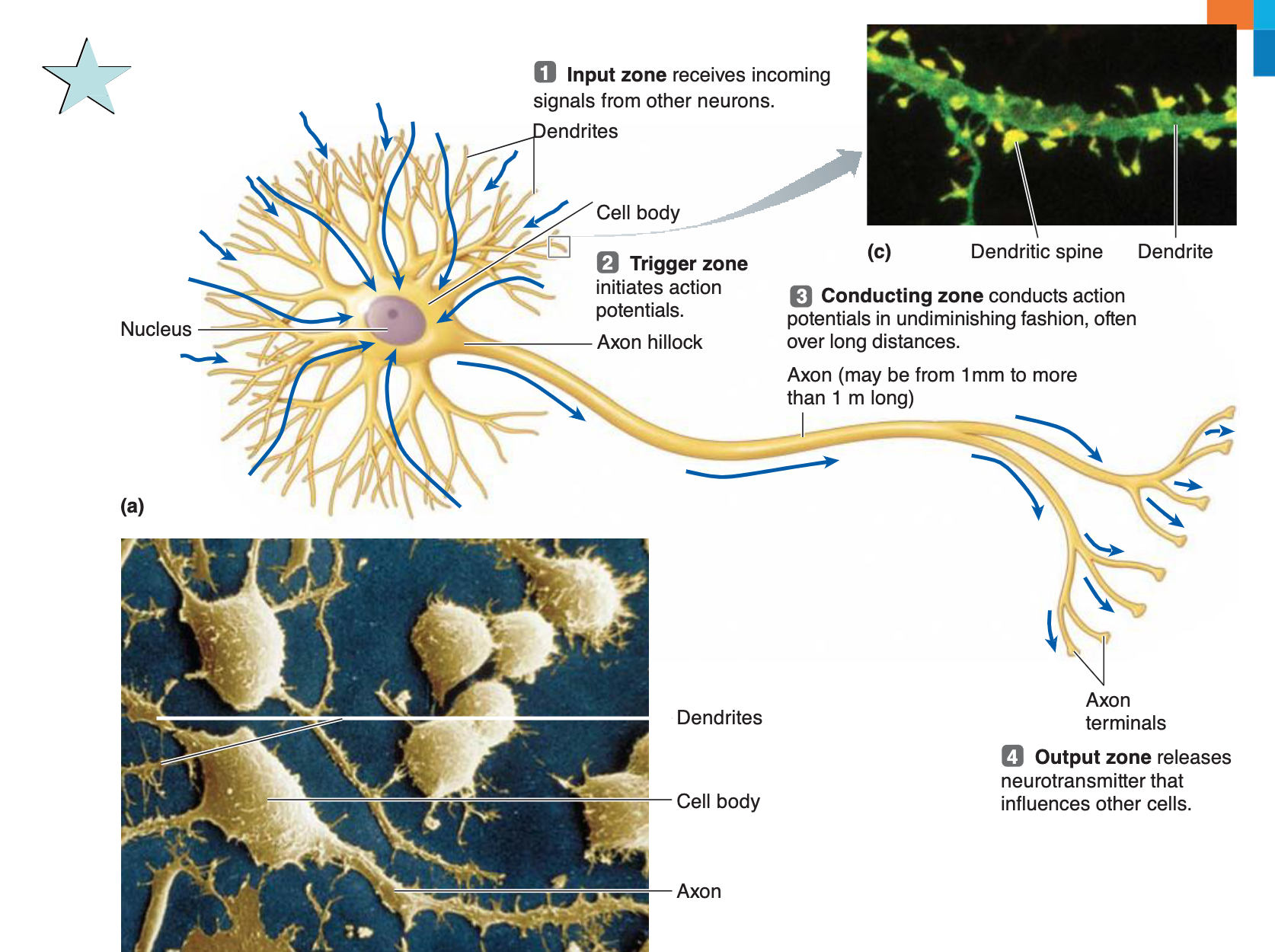
Neuron Structure and Function
Input zone: Dendrites receive incoming neurotransmitter that influences other cells.
Cell body: Contains the nucleus.
Trigger zone: Axon hillock initiates action potentials.
Conducting zone: Axon conducts action potentials in undiminishing fashion, often over long distances (may be from 1mm to more than 1 m long).
Output zone: Axon terminals release signals from other neurons.
Key Concepts
Nerve impulse: Communication between neurons
Resting membrane potential (RMP): Is the voltage (charge) difference across the cell membrane when the cell is at rest. RMP is a product of the distribution of charged particles (ions).
Action potential: Change in RMP that results in action of the cell
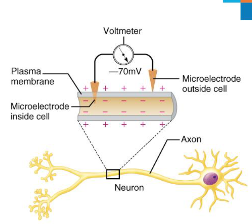
Resting Membrane Potential (RMP)
= the potential (charge) difference across the plasma membrane of a resting neuron
Resting membrane potential of a resting neuron is approximately -70 mV
The actual voltage difference varies from -40 mV to -90 mV
Potential generated by:
Differences in ionic composition of ICF and ECF:
There is a higher concentration of potassium (K^+) and anionic proteins (A^-) inside the neuron
There is a higher concentration of sodium (Na^+) and chloride (Cl^-) outside the neuron.
Differences in plasma membrane permeability
Inside is negatively charged
Outside is positively charged
Membrane Potential Results From
The differences in the concentrations of ions inside and outside of the cell
The permeability of the plasma membrane to these ions
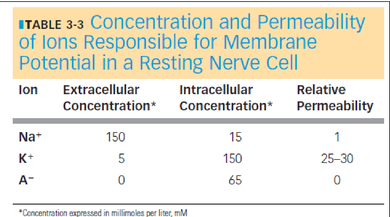
Ion Concentrations and the Na+/K+ Pumps
The differences in the concentrations of ions inside and outside of the cell are established by the Na^+/K^+ Pumps
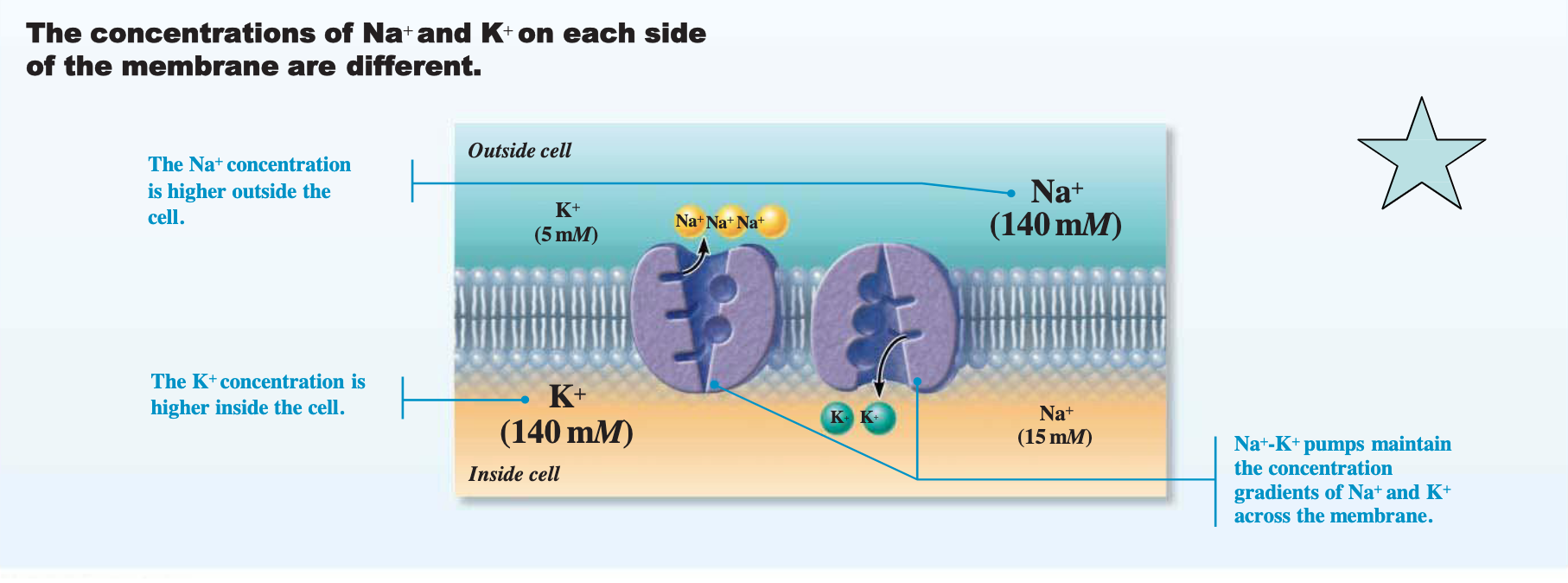
The concentrations of Na^+ and K^+ on each side of the membrane are different.
Outside cell:
K^+ (5 mM)
Na^+ (140 mM)
Inside cell:
K^+ (140 mM)
Na^+ (15 mM)
The Na^+-K^+ pumps maintain the concentration gradients of Na^+ and K^+ across the membrane.
Action Potentials (APs) – Neural Communication
Principal way neurons send signals
Means of long-distance neural communication
Occur only in muscle cells and axons of neurons
Brief reversal of membrane potential with a change in voltage of ~$100 mV$
Action potentials (APs) do not decay over distance
In neurons, also referred to as a nerve impulse
Involves opening of specific voltage-gated channels
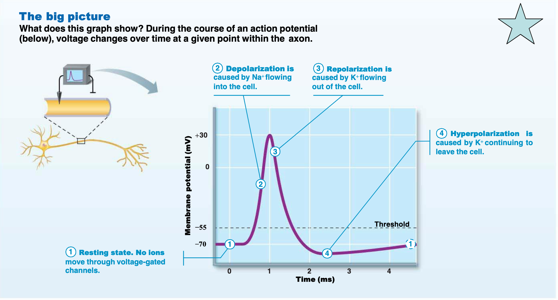
Action Potential Graph
Depolarization is caused by Na^+ flowing into the cell.
Repolarization is caused by K^+ flowing out of the cell.
Hyperpolarization is caused by K^+ continuing to leave the cell.
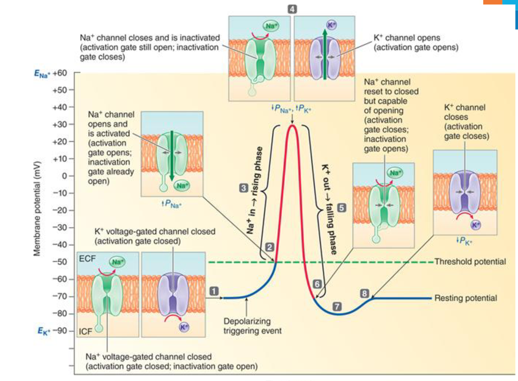
Action Potential Phases and Ion Channel States
Resting potential
Na^+ voltage-gated channel closed (activation gate closed; inactivation gate open)
K^+ voltage-gated channel closed (activation gate closed)
Rising phase
Na^+ channel opens and is activated (activation gate opens; inactivation gate already open)
Peak
Na^+ channel closes and is inactivated (activation gate still open; inactivation gate closes)
Falling phase
K^+ channel opens (activation gate opens)
Na^+ channel reset to closed but capable of opening (activation gate closes; inactivation gate opens)
Return to Resting Potential
K^+ channel closes (activation gate closes)
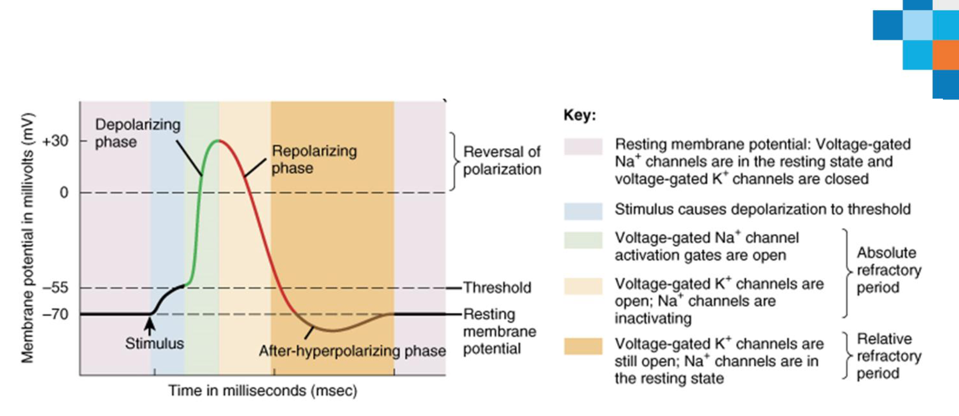
Action Potential Key
Resting membrane potential: Voltage-gated Na^+ channels are in the resting state and voltage-gated K^+ channels are closed
Stimulus causes depolarization to threshold
Voltage-gated Na^+ channel activation gates are open
Voltage-gated K^+ channels are open; Na^+ channels are inactivating
Voltage-gated K^+ channels are still open; Na^+ channels are in the resting state
Four Main Phases of AP
Depolarisation: change in a cell's membrane potential, making it less negative (more positive)
A threshold stimulus (> -55mV)
Change in resting membrane potential
Na^+ channels open → Na^+ rush into the cell → reverse the electrical charge
Positive charge of ICF = a neuron fires = generation of an AP
Repolarization: the process by which a cell membrane returns to its resting potential after depolarization
opening of K^+ channels → outflow of K^+
closing of Na^+ channels → membrane potential returns to resting state
Hyperpolarisation (undershoot): cell's membrane potential becomes more negative than its resting potential
a polarisation even more negative than the resting membrane potential
excessive K^+ efflux
Restoration:
The resting membrane potential is restored
Rules of the Action Potential
caused by voltage-gated channels
all-or-none response
only occur if stimulus exceeds threshold
all action potentials are identical
stronger stimulus = more action potentials
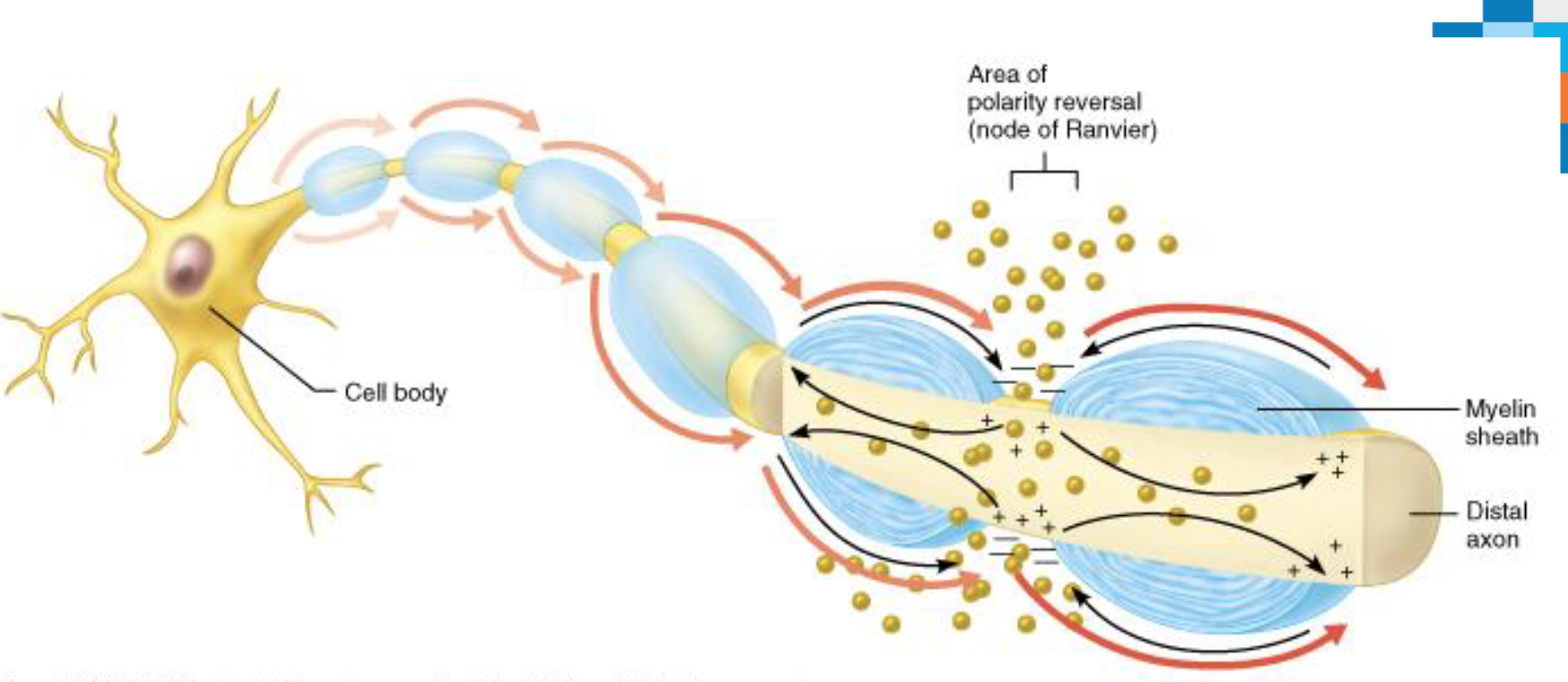
Action Potential Propagation (the action potential, travels down the axon of a neuron)
Action potential causes inside of cell to become positive
Positive charges are attracted to adjacent negative charges
Adjacent areas depolarise → action potential
Action potential propagated down axon – like a “Mexican wave”
Conduction Velocity
APs occur only in axons, not other cell areas
AP conduction velocities in axons vary widely
Rate of AP propagation depends on two factors:
Axon diameter
Larger-diameter fibers have less resistance to local current flow, so have faster impulse conduction
Degree of myelination
Two types of conduction depending on presence or absence of myelin
Continuous conduction (continues)
Saltatory conduction (at nodes)
Myelinated axons conduct impulses faster
Myelination of Axons
Myelin sheath
Composed of myelin, a whitish, protein-lipid substance which covers some axons; formed by glial cells (Schwann cells in the PNS and oligodendrocytes in CNS)
Function of myelin:
Protect and electrically insulate axon
Increase speed of nerve impulse transmission
Myelinated fibers: segmented sheath surrounds most long or large-diameter axons
Nodes of Ranvier – gap between the myelin sheath
Non-myelinated fibers: do not contain myelin sheath and conduct impulses more slowly
Membrane Potential and Ion Channels
Membrane potential is the difference in electrical charge between the inside and outside of a cell generated by different concentrations of Na^+, K^+, Cl^−, and protein anions (A^−) inside and outside of the cell.
Concentrations of K^+ and A^− higher inside the cell and concentrations of Na^+ and Cl^− higher outside the cell.
The resting membrane potential of the neurons is between -45 to -90 millivolts (in average about -70 mV).
Na^+ - K^+ pump maintains the concentration gradient; it constantly expels three Na^+ ions from the cell, and moves two K^+ ions back into the cell.
Ion channels: The membrane pores made of protein permeable selectively to specific types of ions.
Types:
I. Passive, or leakage, channels – always open;
II. Gated channels:
a) Chemically gated channels – open with binding of a specific neurotransmitter;
b) Voltage-gated channels – open and close in response to change in membrane potential;
c) Mechanically gated channels – open and close in response to physical deformation of receptors.
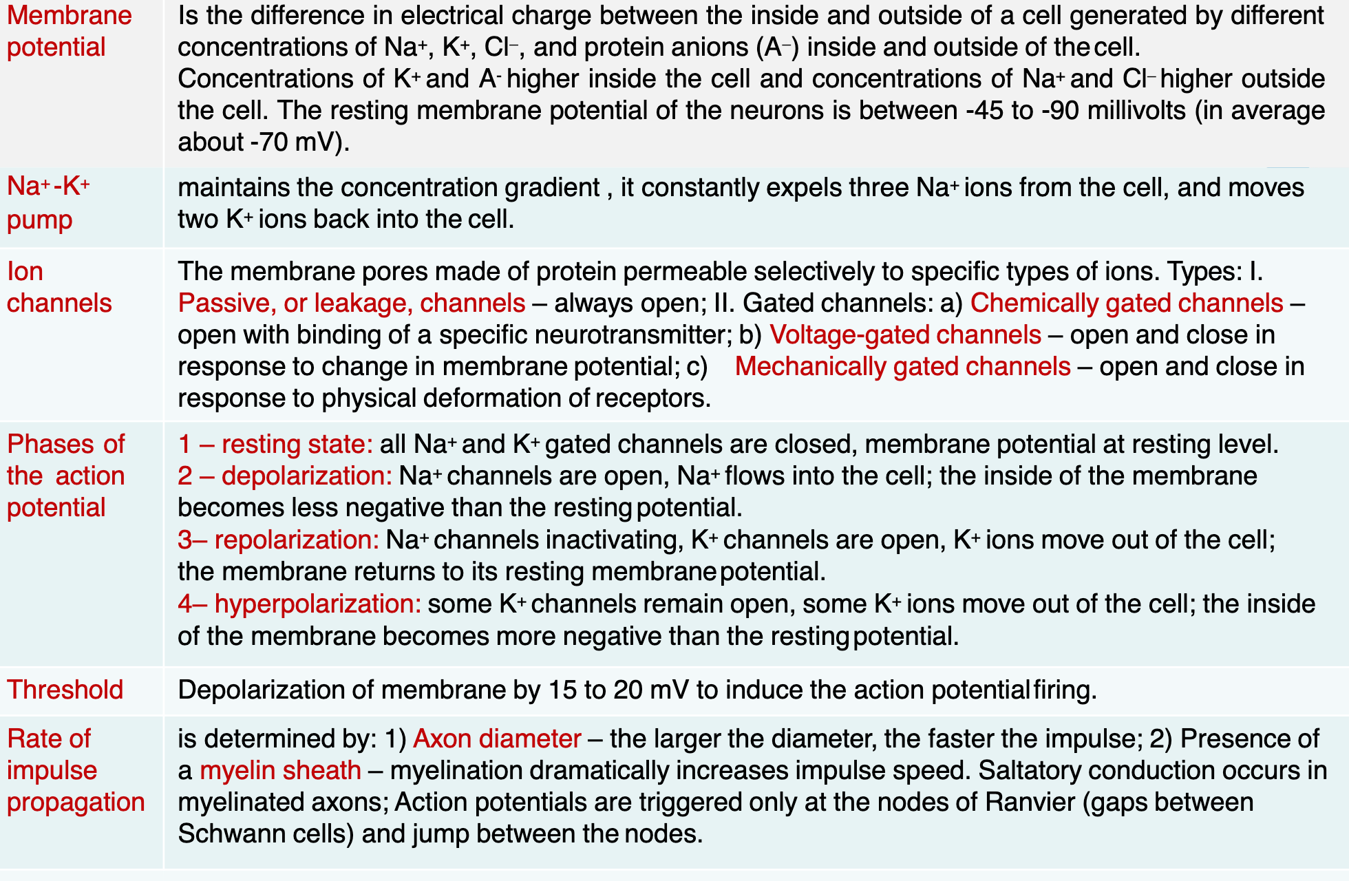
Phases of the Action Potential
Resting state: all Na^+ and K^+ gated channels are closed, membrane potential at resting level.
Depolarization: Na^+ channels are open, Na^+ flows into the cell; the inside of the membrane becomes less negative than the restingpotential.
Repolarization: Na^+ channels inactivating, K^+ channels are open, K^+ ions move out of the cell; the membrane returns to its resting membranepotential.
Hyperpolarization: some K^+ channels remain open, some K^+ ions move out of the cell; the inside of the membrane becomes more negative than the restingpotential.
Depolarization of membrane by 15 to 20 mV to induce the action potentialfiring.
Rate of Impulse Propagation
is determined by:
1) Axon diameter – the larger the diameter, the faster the impulse;
2) Presence of a myelin sheath – myelination dramatically increases impulse speed.
Saltatory conduction occurs in myelinated axons
Action potentials are triggered only at the nodes of Ranvier (gaps between Schwann cells) and jump between the nodes.
Synapses
Definition: A junction that mediates information transfer from one neuron to another neuron and to an effector cell
Presynaptic neuron – conducts impulses toward the synapse
Postsynaptic neuron – transmits impulses away from the synapse
Types of Synapses
Electrical synapse
Chemical Synapse
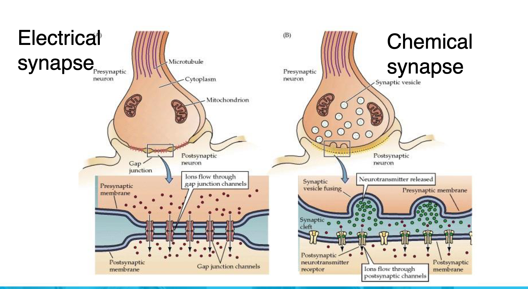
Chemical Synapses - Structure
Most common type of synapse
Specialized for release and reception of chemical neurotransmitters
Typically composed of two parts:
Axon terminal of presynaptic neuron: contains synaptic vesicles
filled with neurotransmitter
Receptor region on postsynaptic neuron’s membrane: receives neurotransmitter; usually on dendrite or cell body
Two parts separated by fluid-filled synaptic cleft
Electrical impulse changed to ‘chemical’ across synapse, then back into ‘electrical’
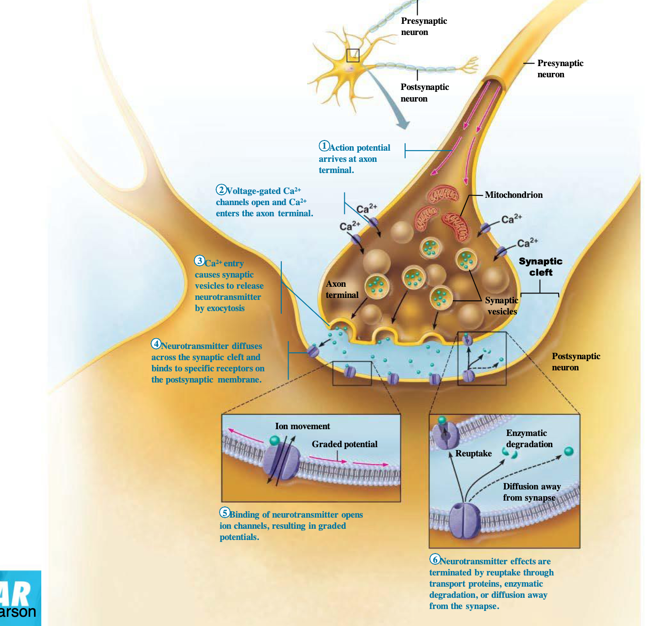
Chemical Synapses - Transmission
Action potential arrives at axon terminal.
Voltage-gated Ca^{2+} channels open and Ca^{2+} enters the axon terminal.
Ca^{2+} entry causes synaptic vesicles to release neurotransmitter by exocytosis
Neurotransmitter diffuses across the synaptic cleft and binds to specific receptors on the postsynaptic membrane.
Binding of neurotransmitter opens ion channels, resulting in graded potentials.
Neurotransmitter effects are terminated by reuptake through transport proteins, enzymatic degradation, or diffusion away from the synapse.
Synapse - Definitions
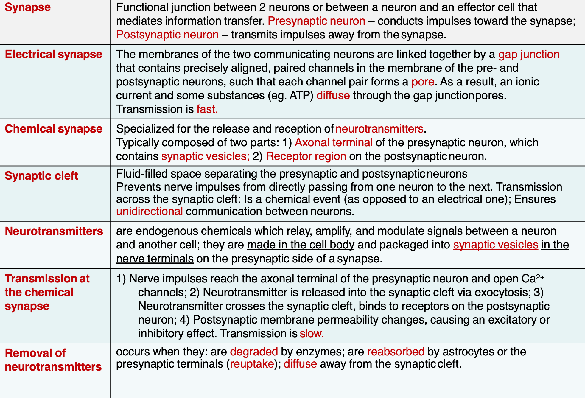
Transmission at the Chemical Synapse
Nerve impulses reach the axonal terminal of the presynaptic neuron and open Ca^{2+} channels
Neurotransmitter is released into the synaptic cleft via exocytosis
Neurotransmitter crosses the synaptic cleft, binds to receptors on the postsynaptic neuron
Postsynaptic membrane permeability changes, causing an excitatory or inhibitory effect. Transmission is slow.
Removal of neurotransmitters occurs when they: are degraded by enzymes; are reabsorbed by astrocytes or the presynaptic terminals (reuptake); diffuse away from the synaptic cleft.
Synapse: Neurotransmitters
Synthesized in neurons, stored in secretory vesicles in pre-synaptic axon terminals
50 or more neurotransmitters have been identified
Most neurons make two or more neurotransmitters
Excitatory receptors depolarize membranes (e.g., glutamate)
Inhibitory receptors hyperpolarize membranes (e.g. GABA)
Some neurotransmitters have both excitatory and inhibitory effects
Determined by the receptor type on the postsynaptic neuron
eg: acetylcholine which is Excitatory at neuromuscular junctions with skeletal muscle and Inhibitory in cardiac muscle
Neuroglia of the CNS main types
Neuroglia Type | Function | Explanation |
|---|---|---|
Astrocytes | Support neurons, maintain blood-brain barrier, regulate nutrients | Star-shaped cells that anchor neurons and control the chemical environment |
Oligodendrocytes | Produce myelin sheaths around CNS axons | Help speed up nerve impulses by insulating axons in the central nervous system |
Microglia | Act as immune cells; remove debris and damaged cells | Small, spider-like cells that protect the CNS by cleaning up waste and invaders |
Ependymal Cells | Line ventricles in brain and spinal cord; produce and circulate CSF | Help form and move cerebrospinal fluid (CSF) using cilia |
major events that occur in an action potential
Event | What Happens | Ion Movement |
|---|---|---|
1. Resting State | The neuron is at rest, membrane potential is ~ -70 mV. | Na⁺ and K⁺ channels are closed; Na⁺ is higher outside, K⁺ higher inside |
2. Depolarization | Stimulus opens voltage-gated Na⁺ channels, Na⁺ rushes into the cell. | Sodium (Na⁺) moves INTO the cell |
3. Peak of Action Potential | Membrane potential reaches about +30 to +40 mV. Na⁺ channels inactivate. | Na⁺ inflow stops; K⁺ channels begin to open |
4. Repolarization | K⁺ channels fully open, K⁺ exits the cell, restoring negative charge inside. | Potassium (K⁺) moves OUT of the cell |
5. Hyperpolarization | K⁺ channels stay open too long; membrane becomes more negative than resting. | Continued K⁺ outflow |
6. Return to Resting Potential | Na⁺/K⁺ pump restores original ion balance (3 Na⁺ out, 2 K⁺ in). | Active transport restores balance |