MICB 212 Chapter 8 Notes
Antigen Processing and Presentation
-antigen presentation is the displaying of peptides derived from pathogens or other proteins on dendritic cell surface that T cells can see
-MHC proteins expressed on cell surface provide physical structure to display antigenic peptides to T cells
Major Histocompatibility Complex (MHC) Proteins
-2 types of MHC proteins
both proteins have grooves that can bind peptides
MHC Class I Proteins
-expressed on all nucleated cells
-consist of transmembrane α chain that is non-covalently associated with a soluble protein (β2-microglobulin)
-peptide bonding groove located within the α chain
binds to nascent MHC class I protein
MHC Class II Proteins
-expressed only on antigen-presenting cells (dendritic cells, macrophages, thymic epithelial cells)
-consist of 2 non-identical transmembrane polypeptide chains (α and β chains)
-peptide bonding groove formed between α and β chains
-peptide of at least 13 amino acids in length binds to MHC class II protein
Antigen Processing
degradation of protein into peptide fragments
Antigen Presentation
binding and display of antigen as peptide fragment bound to MHC proteins on surface of a cell
-MHC class I proteins display peptides derived from proteins in cytoplasm of cell
proteins coming from inside of cell
proteins synthesized by ribosomes
-MHC class II proteins display peptides derived from soluble proteins taken up by a cell via endocytosis or phagocytosis into an endosome or phagosome
proteins coming from outside of cell
proteins can include normal blood proteins, toxins, bacteria or virus particles
Pathways for Processing and Presentation of Protein into Peptide Fragments
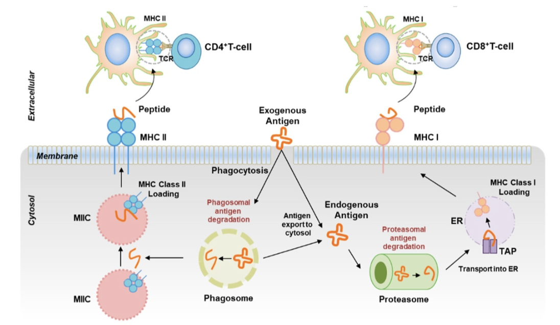
Endogenous Pathway (MHC Class I Peptide Loading)
-intracellular proteins degraded into peptides by proteasome (protease complex located in cytoplasm)
-transporter proteins called TAP proteins move resulting peptides from cytoplasm to lumen of ER
-peptide binds to nascent MHC class I protein
-MHC class I peptide complexes transported to cell surface
Exogenous Pathway (MHC Class II Peptide Loading)
-MHC class II proteins with associated invariant chain (ii) proteins leave ER in vesicles
-after endocytosis or phagocytosis of bacterium/virus/protein, endosome or phagosome fuses with lysosomes that contain lysozyme, proteases and bactericidal substances
-proteases degrade proteins (bacteria, virus, blood) into peptides
-peptides derived from proteins remain inside vesicles
don’t enter cell’s cytoplasm
-vesicles containing MHC class II/invariant complexes fuses with vesicles containing peptide fragments in MIIC region of cell
-invariant chain is degraded to a smaller fragment (CLIP) and occupies peptide-binding site
-CLIP replaced with peptide fragment
-MHC class II peptide complexes are transported to cell surface
Cross-Presentation Pathway
-mix of exogenous and endogenous pathways
-MHC class I proteins display peptides derived from exogenous proteins
-dendritic cells aid with cross-presentation
-peptides derived from antigens taken up by cell in an endosome or phagosome and contents of vesicle are diverted to proteasome
-processing of peptides follows endogenous pathway
dendritic cells present peptides derived from exogenous antigens on both MHC I proteins and MHC class II proteins
MHC Diversity
-goal of MHC protein is to be able to bind and present peptide derived from any protein
especially foreign proteins from a pathogen
-if unable to present peptides from a pathogen → can’t make adaptive immune response against it
-single MHC protein can bind many but not all peptides
HLA-A protein might not be able to bind a particular peptide derived from viral pathogen but same peptide might be able to bind to HLA-C protein
in this case → cells would use HLA-C protein to present viral peptide to T cells
MHC Genes are Polygenic
-several genes that encode proteins that have a similar function
MHC Genes are Codominantly Expressed
-MHC genes from mother and father will be different alleles
2 HLA-A proteins on surface of cells
2 HLA-B proteins on surface of cells
2 HLA-C proteins on surface of cells
-combination of all HLA proteins expressed defines tissue type → important in organ transplants
MHC Genes are Polymorphic
-different alleles for each MHC gene
-at least 5000 versions of HLA-A gene
-polymorphism of MHC genes is important for survival of any species
-for any given pathogen, some portion of population will be able to present peptides from that pathogen and make an immune response to eliminate the pathogen
-reduction in MHC polymorphism may predispose species to infectious disease
Human MHC Class I Proteins
-3 types of MHC Class I proteins — HLA = human leukocyte antigen
HLA-A
HLA-B
HLA-C
-all proteins are expressed on all nucleated cells
-6 different MHC class I proteins
3 (1 HLA-A, 1 HLA-B, 1 HLA-C) from mom
3 (1 HLA-A, 1 HLA-B, 1 HLA-C) from dad
Human MHC CLASS II Proteins
-3 types of MHC class II proteins
HLA-DP
HLA-DQ
HLA-DR
-all proteins only expressed on antigen presenting cells
macrophages
dendritic cells
B cells
thymic epithelial cells
Summary
-MHC proteins expressed on cell surface allows cell to communicate to T cells
-MHC class I proteins consist of membrane-bound chain → non-covalently associated with β2-microglobulin
-MHC class II proteins are dimers of α and β chain
-MHC class I proteins are found on all nucleated cells
-MHC class II proteins are found only on antigen-presenting cells
-antigenic peptides must be processed and loaded onto MHC proteins
MHC class I proteins use endogenous pathway for peptide loading
present peptides derived from proteins made inside cell
MHC class II proteins use exogenous pathway for peptide loading
present peptides derived from extracellular antigens
includes proteins taken up by endocytosis or phagocytosis
-cross-presentation occurs when dendritic cells phagocytose a foreign antigen and present it on MHC class I proteins and MHC class II proteins.