FINAL EXAM REVIEW
KNOW THE FUNCTIONS OF BLOOD
Blood has 3 major functions: Transportation, regulation, and protection.
Transportation: blood carries gases (oxygen & carbon dioxide), nutrients, waste products, hormones, enzymes, and catalysts throughout the body
Regulation: regulates body temperature, blood water concentration, PH values
Protection: provides immunity and promotes blood clotting, essential for hemostasis
KNOW THE CHARACTERISTICS, NUMBER, FUNCTION AND LIFESPAN OF RBCs, WBCs, PLATELETS
Red blood cells
Biconcave dis-shaped structures
NO nucleus
have proteins on the membrane for identification
Number: 4.8 to 5.4 million
Life span: about 120 days to 4 months
Function: carries oxygen around the body
White blood cells
Larger than the red blood cells
YES nucleus
Reproduce on their own
Broken into two headings: ➀ Granulocyte (basophils, eosinophils, neutrophils) ➁ Agranulocytes - no granules (lymphocytes and monocytes): travel to an infection site at a slower rate, increase their #s. Involved with chronic infections. Neutrophils normally get to the infection first then monocytes. ) Lymphocytes-T/B cells, monocyts, Basophil, Enophophils, Neutrophils.
Number: about 5,000 to 10,000
Life span: for hours to days ~ decades
Function: fights infections and microbes in the body
Platelets
Fragments of cells, NOT a whole cell
NO nucleus
Number: about 150,000 to 400,000
Life span: 5 to 9 days
Function: involved with blood clotting and coagulation
DIFFERENTIATE BETWEEN: LEUKEMIA, LEUKOCYTOSIS, LEUKOPENIA
Leukemia: WBC cancer, abnormal WBCs multiply uncontrollably
Leukocytosis: increase in the # of WBCs >10,000
Leukopenia: decrease in the # of WBCs <5,000
DIFFERENTIATE: AGRANULOCYTES VS. GRANULOCYTES
Agranulocytes: non-granulated inside
Examples: lymphocytes and monocytes
Granulocytes: graininess in them
Think of Uncle BEN’s rice: basophils, eosinophils, neutrophils
KNOW THE 3 MAJOR PLASMA PROTEINS
Albumin: “sponge” hold water. most common, works like a sponge and maintains osmotic pressure of water in blood
Globulins: backbone of antibodies in blood, called immunoglobulins that make antibodies
Fibrinogen: involved in coagulation, final step in blood clotting, least common plasma protein
WHAT IS PLASMA?
Plasma is straw-colored liquid function of blood
Plasma makes up 55% of blood
Plasma is 91.5% water
If you took out clotting factor that is called serum
UNDERSTAND HEMOSTASIS AND THE 3 STAGES OF COAGULATION
Hemostasis: process to stop bleeding in small cuts
Vascular spasm/vasospasm: decrease blood vesse. contraction of blood vessels, called vasocontraction. We reduce the diameter of the blood vessel which reduces blood flow.
Platelet plug formation: as platelets strike damaged epithelial lining and collagen fiber, they begin to stick together and get larger and seal up the hole and pull it together.150,000 -400,000, 5-9 days, blood clotting.
Coagulation: has three stages of cascading chemicals. Coagulation.a) Formation of prothombinase. b) protrombin→prothrombinase→thrombin. c) fibrogen→thrombin—>fibrin threads.
Formation of prothrombinase
Conversion of prothrombin into thrombin using prothrombinase
Conversion of fibrinogen (plasma protein) into fibrin threads using thrombin
UNDERSTAND BLOOD TYPING: SURFACE PROTEINS, ISOANTIBODIES, COMPATIBILITY
Blood types: A, B, AB, O
Surface proteins:
Type A has A proteins on cell
Type B has B proteins on cell
Type AB has both AB proteins on cell
O does not have any proteins on cell
Isoantibodies: in plasma,
Type A has anti-B in plasma
Type B has anti-A in plasma
Type AB does not have any in plasma
Type O has both anti-A and anti-B in plasma
Compatibility:
Type A: can receive Type A and O blood
Type B: can receive Type B and O blood
Type AB: can receive Type AB, A, B, O blood (universal receiver)
Type O: can receive Type O blood (universal donor)
RH proteins: another protein on cell
can either have RH proteins on cell (RH-positive) or not have RH proteins on cell (RH-negative)
If RH-positive, you cannot have anti-RH antibodies in plasma or WBCs would attack RBCs
If RH-negative, you are not born with anti-RH antibodies but will make them if exposed to RH-positive blood
Only time it causes problems is in hemolytic disease of newborns - if RH-positive man and RH-negative woman have baby that is RH-positive so mother makes anti-RH antibodies which could affect next RH-positive baby. Mother is given Rogam injections that blocks anti-RH.
KNOW THE COVERINGS SURROUNDING THE HEART
Pericardium: fibrous and inner double serous layers enclose and protect heart.
KNOW THE 3 LAYERS OF THE HEART WALL
Epicardium/visceral layer of the serous pericardium: epicardium= vessels larger of sereous pericardium.
Myocardium: cardiac muscle layer of heart, most substantial layer
Endocardium: deep thin layer that lines the insides of chambers
UNDERSTAND THE 4 CHAMBERS OF THE HEART, 4 VALVES, CHORDAE TENDINEAE, FOSSA OVALIS AND CIRCULATION THROUGH THE HEART
Atria: 2 smaller upper chambers (between right and left)
smooth inside
separated by interatrial septum
Pumps blood to ventricles (left to left, right to right)
Ventricles: 2 larger lower chambers (right and left)
Separated by interventricular septum
Right ventricle: pumps blood to lungs
Left ventricle: pumps blood to rest of body through aorta
Valves: prevent backflow of blood so only moves in one direction
2 AV (atrioventricular) valves: between atria and ventricles
Right atrium to right ventricle: tricuspid valve
Left atrium to left ventricle: bicuspid or mitral valve
Pulmonary valve: right ventricle to pulmonary trunk
Aorta valve: left ventricle to aorta
Chordae tendinae: little strings, tendinous cords made of CT that attach from valve flaps of AV valves and anchor to the papillary muscles in ventricle
tricuspid and bicuspid valves ONLY
Prevent prolapse of AV valves that are under a lot of pressure
Fossa ovalis: small depression in wall of right atrium in interatrial septum
Remnant of foramen ovale, hole in heart during fetal development that closes up
Circulation through heart: right atrium → tricuspid valve → right ventricle → pulmonary valve → pulmonary trunk → lungs → left atrium → bicuspid/mitral valve → left ventricle → aortic valve → aorta → around body → right atrium
KNOW THE PARTS OF THE CONDUCTION SYSTEM OF THE HEART
SA/sinoatrial node:
in posterior wall of right atrium.
Pacemaker of heart
Goes down to two atria
AV/atrioventricular node:
Bottom of right atrium
Staggers for fraction of section
AV bundle
Right and left bundle branches
Purkinje fibers: allowing heart to contract the lower chambers
WHAT CREATES THE HEART SOUNDS? WHAT IS AN ABNORMAL SOUND CALLED?
The closure of the valves create heart sounds
Lubb = closure of AV valves (tricuspid and bicuspid valves) 1st sound closure.
Dupp = closure of semilunar valves (pulmonary and aortic valves) 2nd sound closure..
Abnormal sound is called heart murmur
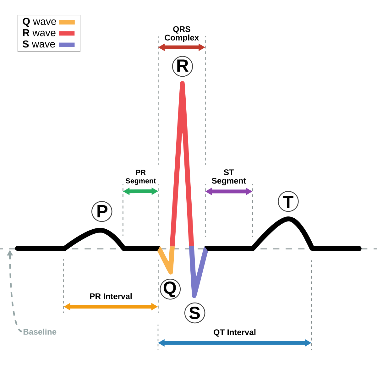
UNDERSTAND WHAT THE 3 DEFLECTION WAVES OF AN EKG INDICATE MATCHING
P-wave: first wave
Can be called atrial contraction, atrial depolarization, and atrial systole (contraction).
QRS complex: ventricular contraction, ventricular depolarization and ventricular systole
T-wave: ventricular relaxation, ventricular repolarization, ventricular diastole
UNDERSTAND THE EFFECTS OF THE SYMPATHETIC AND PARASYMPATHETIC NERVOUS SYSTEM ON THE HEART
Sympathetic nervous system: flight or fight system
Speeds up the heart and increase force of contractions
Uses neurotransmitter norepinephrine/noradrenaline
Parasympathetic nervous system: rest and restore system
Slows down the heart and reduce force of contractions contractions
Uses neurotransmitter acetylcholine
KNOW THE FOLLOWING TERMS: angina pectoris, pernicious anemia, bicuspid valve prolapse, polycythemia, thrombocytopenia
Matching
Angina pectoris: chest pain associated with heart disease due to insufficiency of RBCs
Pernicious anemia: insufficient hemopoiesis from inability of stomach to produce intrinsic factor, needed for absorption of vitamin B in the small intestine
Bicuspid valve prolapse: backflow of blood from the left ventricle into the left atrium,can cause mitral insufficiency. In MVP one or both cusps of the mitral valve protrude into the left atrium during ventricular contraction, when left av pushes back during ventricular systole
Polycythemia: the number of RBCs is abnormally high
Thrombocytopenia: Very low platelet count that results in a tendency to bleed from capillaries.
KNOW THE FOLLOWING TERMS: vaso vasorum, tunica interna, tunica media, tunica externa, elastic arteries, muscular arteries, arterioles, capillaries, veins
MATCHING
Vaso vasorum: blood vessels that supply blood vessels
Tunica interna: deep layer of the blood vessels,
made of endothelium
Internal elastic tissue
Tunica media: middle layer of blood vessels,
made up of smooth muscle
External elastic tissue
Tunica externa: fibrous collagen tissue that supports it
Elastic arteries: large conducting elastic arteries
Example: aorta
Muscular arteries: distributing medium-sized arteries
Have more muscle tissue
Involved with vaso constrction and vaso dilation
Example: radial arteries, ulnar arterieries brachial arteries, femoral arteries,
Tibial arteries, etc (limb arteries)
Arterioles: very very small transitional arteries
Carries blood from the medium-sized arteries to the capillary beds
Capillaries: smallest microscopic blood vessels, major area of diffusion
Found everywhere in the body except in epithelial tissue, cartilage, epidermis, and cornea of the eyes
Veins: drain the blood from the capillary beds coming back to the heart
Have a thinner wall than an artery
NO elastic tissue
NO under pressure
YES valves, when arteries don’t have the valves
UNDERSTAND: vasoconstriction vs. vasodilation
Vasoconstriction: when blood vessels narrow, reduce blood flow, increase blood pressure
Vasodilation: when blood vessels widen, increase blood flow, lower blood pressure
KNOW: which vessels are the most important physiologically
Capillary!!
They don’t have the most blood in them but they are important bc all of our diffusion and active transport mechanisms take place
Where we give off nutrients and pick up waste products
KNOW: which artery is used to check the pulse which artery is used to check the blood pressure
Radial artery - When checking the PULSE
Easy to get to, lateral side of the wrist
Brachial artery - When checking the BLOOD PRESSURE
Next to the biceps
KNOW: the name of the instrument used to take blood pressure
Sphygmomanometer
KNOW: features of the fetal circulation [Matching]
Structures | Blood flow | ||
Umbilical vein | Placenta | → | Fetus |
Umbilical arteries | Fetus | → | Placenta |
Ductus venosus | Bypasses the liver | ||
Ductus arteriosus | Pulmonary trunk | → | Aorta |
Foramen ovale | Right atrium | → | Left atrium |
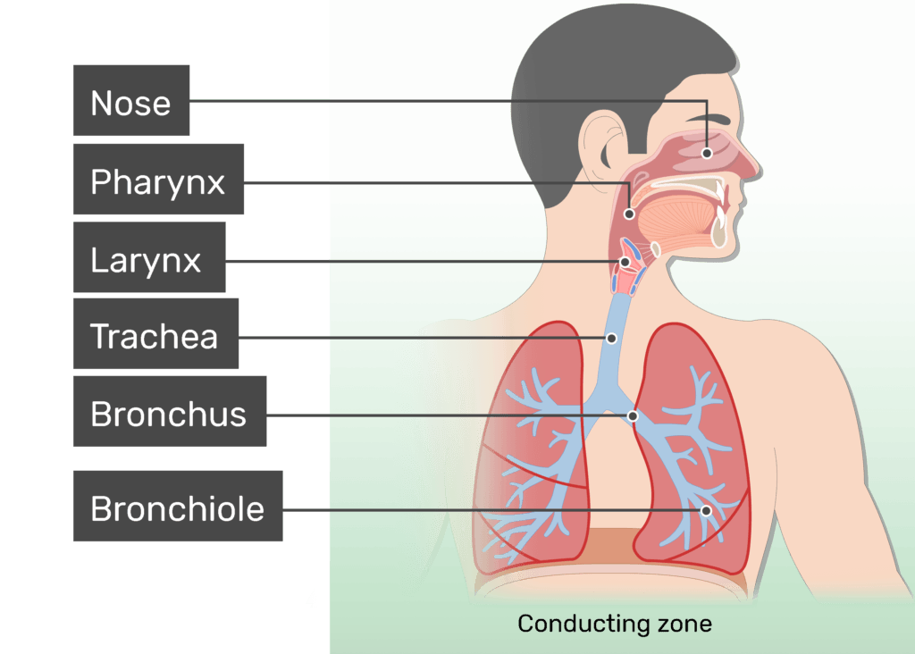
KNOW: the anatomical order of the respiratory system [multiple choice]
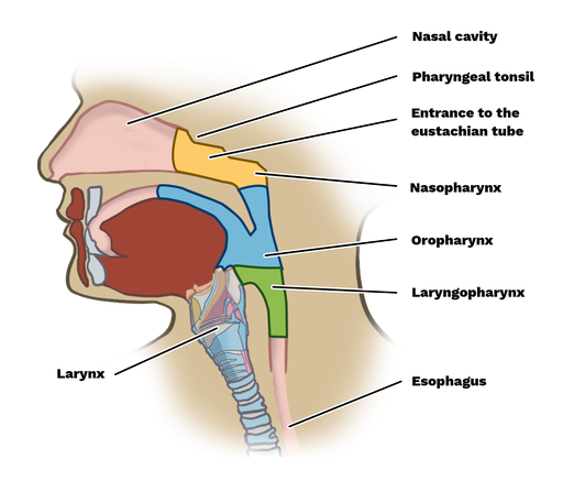
Nose/nasal cavity area
Nasopharynx
Oropharynx (directly posterior to the oral cavity)
Laryngopharynx (most inferior part of the pharynx)
Larynx (voice box)
Trachea
Primary (main) bronchi
Secondary (lobar) bronchi
Tertiary (segmental) bronchi
Bronchioles
Terminal bronchioles
Respiratory bronchioles
Alveolar ducts
Alveolar sacs
KNOW: the functions of the nose
1. Warms, moistens and filters the air
2. Olfaction = sense of smell
3. Vocal resonance (sound of voice comes from the shape of the nose/nasal cavity, sinuses)
KNOW: the parts of the nasal septum
Perpendicular plate of ethmoid
Vomer
Septal cartilage
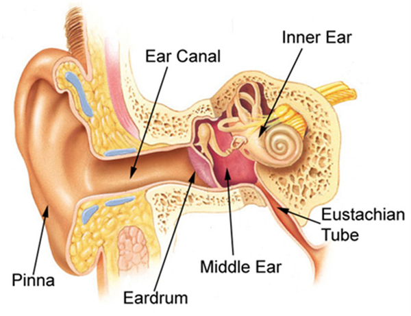
KNOW: where the auditory tube location and function
Auditory tube:
Also known as Eustachian tube
Connects the nasopharynx to middle ear
Allows us to equalize the air pressure on both sides of the eardrum
If you travel in mountains, there is less atmospheric pressure and the eardrum wants to bulge out slightly. If you swallow, it allows atmospheric pressure to travel up the auditory tube to the inner side of the eardrum and it equalizes.
KNOW: cartilage structures in larynx / structure & function
(1) epiglottis: moveable cartilage flap that seels off the larynx when we are swallowing so we don't aspirate liquids or solids into our respiratory system
(2) thyroid cartilage: Adam’s apple which is largest cartilage structure. It is a landmark structure that lets us know where the larynx is
(3) Cricoid cartilage: first true cartilage ring that we feel below thyroid cartilage and it indicates junction of larynx and trachea.
KNOW: the control center of normal respiration
Medullary rhythmicity center - found in medulla oblongata in brainstem - lowest most primitive part of the brain
Pons - allows us to take prolonged inhalation or exhalation or hold our breath.
KNOW: the chemical control mechanism of respiration
Carbon dioxide is the main chemical control because as CO2 l in evels go up, our blood pH will go down. Blood has to have pH of 7.35-7.5 so if you hold your breath, you affect pH of blood which signals brain to breathe faster and get CO2 out and O2 in and shift pH levels.
KNOW: anemic hypoxia, stagnant hypoxia, histotoxic hypoxia, hypoxic hypoxia
Matching
Hypoxia: medical term for a decrease delivery or availability of oxygen to tissues
Anemic hypoxia: oxygen is available but RBC issue - decreased number of RBC or decreased number of functional RBC. Oxygen is available, but RBCs can’t bring them
Stagnant hypoxia: oxygen and RBC is available but there is something going on with the cardiovascular system so can’t pump blood to lungs or out the aorta to the rest of body
Ex: heart attack, major blood loss,
Histotoxic hypoxia: inability of cells to use oxygen
Ex: cyanide poisoning which blocks the cells from using oxygen
Hypoxic hypoxia: decreased availability of oxygen due to decreased availability of oxygen
High altitude which has less oxygen
KNOW: parts of the lymphatic system; function of the lymphatic system; different types of lymphatic tissue MATCHING
Parts:
Lymph - fluid interstitial intercellular fluid found in lymph vessels
Lymph vessels - like veins, thinly muscled walls, have valves, do NOT transport lymph anywhere, pick lymph from capillary beds and carry it back to bloodstream, returning leaked intercellular fluid and plasma proteins out cleansing it and returning it to blood
Lymph organs and structures: spleen (largest lymphatic tissue), thymus gland, lymph nodes, tonsils, diffuse loose lymphatic tissue in digestive system, bone marrow
Function:
Cleanses and returns leaked plasma proteins from capillary beds back to blood,
Cleanses and returns interstitial plasma fluid back to bloodstream
transportation of dietary fats,
Protection and immunity in both specific and non-specific immune resistance - involved in manufacture of WBCs
KNOW: methods of non-specific disease resistance
Mechanical barrier: skin, intact mucous membranes, epiglottis, urination, defecation
Chemical: sebum on skin, acids in vagina, lacrimal apparatus (tears), gastric juice
Antimicrobial agents: complement, interferons, properdins
Phagocytosis: eating cells
Natural Killer (NK) Cells
Inflammation
Fever
DIFFERENTIATE BETWEEN: IgG, IgA, IgM, IgD, IgE
Matching
Ig: immunoglobins or antibodies
IgG: involved in protecting against in bacteria and viruses by enhancing phagocytosis
IgA: found in secretions of body - tears, saliva, mucous, breastmilk, blood
IgM: involved in causing microbes in agglutination and cytolysis (punching hole in cell membrane)
IgD: activates more B-cells to be converted into plasma cells to make more antibodies
IgE: found on outside of mass cells or basophils and involved in allergic reactions
KNOW: cells produced upon exposure to an antigen, plus their functions
T cells: directly attack the foreign invaders, develop from the thymus gland
Helper T Cells
Cytotoxic T Cells
Memory T cells
B cells: produce antibodies to attack the foreign proteins, develop from the gut and spleen, and differentiate into plasma cells
Memory cells: BOTH T and B cells produce MEMORY CELLS are programmed to recognize the original antigen, with a SECOND EXPOSURE there is a faster reaction time, conferring immunity
Helper Cells: When a person is infected with HIV(human immunodeficiency virus), it will affect and infect helper cells, preventing both T cells and B cells from functioning properly
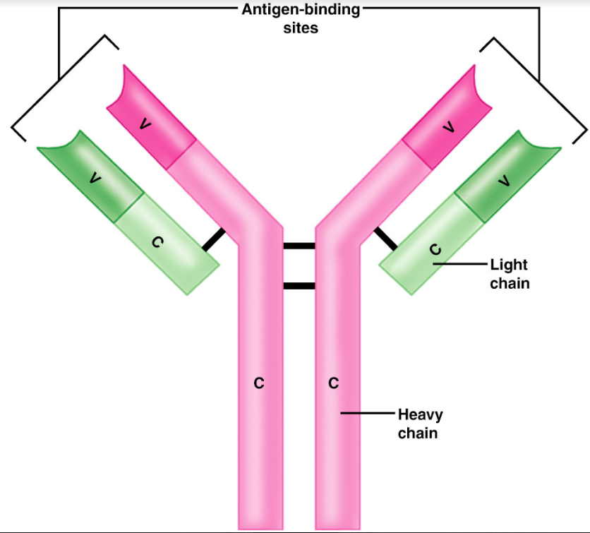
KNOW: the structure of an antibody
Y-shaped protein substances that have a pair of heavy chains and a pair of light chains
Constant portion
determines the antibody class (IgA, IgG, IgM, IgD, etc)
Variable portion
antigen-binding site(ABS) is found on the tip of the variable portion, which changes to match each specific antigen (foreign protein) the antibody is exposed to
KNOW DEFINITION OF: dyspnea, bradycardia, tachycardia MATCHING
Dyspnea
Difficulty or labored breathing, shortness of breath
Bradycardia
Abnormally slow heart rate, typically below 60 beats per minute
Tachycardia:
Abnormally fast heart rate, generally above 100 beats per minute