Biological Bases of Behavior
The Nervous & Endocrine Systems
Nervous system - all nerve cells and electrochemical communications
- Central nervous system (CNS) - brain and spinal cord
- Peripheral nervous system - connects CNS to the rest of the body
- Types of Neurons:
- Sensory/afferent - carries signals from the body’s sensory receptors to the brain
- Motor/efferent - carries signals from the brain to the body
- Interneurons - within the brain they process info
Peripheral Divisions:
- Somatic - enables control of skeletal muscles
- Autonomic (ANS) - controls functions like gland activity, digestion, heartbeat, etc
- usually not consciously aware of it
- Sympathetic - amps you up in response to a stressor
- eg. raises heart rate, blood pressure, sugar, etc
- Parasympathetic - calms you down when a stressor is over
Neural Network - groups of neurons that are near each other. work together as we learn and reinforce tasks/skills
- when learning a new skill you’re forming new brain networks
Spinal Cord reflex arc: sensory info enters the body → spinal cord intercepts signal and reacts → motor neurons receive commands from spinal cord → pain stimulants enter the brain
The Endocrine System - a set of glands that secrete hormones into the bloodstream
- Hormones - chemical messengers that act on body tissues
- Adrenal glands - near the kidneys, release (nor)epinephrine that gives us fight or flight response; feel effects for a while after event
- Pituitary gland - releases hormones related to growth but tells the other glands to release sex and stress hormones
Studying the Brain
The tools of discovery:
- lesion - tissue destruction
- naturally or experimentally caused by the destruction of brain tissue
- (EGG) electroencephalogram - an amplified recording of the waves of electrical activity sweeping across the brain’s surface
- the waves are measured by electrodes placed on the scalp
- CT scan - a series of x-ray photos taken from different angles and combined by a computer into a composite representation of a slice of the brain’s structure (aka CAT scan)
- PET scan - a visual display of brain activity that detects where a radioactive form of glucose goes while the brain performs a given task
- MRI - a technique that uses magnetic fields and radio waves to produce a computer-generated image of the soft tissue
- fMRI - a technique for revealing blood flow and brain activity by comparing successive MRI scans
Neurotransmitters
Acetylcholine (ACh) - involved in all body movements, slows heart rate, contracts smooth muscles
- Excess: muscle weakness, cramps, paralysis
- Shortage: dementia, Alzheimer’s
Dopamine - a feel-good hormone, gives pleasure feeling (nostalgia)
- Excess: ADHD
- Shortage: Parkinsons
Serotonin (5HT) - mood & sleep, produces melatonin; carries messages from nerve cells to brain; influence digestion, nausea, etc
- Excess: severe serotonin syndrome
- Shortage: depression, anxiety
Norepinephrine - increases alertness, constricts blood vessels, affects the sleep-wake cycle
- Excess: pheochromocytoma
- Shortage: ADHD
GABA - primary inhibitory, blocks impulses for nerve cells
- Excess: hypersomnia
- Shortage: epilepsy, autism, schizophrenia
Glutamate - learning and memory, the energy source for brain cells
- Excess: death, Parkinsons, Alzheimers
- Shortage: insomnia, concentration problems, mental exhaustion
The Cerebral Cortex
Cerebral Cortex - the intricate fabric of interconnected neural cells covering the cerebral hemisphere; the body’s ultimate control and information processing center
Structure of the Cortex:
- glial cells - cells in the nervous system that support, nourish, and protect neurons; they also play a role in learning and thinking
- 4 lobes:
- Frontal- involved in speaking, muscle movement, thinking, planning, making judgments, and inhibiting behavior
- location: behind the forehead
- Parietal - involved in the processing of physical touch, helps give us our sense of body positions, helps facilitate language
- Location: top rear of the skull
- Occipital - involved in visual processing
- location: far back of the skull
- Temporal - responsible for processing hearing, involved in facial recognition
- location: right inside and above the ears
Functions for the Cortex:
- motor cortex - an area at the rear of the frontal lobes that controls voluntary movements
- somatosensory cortex - area at the front of the parietal loves that registers and processes body touch and movement sensations
- association areas - areas of the cerebral cortex that are not involved in primary motor sensory functions; rather, they are involved in higher mental functions such as learning, remembering, thinking, and speaking
The Brain’s Plasticity:
- plasticity - the brain’s ability to change, especially during childhood, by reorganizing after damage or by building new pathways based on experience
- 2 hard facts about the effects of brain damage:
- severed neurons usually don’t regenerate (paralysis)
- some brain functions seem preassigned to specific areas
- neurogenesis - the formation of new neurons
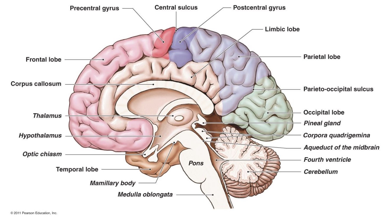
Other Parts of the Brain
Medulla - the base of the brainstem; controls heartbeat and breathing
- location: where the spinal cord enters the brain
Pons - damage here can cause a coma
- location: above the medulla
Reticular formation - a relay network where neurons cross and are responsible for arousal (attention)
- location: stretches from the spinal cord through the brainstem and thalamus
Thalamus - sends signals to other parts of the brain; doesn’t have an actual function other than that
- location: atop the brainstem in the core of the brain
Hypothalamus - contains pleasure centers (releases dopamine), responsible for 4 F's: fighting, fleeing, feeding, and mating
- location: below the thalamus
Cerebellum - processes sensory inputs, enables motor coordination and associated with non-verbal learning, sense of balance
- Location: rear of the skull behind the brainstem, below occipital lobe
Amygdala - associated with emotion and aggression
- location: in the core
Hippocampus - processes conscious memories, translates immediate experiences, and transfers them to long-term memory
- location: near thalamus
Corpus callosum - connects the hemispheres and transfers messages between them
Motor cortex - controls voluntary "big" movements
- location: rear of frontal lobes
Somatosensory cortex - processes sensory inputs (especially touch)
- location: front of parietal lobes
Association areas - involved in cognitive tasks, learning, memory, reasoning, speaking, etc
- Location: all over the cerebral cortex
Broca’s Area - responsible for language production (written, spoken, signed)
- Location: near the rear of the left frontal lobe
Wernicke’s Area - responsible for language comprehension
- Location: in left temporal lobe
Agonists & Antagonists
Agonists - chemicals that bind to NT receptor sites and activate a response
- full agonists - activate receptor sites at full efficiency (eg. morphine)
- partial agonists - only activate receptor sites at partial efficiency (eg. buprenorphine)
Antagonists - chemicals that bind to NT sites and block a response
- they have to be similar enough to NTs to fit in the receptor, but not so similar as to cause a response