The Somatosensory System
Main Somatosensory Pathways
Component | Function | Pathway Characteristics |
Posterior Column-Medial Lemniscal Pathway | Fine touch, vibration, proprioception | Ascends in the dorsal column without decussating until the medulla, then crosses and ascends to the thalamus and cortex |
Anterolateral Pathway | Crude touch, temperature, pain (localisation and nature) and pressure | Composed of multiple tracts that decussate in the spinal cord shortly after entry, then ascend to higher brain structures for processing (e.g., thalamus, cortex, reticular formation) |
Neuospinothalamic Tract (Lateral Pain System) | Fast, sharp and localisable pain and temperature | Decussates in the spinal cord, ascends to the thalamus and cortex |
Paleospinothalamic Tract (Medial Pain System) | Slow, dull, aching and poorly localisable pain and crude touch (also emotional aspects) | Decussates in the spinal cord, ascends to the thalamus and cortex |
Spinoreticular Tract | Emotional response and arousal to pain | Ascends to the reticular formation in the brainstem |
Spinomesencephalic Tract | Pain modulation | Ascends to the periaqueductal grey (PAG) in the midbrain |
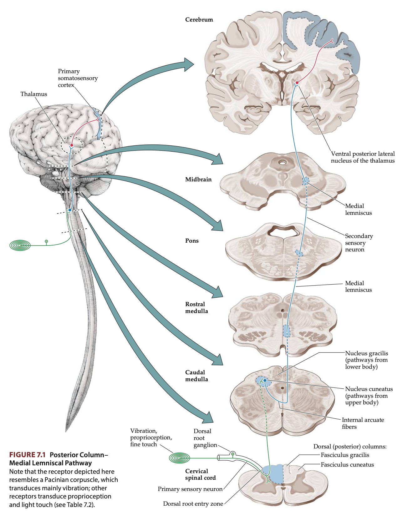
Posterior Column-Medial Lemniscal Pathway
Posterior Column-Medial Lemniscal Pathways: Sensory pathways in the spinal cord that carry information about fine touch, vibration, pressure, and proprioception (sense of body position) from the peripheral nervous system to the central nervous system.
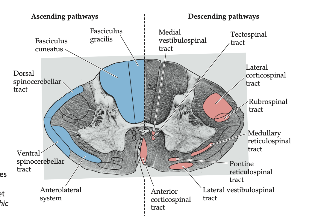
Gracile Fasciculus: Medial portion of the posterior column that carries information from the legs and lower trunk.
Cuneate Fasciculus: Lateral portion of the posterior column that carries information from the upper trunk, arms and neck.
Pathway:
First-Order Neurons (Peripheral to Spinal Cord): Sensory receptors detect sensory information and send their axons through the dorsal roots of the spinal nerves and enter the ipsilateral posterior columns of the spinal cord to ascend to the dorsal column nuclei of the medulla.
Second-Order Neurons (Spinal Cord to Medulla): The axons of the first-order neurons synapse with second-order neurons in the medulla. These second-order neurons then cross to the opposite side (decussate) in the medulla as internal arcuate fibres to form the medial lemniscus and continue to ascend.
Third-Order Neurons (Medulla to Thalamus and Cortex): The medial lemniscus axons terminate in the ventral posterolateral nucleus (VPL) of the thalamus where they synapse to the third-order neurons which send their axons to the primary somatosensory cortex (S1) in the parietal lobe trough the posterior limb of the internal capsule.
Functions:
Fine Touch (Tactile Discrimination): The pathway enables the ability to detect small, detailed textures and identify objects by touch.
Vibration Sense: It allows the detection of vibration on the skin, such as the sensation felt when holding a vibrating object.
Pressure Sensation: It carries information about deep pressure applied to the skin.
Proprioception (Sense of Body Position): The pathway conveys information about the position and movement of different parts of the body, contributing to balance and coordination.
Somatotopic Organisation: In the posterior columns the feet are medial to the arms (think of fibres adding on laterally from higher levels as the posterior columns ascend) while for the medial lemniscus the feet are lateral to the arms (humunculus lying down)
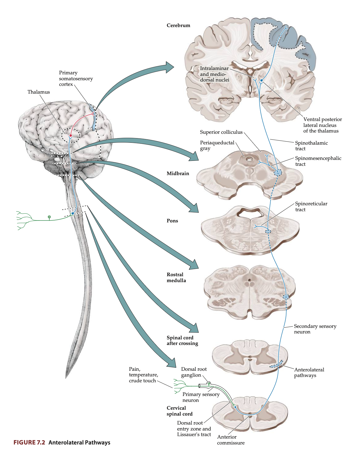
Anterolateral Pathways
Anterolateral Pathways: Sensory pathways in the spinal cord that carry information related to pain, temperature, crude touch, and pressure from the peripheral nervous system to the central nervous system.
Spinothalamic Tract: Mediates discriminative aspects of pain and temperature sensation
Neospinothalamic Tract (Lateral Pain System):
Carries sharp, pricking, localisable pain and temperature information.
Critical for detecting noxious stimuli that can cause injury.
Paleospinothalamic Tract (Medial Pain System):
Transmits information about slow burning, aching pain, crude touch and pressure.
The touch information carried by this tract is less precise compared to the posterior column pathway.
Spinoreticular Tract:
Terminates in the medullary-pontine reticular formation in the brainstem, involved in alertness and the emotional response to pain.
Helps the brain process the wakefulness and behavioural arousal associated with painful stimuli by projecting diffusely to the entire cerebral cortex.
Spinomesencephalic Tract:
Projects to the periaqueductal grey (PAG) and superior colliculi in the midbrain, which plays a role in pain modulation and can activate descending pain inhibition pathways.
Pathway:
First-Order Neurons (Peripheral to Spinal Cord):
Sensory receptors detect sensory information and send signals via neurons which send their axons through the dorsal roots to enter the spinal cord.
Second-Order Neurons (Spinal Cord to Thalamus):
These axons immediately synapse with second-order neurons in the dorsal horn of the spinal cord which then decussate in the anterior commissure of the spinal cord within a few segments of entry. After crossing, the fibres ascend in the anterolateral white matter of the spinal cord as part of the spinothalamic tract.
Third-Order Neurons (Thalamus to Cortex): The second-order neurons terminate in the ventral posterolateral nucleus (VPL) of the thalamus. The thalamus then relays the information to the primary somatosensory cortex (S1) in the parietal lobe, where conscious perception of the sensory stimuli occurs. Some fibres also project to other thalamic nuclei and regions involved in the emotional and autonomic aspects of pain, such as the reticular formation and periaqueductal grey (PAG).
Functions:
Pain Sensation (Nociception): Transmit information about tissue damage, allowing the body to perceive pain and initiate protective responses.
Temperature Sensation: Convey information about changes in temperature, such as detecting heat or cold.
Crude Touch and Pressure: Transmit sensations of general touch and pressure that lack fine discrimination.
Pain Modulation: Certain tracts within the system, such as the spinoreticular and spinomesencephalic tracts, are involved in the modulation of pain and the emotional and autonomic responses to painful stimuli.
Somatotopic Organisation: In the anterolateral pathways the feet are most lateral (picture fibres from the anterior commissure adding on medially as the anterolateral pathway ascends in the spinal cord)
Example: If you step on a thumbtack with your left foot:
your spinothalamic tract (neo division) enables you to realise ‘something sharp is puncturing the sole of my left foot’
your spinothalamic intralaminar projections (paleo division) and spinoreticular tract cause you to feel ‘ouch, that hurts’
and your spinomescencephalic tract leads to pain modulation, allowing you to eventually think ‘ahh, that feels better’
Thalamus
Thalamus: Pair of structures located deep in the brain, serving as the brain's sensory relay station by processing and transmitting sensory information to the cerebral cortex
Each sensory modality - except for olfactory input - has a different nuclear area where synapses occur before the information is relayed to the cortex (reciprocal circuits):
Ventral Posterolateral (VPL) Nucleus: Processes somatosensory information from the body (fine touch, proprioception, pain, temperature) and projects to the primary somatosensory cortex (S1). Receives input from both the spinothalamic tract and poterior columns-mendial lemniscal pathway, however, to separate neurons
Lateral Geniculate Nucleus (LGN): Relays visual information from the retina to the primary visual cortex (V1)(Think lateral light).
Medial Geniculate Nucleus (MGN): Transmits auditory information from the ear to the primary auditory cortex (A1) (Think medial music).
Pulvinar Nucleus: Involved in visual attention and integrates sensory information for higher-level processing.
Reticular Nucleus: Thin layer of inhibitory neurons (GABAergic neurons) that surrounds the thalamus like a shell. It is unique in that it does not send signals directly to the cerebral cortex but instead communicates exclusively with other thalamic nuclei. It receives input from both the thalamus and the cerebral cortex, allowing it to act as a regulatory filter for information travelling through the thalamus.
Role in Sensory Processing:
Relay Function: The thalamus receives sensory inputs from the peripheral nervous system and lower brain regions, relaying them to the primary cortical areas for conscious perception (e.g., visual, auditory, somatosensory).
Modulation and Filtering: It filters and prioritises sensory signals, enhancing important stimuli and suppressing irrelevant ones based on attention and context.
Multimodal Integration: The thalamus integrates sensory inputs across different modalities, ensuring a coherent perception of the environment.
Functions Beyond Sensory Relay:
Conscious Perception: The thalamus helps make sensory information available to conscious awareness.
Attention and Arousal: It modulates sensory input based on focus and arousal levels, influencing what reaches conscious awareness.
Motor Control: Through connections with the basal ganglia and cerebellum, it aids in coordinating movement.
Damage to the Thalamus: When the thalamus is damaged, particularly the bilateral thalamus (affecting both sides), it can result in a coma. This is because the disruption impairs the relay of sensory and arousal signals to the cortex, preventing the brain from achieving a state of consciousness.
Nuclei | Main Input | Main Output | Functions |
Ventral Posteriorlateral Nucleus (VPL) | Medial lemniscus and spinothalamic tract | Somatosensory cortex | Relays somatosensory spinal input to cortex (body) |
Ventral Posteriormedial Nucleus (VPM) | Trigmental lemniscus and trigmentothalamic tract | Somatosensory and taste cortex | Relays somatosensory cranial nerve inputs and taste to cortex (face) |
Lateral Geniculate Nucleus (LGN) | Retina | Primary visual cortex | Relays visual input to cortex |
Medial Geniculate Nucleus (MGN) | Inferior colliculus | Primary auditory cortex | Relays auditory input to cortex |
Pulvinar | Tectum and other sensory inputs | Parietotemporo-occcipital association cortex | Behavioural orientation toward relevant visual and other stimuli |
Reticular Nucleus | Cerebral cortex, thalamic relay and intrathalaminar nuclei, ARAS | Thalamic relay and intralaminar nuclei | Regulates state of other thalamic nuclei |
Pain Modulation
Types of Pain
Nociceptive Pain: Type of pain that occurs in response to actual or potential tissue damage. It results from the activation of nociceptors (A-delta and C fibres) in response to harmful stimuli which generate action potentials that travel to the spinal cord via A-delta (sharp, fast pain) or C fibres (dull, slow pain). From there, signals are relayed trough the spinothalaic tract and thalamus to the somatosensory cortex. This type of pain can be classified as somatic (sharp and localized → neo) or visceral (deep and poorly localized → paleo), depending on the source.
Mechanical: Physical trauma, pressure, or stretching.
Thermal: Exposure to extreme heat or cold.
Chemical: Inflammatory mediators like prostaglandins or bradykinin released during tissue damage.
Neuropathic Pain: Type of pain caused by abnormal neural signaling due to damage or dysfunction of the nervous system—either the peripheral nerves or the central nervous system (CNS).
Ectopic activity: Spontaneous nerve firing in damaged neurons.
Sensitization: Heightened response to stimuli in the nervous system.
Loss of inhibitory control: Reduced ability of the CNS to suppress pain signals.
Descending Pain Modulation Systems - Cortical Level
There are three major descending systems that assist in the modulation and suppression of pain stimuli:
Prefrontal Cortex: The prefrontal cortex (PFC), especially the dorsolateral and anterior cingulate regions, is responsible for the higher-order regulation of pain. It processes the emotional and cognitive aspects of pain, allowing individuals to evaluate the meaning or context of a painful experience. It influences pain perception trough top-down control mechanisms:
Cognitive Reappraisal: The PFC modulates the emotional experience of pain by reframing or shifting focus away from the pain stimulus.
Activation of Descending Pathways: The PFC also modulates pain perception by activating the PAG, which then initiates descending inhibitory pathways
Additionally, it works closely with the limbic system to regulate the affective dimension of pain, such as fear or distress.
Periaqueductal Gray (PAG): The PAG projects to the raphe nuclei in the medulla, which release serotonin into the spinal cord to suppress pain signals. Serotonin inhibits pain by activating spinal inhibitory interneurons and directly reducing the excitability of nociceptive neurons. The PAG is also rich in opioid receptors, making it a critical site for both endogenous (endorphins) and exogenous (e.g., morphine) opioid-mediated pain relief.
Locus Coeruleus: The locus coeruleus (LC) is a major source of norepinephrine in the central nervous system, which acts to suppress pain at the spinal cord level. It sends descending fibres to the dorsal horn of the spinal cord, where norepinephrine reduces pain transmission by two mechanisms:
Directly inhibiting nociceptive neurons by suppressing the release of excitatory neurotransmitters
Activating inhibitory interneurons that release GABA or glycine, further dampening pain signals.
The LC’s activity is also influenced by stress and arousal, helping to reduce pain perception during situations requiring heightened focus or survival responses.
Gate Control Theory - Spinal Level
Gate Control Theory: Theory of pain modulation that proposes that a gate in the spinal cord regulates the transmission of pain signals to the brain. Pain signals from A-delta and C fibres open the gate, increasing pain peception, while non-painful stimuli from A-beta fibres can close the gate, inhibiting pain.
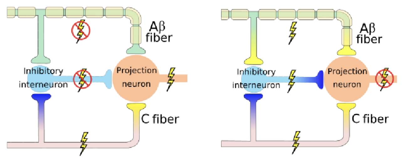
Fibre Type | Conduction Speed | Myelination | Function |
A-delta Fibres | Medium | Lightly | Transmission of sharp, localised pain and detection of coldness and pressure |
A-beta Fibres | Fast | Heavily | Transmission of touch, vibration and proprioception (Important for non-painful sensory input) |
C Fibres | Slow | No | Transmission of dull, poorly-localised pain and detection of warmth |