knowt logo

Pediatric Oral Surgery 2019

Introduction

  • Diagnosis

    • Med history, x-rays

    • Assess ability to cooperate

  • General principles ~ to adult

    • Profound anesthesia, aseptic technique, visibility and surgical site stability, protect airway

Equipment Used

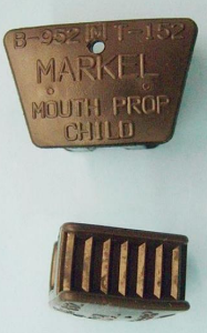
McKesson Bite Block
  • Degree of opening controlled by size selected and placement

  • Attach floss and knot

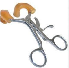
Molt Mouth Prop
  • Utilizes a ratchet-type mechanism

  • Prevents iatrogenic (treatment-induced) injury to the patient

Indications for Simple Exodontia

  • Nonrestorable teeth

  • Fracture of crown/root

  • Prolonged retention due to improper root resorption or ankylosis

  • Impacted teeth

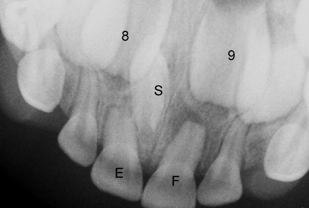
  • Supernumerary teeth

Caveats

  • Concepts that may dictate modifications

    • Proximity of primary tooth to succedaneous tooth

    • Roots on primary teeth with nonresorbed roots will be long and slender

Steps for Tooth Extraction

  1. Preparation

    • Place gauze screen

    • Separate attachment

    • Elevation - concave blade placed against tooth being extracted

  2. Elevation Technique

    • Elevator turned so that the blade resting on alveolus acts as a fulcrum & coronal part of blade rotates toward tooth being extracted.

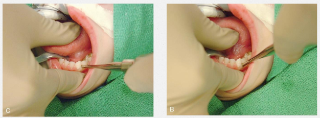
  3. Forceps Extraction

    • Beaks should adapt of root surface

    • Beaks should be parallel to long axis

    • Size of beaks should not impinge on adjacent teeth

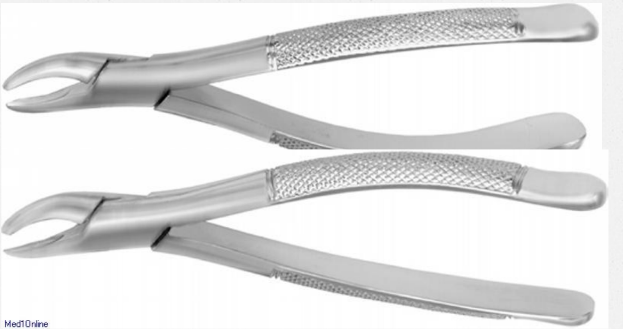
Pediatric Forceps

  • These stainless steel forceps are designed especially for use with primary teeth.

  • Beak & handle are accurately proportioned

  • Forceps can be hidden in the palm, reducing patient stress

  • Allows greater field of view in child’s mouth

  • 5 different forceps are available

  • Cowhorn’s are contraindicated in primary exodontia

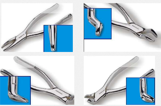
Universal Pediatric Forceps

  • 150s and 151s

  • Denovo Dental

Exodontia Technique

  • First force is apical

  • Apical center of rotation decreases the translational movement of the root

    • Decreased root fracture

    • Disrupts PDL

  • Tooth is SLOWLY luxated to buccal/lingual

  • Movement is in one direction & then stopped allowing the alveolus to expand

  • With each movement force is increased causing expansion of alveolus

  • May position opposite hand so that thumb or index finger can feel expansion

  • Finally slight coronal traction forces are applied

Extraction Techniques

Maxillary and Mandibular Anterior Teeth
  • Accomplished with a rotational movement due to conical single roots

  • Care must be taken not to loosen adjacent teeth

Erupted Maxillary and Mandibular Molars
  • Complications

    • Root fracture not uncommon

    • Inadvertent extraction or dislocation of succedaneous tooth

  • Technique

    • slow palatal/lingual and buccal force to expand the alveolar bone

    • Support mandible to protect TMJ

    • Consider sectioning when primary molar roots encirle the successor's crown

Fractured Primary Tooth Roots

  • Inform perform you perform

  • Dilemma: removing root tip may damage permanent tooth, while leaving may cause infection, delay or deflect permanent tooth eruption

  • Rule of thumb:

    • if tooth root is easily removed do it

    • if tooth root is very small, deep in the socket, close to permanent successor, or unable to remove after several attempts let it be

Ankylosis

  • Periodic monitoring for timing of extraction if space loss, supereruption

    • Wait too long results in tipping of adjacent teeth and creating a difficult extraction

  • Show no signs of mobility despite amount of root present and exhibit a distinctive sound when percussed
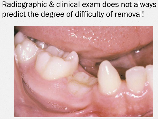
Standard of Care

  • Timing to optimize health and minimize risks

  • Optimal bone healing with improvement of intrabony defects on 2nd molars occurs when surgery is done on individuals less than 25 years of age

  • Associated risks of surgery have been shown to be greater in individuals 25 years and older

Other Impactions

  • Most common is 3rd molar

  • Maxillary canine > second premolar > mandibular second molar > maxillary incisors

  • Impactions of primary teeth associated with patholory

    • Supernumerary teeth

    • Odontomas

      • seen frequently

Soft Tissue Lesions

  • Mucoceles

  • Ranulas

  • Fibromas

  • Pyogenic Granulomas

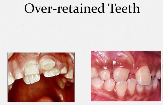
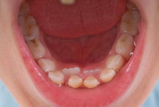
Over-retained Teeth

MD

Pediatric Oral Surgery 2019

Introduction

  • Diagnosis

    • Med history, x-rays

    • Assess ability to cooperate

  • General principles ~ to adult

    • Profound anesthesia, aseptic technique, visibility and surgical site stability, protect airway

Equipment Used

McKesson Bite Block
  • Degree of opening controlled by size selected and placement

  • Attach floss and knot

Molt Mouth Prop
  • Utilizes a ratchet-type mechanism

  • Prevents iatrogenic (treatment-induced) injury to the patient

Indications for Simple Exodontia

  • Nonrestorable teeth

  • Fracture of crown/root

  • Prolonged retention due to improper root resorption or ankylosis

  • Impacted teeth

  • Supernumerary teeth

Caveats

  • Concepts that may dictate modifications

    • Proximity of primary tooth to succedaneous tooth

    • Roots on primary teeth with nonresorbed roots will be long and slender

Steps for Tooth Extraction

  1. Preparation

    • Place gauze screen

    • Separate attachment

    • Elevation - concave blade placed against tooth being extracted

  2. Elevation Technique

    • Elevator turned so that the blade resting on alveolus acts as a fulcrum & coronal part of blade rotates toward tooth being extracted.

  3. Forceps Extraction

    • Beaks should adapt of root surface

    • Beaks should be parallel to long axis

    • Size of beaks should not impinge on adjacent teeth

Pediatric Forceps

  • These stainless steel forceps are designed especially for use with primary teeth.

  • Beak & handle are accurately proportioned

  • Forceps can be hidden in the palm, reducing patient stress

  • Allows greater field of view in child’s mouth

  • 5 different forceps are available

  • Cowhorn’s are contraindicated in primary exodontia

Universal Pediatric Forceps

  • 150s and 151s

  • Denovo Dental

Exodontia Technique

  • First force is apical

  • Apical center of rotation decreases the translational movement of the root

    • Decreased root fracture

    • Disrupts PDL

  • Tooth is SLOWLY luxated to buccal/lingual

  • Movement is in one direction & then stopped allowing the alveolus to expand

  • With each movement force is increased causing expansion of alveolus

  • May position opposite hand so that thumb or index finger can feel expansion

  • Finally slight coronal traction forces are applied

Extraction Techniques

Maxillary and Mandibular Anterior Teeth
  • Accomplished with a rotational movement due to conical single roots

  • Care must be taken not to loosen adjacent teeth

Erupted Maxillary and Mandibular Molars
  • Complications

    • Root fracture not uncommon

    • Inadvertent extraction or dislocation of succedaneous tooth

  • Technique

    • slow palatal/lingual and buccal force to expand the alveolar bone

    • Support mandible to protect TMJ

    • Consider sectioning when primary molar roots encirle the successor's crown

Fractured Primary Tooth Roots

  • Inform perform you perform

  • Dilemma: removing root tip may damage permanent tooth, while leaving may cause infection, delay or deflect permanent tooth eruption

  • Rule of thumb:

    • if tooth root is easily removed do it

    • if tooth root is very small, deep in the socket, close to permanent successor, or unable to remove after several attempts let it be

Ankylosis

  • Periodic monitoring for timing of extraction if space loss, supereruption

    • Wait too long results in tipping of adjacent teeth and creating a difficult extraction

  • Show no signs of mobility despite amount of root present and exhibit a distinctive sound when percussed

Standard of Care

  • Timing to optimize health and minimize risks

  • Optimal bone healing with improvement of intrabony defects on 2nd molars occurs when surgery is done on individuals less than 25 years of age

  • Associated risks of surgery have been shown to be greater in individuals 25 years and older

Other Impactions

  • Most common is 3rd molar

  • Maxillary canine > second premolar > mandibular second molar > maxillary incisors

  • Impactions of primary teeth associated with patholory

    • Supernumerary teeth

    • Odontomas

      • seen frequently

Soft Tissue Lesions

  • Mucoceles

  • Ranulas

  • Fibromas

  • Pyogenic Granulomas

Over-retained Teeth

robot