BOTANY- LAB -NOTES
EXERCISES:
1 THE PLANT CELL
2 PLANT MITOSIS
3 PLANT MEIOSIS IN FLOWERING PLANTS
4 PLANT TISSUE SYSTEM
1 THE PLANT CELL
The cell is generally known as the basic structural and functional unit of life. The cells vary greatly in size and shape.
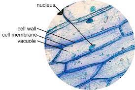
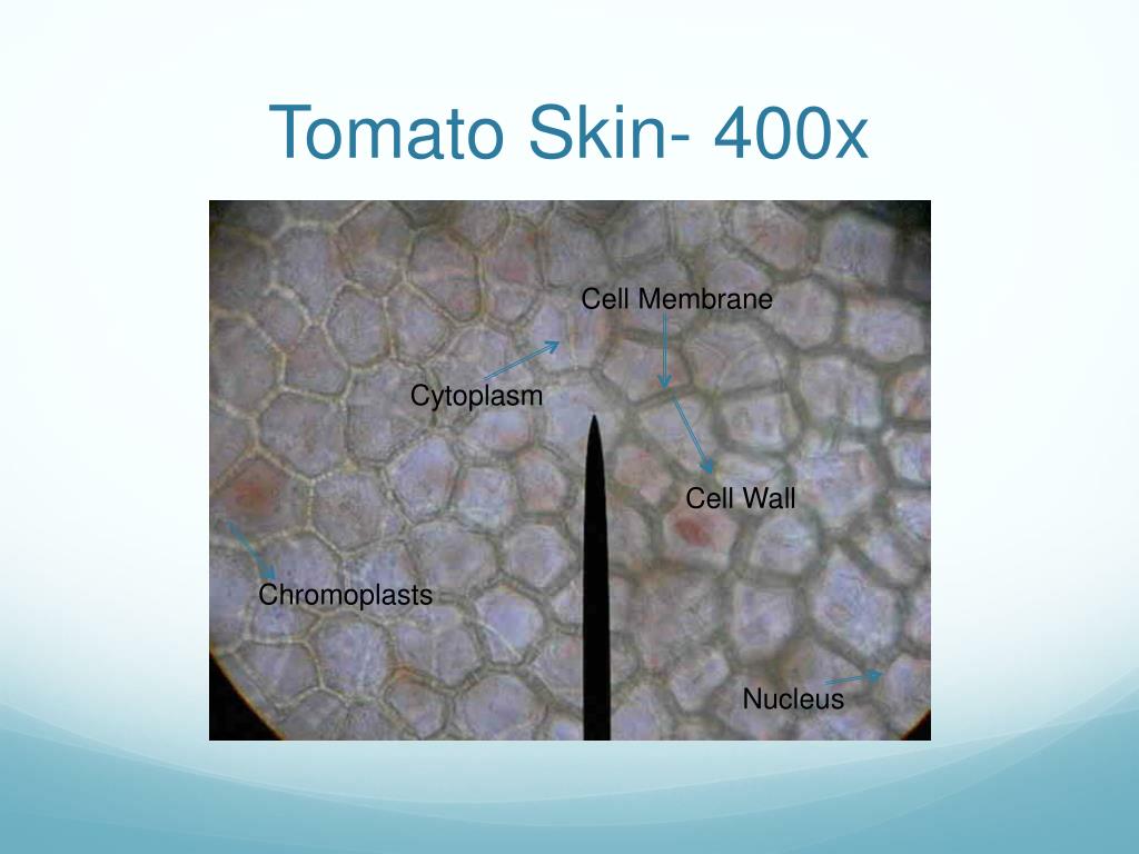
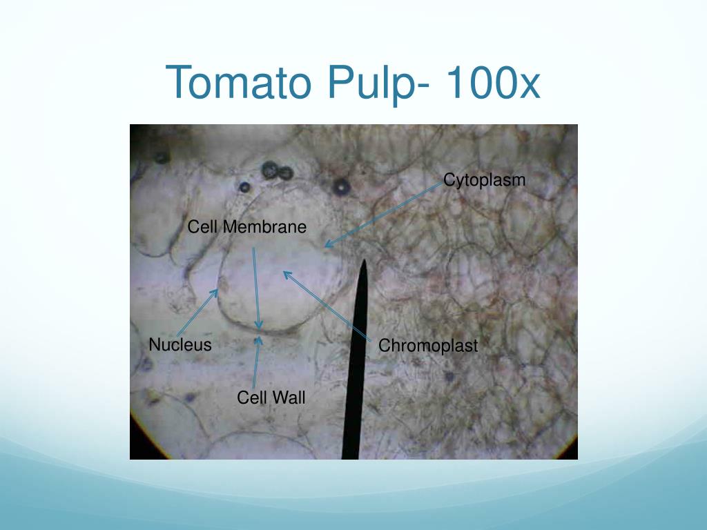
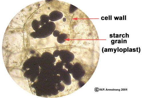
Cell wall- the outermost layer of the cell which distinguishes plant cells from animal cells. It limits the size of the protoplast and prevents rupture of the plasma membrane when the protoplast enlarges following the uptake of water by the cell.
Protoplast- composed of the nucleus and the cytoplasm where the organelles are suspended.
Nucleus- is often the most prominent structure within the protoplast of the eukaryotic cells. It is usually spherical in shape.
Nucleolus- spherical structure within the nucleus.
Cytoplasm- Inside the cell that is a jelly-like material.
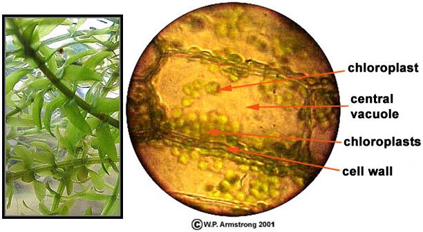
The clear area (vacuole) is filled with fluid (cell sap).
The nucleus, a colorless, somewhat spherical structure could be located by careful observation.
Iodopotassium iodide (IKI) solution**-**Used to stain mounts.
Chloroplastids- The oval green bodies inside the cells.
Cyclosis**-**Streaming protoplast.
Vacuole pigment- Anthocyanins are water soluble pigments. These are present in the vacuoles. These appear in different color like red, blue or purple depending on the pH. Xanthophylls are the typical yellow pigments of leaves.
Plastid pigment- double-membrane organelles that contain the pigments used in photosynthesis and manufacture and store the important chemical compounds used by the cells. These pigments give the colour of the cell.
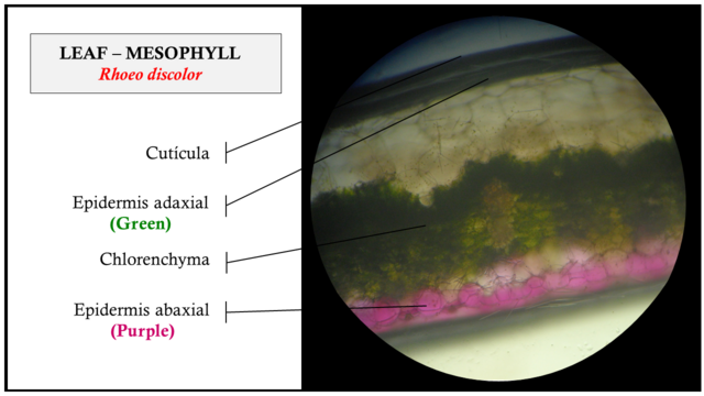
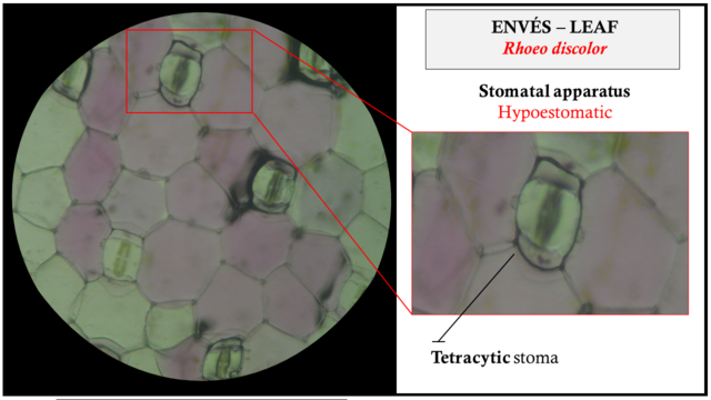
Guard cells- bean-shaped cells scattered all over the area of the leaf blade.
Primary wall- bounded by a single wall.
Middle lamella- an intercellular layer between the primary walls of adjacent cells.
Ergastic substances- are passive products of the protoplast; some are storage products; others are waste products. These substances may appear and disappear at different times in the life of the cell.
Crystals- are usually composed of calcium compounds like calcium carbonate. They vary in shape and size and may be found singly or in clusters.
Calcium oxalate crystals- The shape can either be raphide, druse, prismatic, and sand crystals.
Calcium carbonate crystals – cystoliths. These are outgrowths of the cell wall encrusted with calcium carbonate. They are like bunch of grapes found either in the epidermis, hypodermis or in ground parenchyma cells.
ONION EPIDERMAL CELL (Allium cepa L.)
DIGMAN LEAF CELLS (Hydrilla verticellata)
CELLS OF TOMATO FRUIT (Lycopersicum esculentum L.)
BANGKA-BANGKAAN LEAF CELLS (Rhoeo spathacea)
CELLS OF COCONUT SHELLS
Petiole of gabi-gabi (Calocasia esculenta)
Midrib of dumb cane (Diffenbachia sp.)
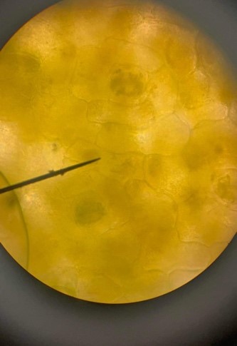
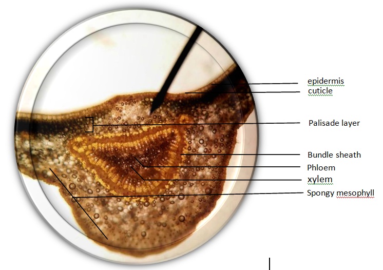
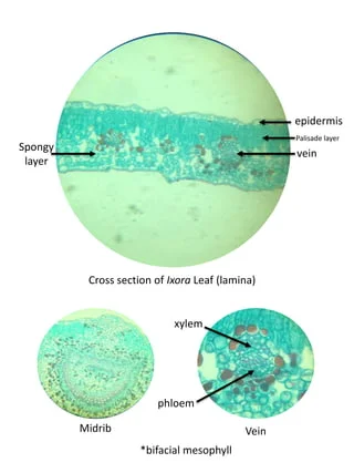
santan leaf (lxora sp.)
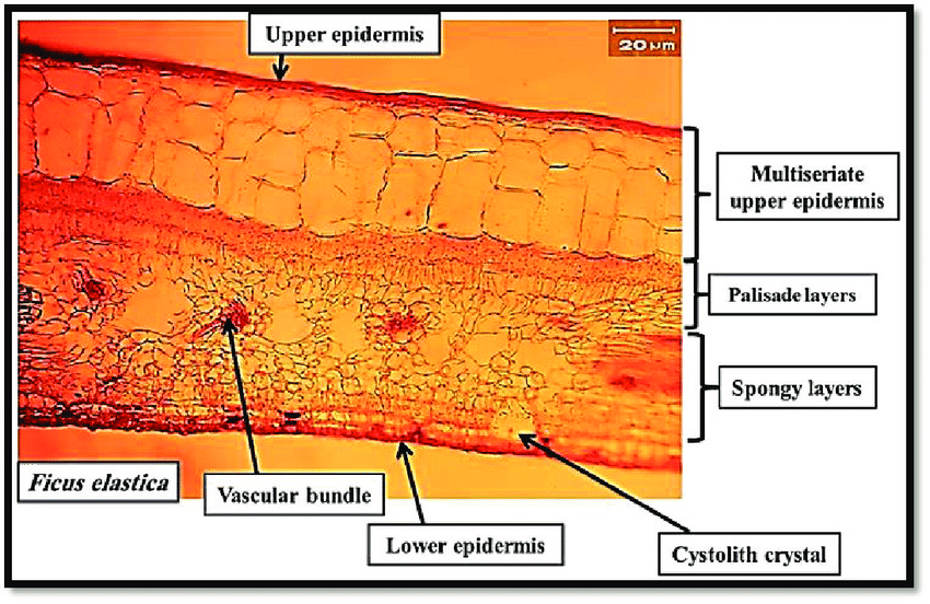
Indian rubber tree (Ficus elastica)
potato
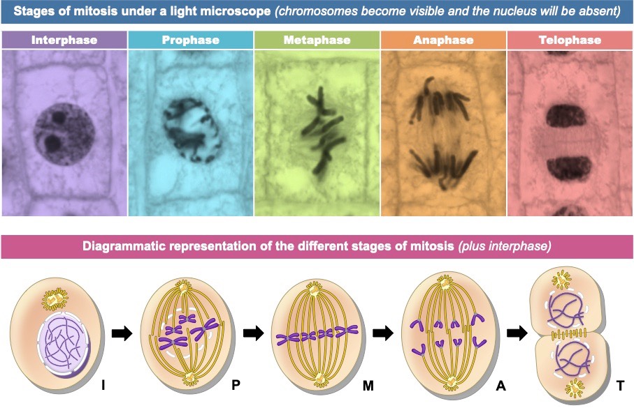
2 PLANT MITOSIS
Mitosis- All cells arise from pre-existing cells by a process of cell division.
two stages:
karyokinesis or mitosis (nuclear division) and
cytokinesis (division of the cytoplasm).
Cell division in plants mainly occurs in special meristematic tissues (regions in the plant in which cells retain the capacity for repeated division).
Meristematic region- the region of rapidly dividing cells.
INTERPHASE- the stage in which the cell is p__reparing for the incoming cell division or mitosis.__
PROPHASE- This is characterized by dark-staining thread-like structures called chromosomes that are randomly distributed throughout the cytoplasm. The nuclear membrane and nucleoli disappear.
METAPHASE- the chromosomes have lost their random orientation and have arranged themselves near the center of the cell. Very thin strands called spindle fibers may be seen extending from the chromosomes to opposite ends of the cell.
ANAPHASE- The duplicates of the chromosomes are separated and are moved towards the opposite poles of the spindle. Spindle fibers are evident between the migrating chromosomes.
TELOPHASE- the chromosomes elongate and become indistinct. A developing cell plate appears across the center of the parent cell separating the two developing nucleus. In here, the nuclear membrane and nucleoli begin to form. The completion of the cell wall ends the cell division.
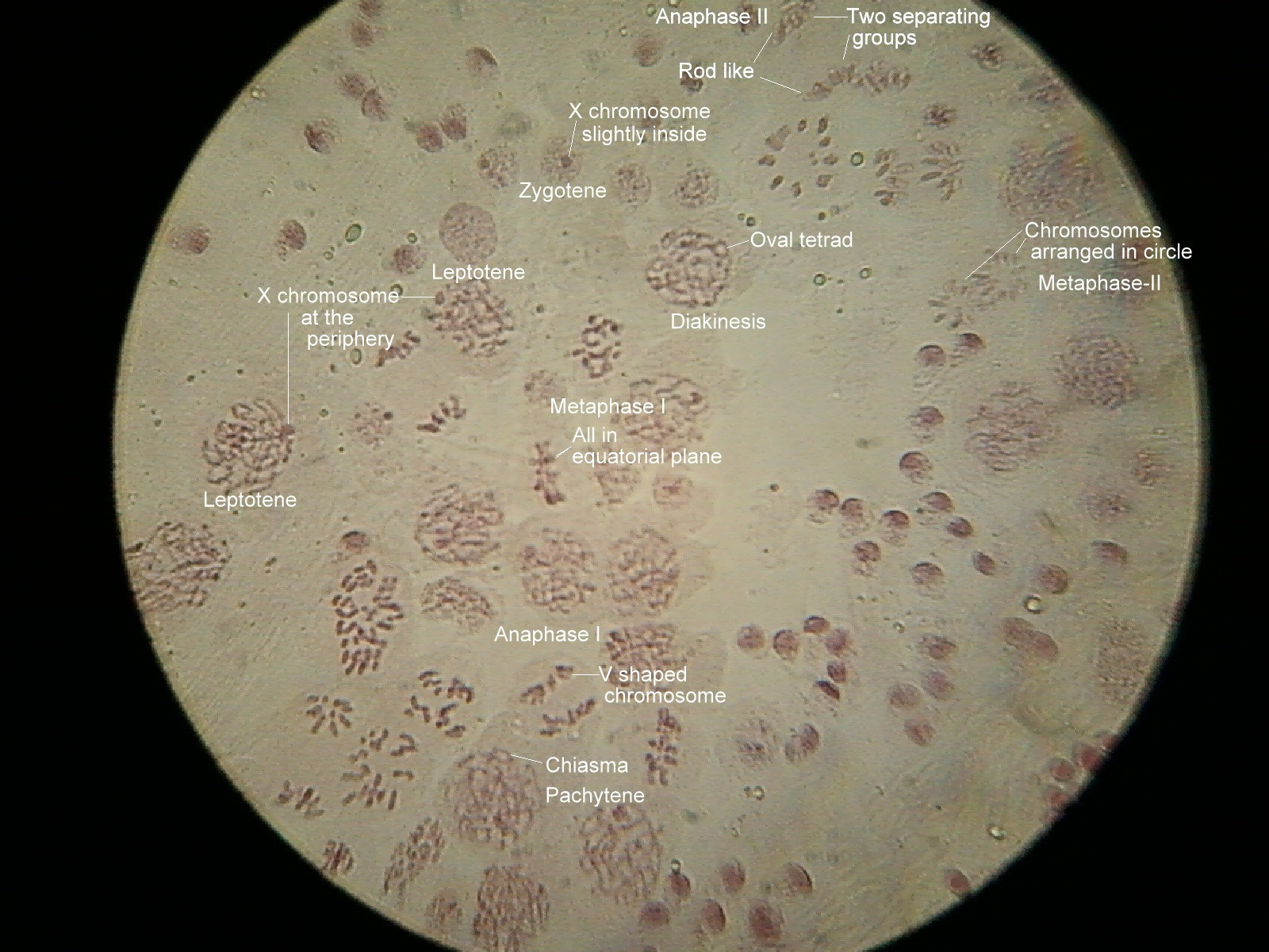
3 PLANT MEIOSIS IN FLOWERING PLANTS
MEIOSIS- It is a special kind of cell division that ensures the constancy of the chromosome number in the cells of the succeeding generations of the organism.
The diploid (2N) number of chromosomes during cell division is reduced into half thus, the daughter cells produced, or gametes are haploid (N), half the number of the parent cells.
During fertilization, the zygote becomes diploid again.
Meiosis occur in the anther and pistil, the male and female reproductive organs of the plant respectively.
Anther- produces male spores (microspores) which will develop into pollen grains.
Ovary- The basal enlarged part of the pistil, are embryonic structures called ovules which will produce megaspores.
When these gametes fertilized, the ovary will develop into fruit and the ovules will mature into seeds.
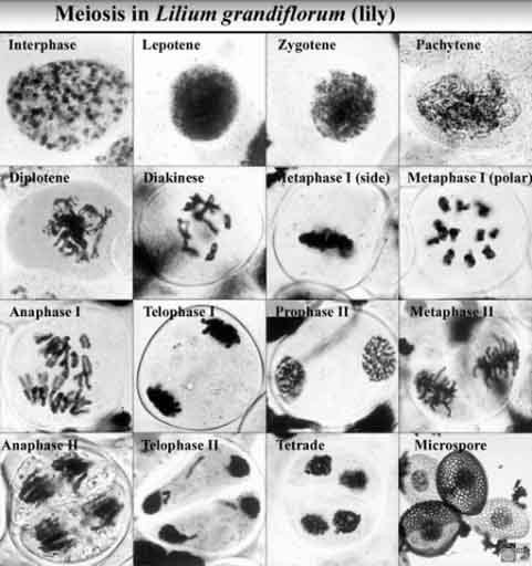
Stages of Meiosis
Meiosis I
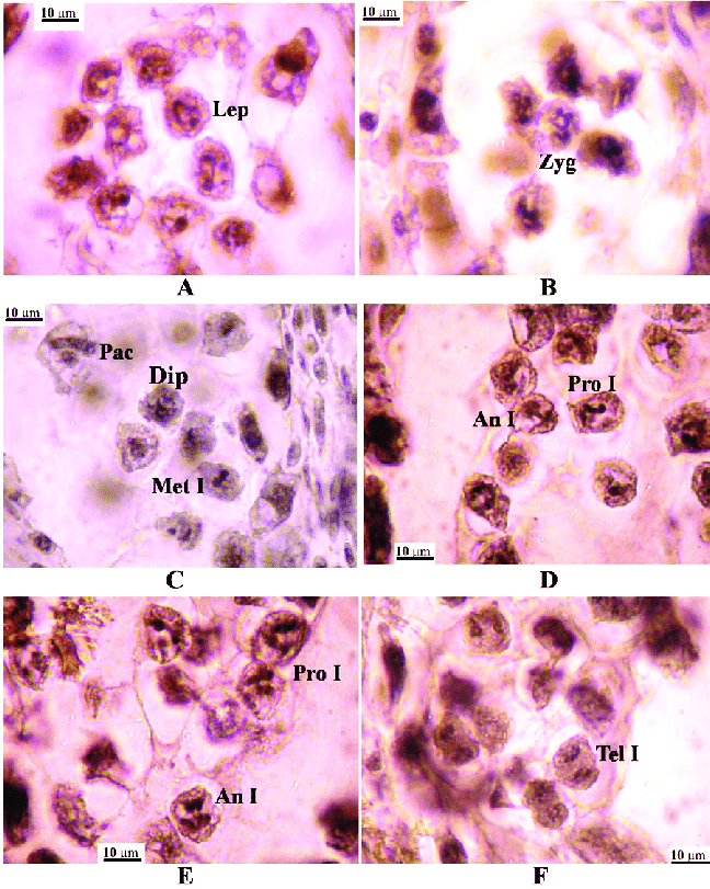
Prophase I. This stage is divided into substages.
Leptotene: The chromosomes have just began to shorten and thicken and forming into a rod-like structures called chromosomes.
Zygotene: The homologous chromosomes appear to attract each other and enter into a very close longitudinal or zipper-like pairing called synapsis.
Pachytene: It is a stage of progressive shortening and coiling of the chromosomes. The double chromosomes, each half now called chromatid, remain attached at the centromere. The two sister chromatids of one are associated with the two sister chromatids of their homologous partner.
This group of four chromatids is known as a tetrad.
Each tetrad closely intertwines and undergoes a series of exchange of genetic material between the non-sister homologous chromatids.
Such exchanges signify the genetic mechanism of crossing-over or recombination.
Diplotene: Chromosomes now act as though they are repelling their closely paired homologue.
Distinctly visible separation occurs between homologous chromosomes except for specific region where an actual physical crossing-over appear to have taken place between the homologous chromatids.
These crosses areas or chiasmata (singular chiasma), an X-shaped attachments between the chromosomes and seem to be the only remaining force holding each bivalent (tetrad) together.
Diakinesis: coiling and contraction of the chromosomes continue until they are thick, heavy-staining bodies.
The centromeres of the original pair of homologous chromosomes are no longer close together but the t__wo chromatids derived from each chromosome of a pair still joined at their centromeres.__
The nucleolus disappears and nuclear membrane dissolved, and the bivalent attach themselves by their centromere to the rapidly formed spindle.
Metaphase I.
At the onset of metaphase I, the synapsed chromosome pair (bivalent) move together or positioned to the equatorial plane of the spindle in such a way that the homologues of each can eventually move to opposite poles.
Each of the chromosome pair is attached to a s__ingle spindle fiber by its centromere,__ and in this process, each bivalent acts independently of the others so that it results in random shuffling of the chromosomes into the daughter cells.
Anaphase I.
During this phase, the entire chromosomes of each homologous pair (having been modified by crossing-over) s__eparate so that each pole receives either a paternal or maternal longitudinally double chromosome of each pair.__
There is no division of the centromere.
A cell plate can be observed in the late stage.
Telophase I.
This phase is marked by the arrival of the chromosomes at the poles of the spindle and a cell plate is formed.
Cytokinesis is completed forming two unlike daughter cells, each has haploid (n) number of chromosomes but still in the duplicated state.
Interkinesis.
It is a brief stage between two successive divisions in meiosis. It is generally similar to the interphase of mitosis except that, there is no replication of genetic material and therefore no new chromatids are formed. Each daughter cell prepares for the second division.
Meiosis II
Prophase II.
The second meiotic division is essentially an ordinary mitotic division.
In this stage, there is no synapsis nor crossing-over as in Prophase I. The double stranded member of the homologous pair reappears and start moving toward its cell’s equator.
Metaphase II.
Each double-stranded chromosome move into the spindle independently and their centromeres attach to a spindle fiber until they become arranged in the equatorial plane.
Anaphase II.
The centromere divides lengthwise and the sister chromatids of each chromosomes are separated from each other____. The newly separated single-stranded chromosomes migrate toward opposite poles of the spindle. The arrival at the poles signals the close of this phase.
Telophase II.
The single stranded chromosomes return to their long, r__eticulate configuration.__
The nuclear membrane reappears and the nucleoli reform.
The new single stranded chromosomes in the nucleus contain only half of the number of chromosomes as in Prophase. Four progeny cells or daughter cells resulted when cytokinesis is completed.
4 PLANT TISSUE SYSTEM
Some of the tissues are composed mostly of a single cell type, these are called simple tissue.
While some tissues that are composed of several types of cells are complex tissue.
The tissues in plant are group into three tissue system : dermal tissue system, ground or fundamental and the vascular tissue system.
I. DERMAL TISSUE SYSTEM.
Covers and protects the plant surface.
Epidermis - the outermost layer of the cells of the primary plant body. Several structures can be observed in the epidermis.
Periderm- replaces the epidermis of the roots and stems of woody plants as they age. Compared with the epidermis, this tissue is made up of s__everal layers which makes it complex.__ The outermost layer is the protective cork or phellem, followed by phellogen (cork cambium) and the phelloderm respectively.
II. GROUND TISSUE SYSTEM (FUNDAMENTAL TISSUE).
Comprises almost the bulk of the plant body, for support, strength and storage.
The ground tissues system consists of three simple tissues – parenchyma, collenchyma, and sclerenchyma-sclereids.
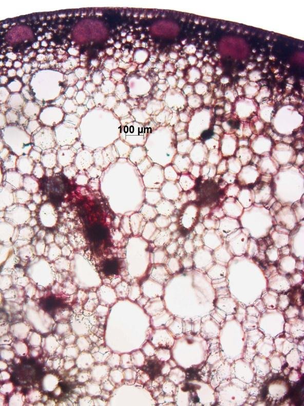
Parenchyma tissue – is composed of parenchyma cells. These cells commonly occur as a c__ontinuous mass in the cortex of stem and roots, in the pith of stem, in leaf mesophyll and flesh of fruits__. In other words, these tissue is found almost throughout the primary plant body.
Collenchyma tissue – is composed of collenchyma cells. The tissue commonly occurs in discreet or a continuous cylinder beneath the epidermis. It is composed to three of four layers of cells with unevenly thickened walls.
Sclerenchyma tissue – the tissue is composed of sclerenchyma cells. There are two types of sclerenchyma cells, the s__clereids (stone cells) and fibers__. You already studied these tissues from your previous experiment (The Plant Cell).
III. VASCULAR TISSUE SYSTEM (Conducting tissue)
The vascular tissue system is composed of two conducting tissues, the xylem and the phloem which are responsible for the distribution of water and solutes in the plant body respectively. These are complex tissues.
cross-section of monocot and dicot stem cross-section of monocot and dicot root santan leaf or any dicot leaf.