PM371 Encoding Part 1 & Language Cortical Areas - Lecture 3
Encoding Part 1: Association Cortices
Presented by Prof Phil Newton, PM371 Learning, Memory and Cognition.
Encoding
Key processes involved in encoding include:
Sensory Memory: Initial stage that holds sensory information briefly.
Short-term memory: Temporary storage system for holding limited information.
Attention: Focusing awareness on specific stimuli or aspects of the environment.
Working memory: Actively holding and manipulating information in mind.
Retrieval: Accessing and bringing stored information back into consciousness.
Learning: Acquiring new information or skills through experience, study, or being taught.
Long-term memory: Storage of information over an extended period.
Storage: Maintaining information in memory over time.
Ascending Sensory Pathway
The ascending sensory pathway involves:
First-order neuron: Detects the stimulus at the periphery.
Second-order neuron (Spinothalamic tract): Transmits the signal from the spinal cord to the thalamus.
Third-order neuron (Thalamocortical projection): Projects the signal from the thalamus to the cortex.
The signal travels from the spinal cord to the thalamus and then to the cortex for processing.
Visual System Basics
Visual information flow:
Left visual field projects to the right hemisphere, and vice versa, enabling contralateral processing.
Temporal retina receives information from the nasal visual field.
Nasal retina receives information from the temporal visual field, aiding in depth perception and spatial awareness.
Pathway: Eye -> Optic nerve -> Optic chiasma (where nasal retinal fibers cross) -> Lateral geniculate nucleus (LGN) in the thalamus -> Primary visual cortex (V1).
Cortical Lobes and Their Functions
Frontal Cortex: Deals with "What do I do about it?" - higher-order cognitive processes, decision-making, and motor control.
Parietal Cortex: Deals with "Where is it?" - spatial processing, attention, and sensory integration.
Temporal Cortex: Deals with "What is it?" - object recognition, auditory processing, and memory.
Occipital Cortex: Primarily involved in vision and visual processing.
Learning and Memory - Information Flow
The flow of information in learning and memory involves several brain structures:
Sensory information enters through the thalamus, which acts as a relay station.
Cortical association areas process the information, integrating it with existing knowledge.
Parahippocampal and rhinal cortices are involved in the formation of new memories.
The hippocampus plays a crucial role in encoding new memories and spatial navigation.
The fornix connects the hippocampus to the mammillary bodies (hypothalamus), which are important for memory consolidation.
Neuroanatomy: Core Principles
Key anatomical terms:
Cortex vs. Lobe: Cortex is the outer layer of the brain; lobes are divisions of the cortex.
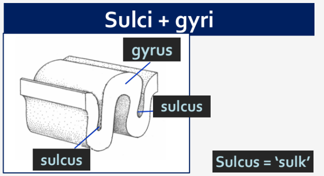
Sulcus (plural: Sulci): A groove or furrow on the surface of the brain that increases surface area.
Gyrus (plural: Gyri): A ridge or fold between two clefts on the cerebral surface in the brain.
Brodmann Area: A region of the cortex defined by its cellular organization and function.
White Matter Tracts: Bundles of myelinated axons connecting different brain areas, facilitating communication.
Laterality: The specialization of function between the two hemispheres of the brain.
Sulci and Gyri
Sulci are infoldings ('sulk') that increase the surface area of the cortex, allowing for more neurons to be packed into the brain.
Gyri are the ridges between the sulci, forming the visible convolutions of the brain.
Key Sulci
Central Sulcus: Separates the Parietal and Frontal Cortices, delineating motor and sensory areas.
Lateral Sulcus (or Sylvian Fissure): Separates the Temporal Lobe from the Frontal and Parietal Lobes; important for auditory processing.
Insula (Insular Cortex)
The insula is a region of the brain located deep within the lateral sulcus, involved in consciousness, emotion, and homeostasis.
Brodmann Areas
Brodmann Areas: Map of the cortex based on histology (cellular structure), created by Korbinian Brodmann in 1909.
There are 52 regions, some subdivided, each associated with specific functions.
Still widely used today for localizing brain functions.
Examples of Brodmann Areas
Primary Visual Cortex: Brodmann Area 17, responsible for initial visual processing.
Fusiform Face Area: Part of Brodmann Area 37, involved in facial recognition.
Association Cortices
Association cortices are not primary cortices; they integrate information from multiple sensory modalities and higher-order cognitive processes.
Posterior Parietal Cortex
Function: Attending to stimuli; spatial awareness ("Where is it?").
Brodmann areas: 5, 7, 39, 40.
Important for attention, especially spatial attention, and integrates visual, auditory, and somatosensory information.
Damage can result in "neglect", a condition where patients ignore one side of their visual field or body.
Hemineglect
Hemineglect is a sign of posterior parietal cortex damage, characterized by neglect of one side of space or the body.
Sensory neglect: Ignoring incoming sensory information from the contralateral hemispace.
Conceptual neglect: Neglect of the body and external world in the contralateral hemifield.
Hemiasomatognosia: Patient denies that the affected side of the body belongs to them.
Motor neglect: Fewer movements in the contralateral space.
Temporal Cortex
Function: Identifying the nature of stimuli ("What is it?") and processing auditory information, memory, and object recognition.
Agnosia
Agnosia is the inability to recognize sensory stimuli, despite intact sensory processing.
Temporal Cortex Damage
Inferior Temporal Cortex: Visual agnosia (“Psychic blindness”); patient can see but not identify objects.
Prosopagnosia (Face Blindness): Inability to recognize individuals from their faces; associated with the fusiform gyrus. Patients can describe facial parts but cannot identify the subject by voice or clothes.
Middle Temporal Cortex: Movement agnosia; inability to distinguish between moving and stationary objects.
Integration of Sensory Streams
Integration allows us to assemble a coherent perspective on the world, combining inputs from different senses to create a unified experience.
Consciousness
The brain predicts a view of the world based on perception and prior knowledge, constructing a subjective reality.
The McGurk Effect
The McGurk Effect demonstrates the integration of auditory and visual information in speech perception, where visual cues can alter auditory perception.
Vision can dominate audition, illustrating the multisensory integration in the brain.
The effect varies among individuals and is reduced in dyslexia and autism, suggesting differences in sensory integration.
Auditory Cortex
Located in the Temporal Lobe (Brodmann areas 41 and 42), responsible for processing auditory information.
Posterior Superior Temporal Gyrus: Involved in integrating audio and visual information in speech processing; crucial for language comprehension.
Frontal Cortex
Function: Selecting and planning an appropriate response ("What to do about it?"), involving higher-order cognitive functions and motor control.
Prefrontal Cortex
Rostral to the primary motor cortex, the prefrontal cortex is very large in humans and develops late (20-30 years old), playing a key role in executive functions.
Dorsal: Thoughts and attention, including working memory and cognitive flexibility.
Ventral: Emotion and social behavior, influencing decision-making and emotional regulation.
Functions of the Prefrontal Cortex
Restraint: Judgement, foresight, inhibiting inappropriate actions, and concentration.
Initiative: Drive, creativity, curiosity, personality, and flexibility.
Order: Planning, abstract reasoning, working memory, and attention.
Frontal Cortex Damage Symptoms
Difficulty planning sequences to complete tasks, indicating impaired executive function.
Loss of spontaneous interactions, reflecting changes in social behavior.
Loss of flexibility in thought, resulting in rigid and inflexible thinking patterns.
Perseveration: Persistence of a single thought or action, showing impaired cognitive control.
Inability to focus on the task in hand, suggesting deficits in attention and concentration.
Emotional lability, characterized by rapid and unpredictable mood changes.
Abulia: Passivity, apathy, and a lack of motivation.
Socially inappropriate behavior, indicating impaired social cognition.
Personality change, reflecting alterations in character and behavior.
Difficulty with problem-solving, showing deficits in executive function.
Expressive aphasia, affecting the ability to produce speech.
Hemiplegia, causing paralysis on one side of the body.
Executive Function
Executive function resides in the prefrontal cortex and involves:
Long-term planning, setting goals, and strategizing.
Withholding impulsive behavior, showing self-control and regulation.
‘Cognitive control,’ enabling flexible and goal-directed behavior.
Important in pathologies like addiction, personality disorders, and dementia, and in everyday behaviors, influencing decision-making and adaptive behavior.
White Matter Tracts
Connect the association cortices and different parts of the brain, facilitating communication between brain regions.
Composed of myelinated neurons, which enhance the speed and efficiency of neural transmission.
Properties of White Matter Tracts
Not visible by physical exam, CT, or static MRI; require specialized imaging techniques for visualization.
Use Diffusion Tensor Imaging to map the direction and integrity of white matter tracts.
Association fibres: Connect cortical areas in the same hemisphere, supporting intrahemispheric communication.
Commissural fibres: Connect across hemispheres, enabling interhemispheric communication.
Projection fibres: Connect cortex to other brain regions, such as the spinal cord and thalamus.
Fasciculus means ‘bundle,’ referring to a collection of nerve fibers.
Examples of White Matter Tracts
Superior Longitudinal Fasciculus: An Association tract connecting frontal, parietal, and temporal lobes; important for language and attention.
Corpus Callosum: A Commissural tract that connects the hemispheres, allowing for the transfer of information between them.
Corticospinal (motor) and Corona radiata: Projection tracts that carry motor and sensory information, respectively.
Language
Language involves cortical areas working together, including Broca's area, Wernicke's area, and the arcuate fasciculus.
Speech Difficulties
Dysarthria: Difficulty moving the muscles of the face and tongue that mediate speaking, affecting articulation and speech clarity.
Aphasia: Difficulty in naming objects and impaired repetition of words; difficulty with language production or comprehension.
Wernicke's Area
Located in the Temporal Cortex (Posterior Superior Temporal Gyrus, Brodmann Area 22), responsible for understanding language.
Wernicke's Aphasia
Unable to understand language, resulting in impaired comprehension.
Fluent speech, but makes no sense, characterized by nonsensical content.
Little repetition, showing reduced ability to repeat spoken words.
Adequate syntax and grammar, but the content is meaningless.
Contrived or inappropriate speech, indicating impaired language processing.
Damage to Wernicke’s area is often a result of a stroke affecting branches of the Middle Cerebral Artery, disrupting blood supply to the region.
Also called fluent, sensory, or receptive aphasia, reflecting the nature of the language deficit.
Broca's Area
Located in the Inferior Frontal Gyrus (Brodmann Areas 44 and 45), responsible for creating language.
Broca's Aphasia
Few problems understanding language, assuming Wernicke's Area is intact, indicating preserved comprehension.
Difficulty constructing their own speech, resulting in impaired language production.
Halting speech – makes sense?, characterized by effortful and fragmented speech.
Repetitive, showing perseveration of words or phrases.
Disordered syntax and grammar, affecting sentence structure.
Disordered structure of individual words, leading to errors in word formation.
Also called non-fluent, motor, expressive, or production aphasia, reflecting the nature of the language deficit.
Damage to Broca’s area is often a result of a stroke affecting different branches of the Middle Cerebral Artery, disrupting blood supply to the region.
Broca vs Wernicke Aphasia
Broca's Aphasia (Frontal):
Understand language, with relatively preserved comprehension.
Cannot construct their own, resulting in impaired language production.
Halting speech – makes sense?, characterized by effortful and fragmented speech.
Repetitive, showing perseveration of words or phrases.
Disordered syntax, grammar, and structure of individual words, affecting sentence structure and word formation.
Wernicke's Aphasia (Temporal):
Unable to understand language, resulting in impaired comprehension.
Fluent speech, but makes no sense, characterized by nonsensical content.
Little repetition, showing reduced ability to repeat spoken words.
Adequate syntax and grammar, but the content is meaningless.
Contrived or inappropriate speech, indicating impaired language processing.
General Points About Aphasias
Recognition of ‘conversation cues’ seems OK, indicating some preserved social communication skills.
Affects other forms of language, including reading, writing, and sign language, showing a global impact on language abilities.
Many other forms of aphasia exist, reflecting the complexity of language processing in the brain.
Arcuate Fasciculus
Connects Broca's and Wernicke's areas, enabling communication between language production and comprehension regions.
Comprised of Association Fibres, facilitating the flow of information between different cortical areas within the same hemisphere.
Summary
Association cortices process sensory and learned information, integrating inputs from different modalities.
Posterior Parietal Cortex: Where is it? - Spatial awareness