Biopsychology
Nervous System
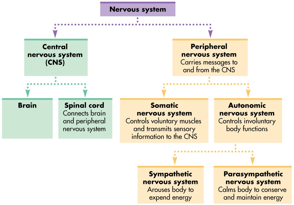
- Collects information, processes it and takes action by directing organs and muscles
- CNS- provides complex processing
- Brain- all conscious and most unconscious processing
- Spinal cord- receives and transmits info to and from brain and body, can also perform basic motor reflexes
- PNS- body-wide network of messenger neurons that deliver instructions to body from CNS
- Somatic nervous system- controls skeletal muscles for movement, voluntary system
- Autonomic nervous system- controls internal organs and glands, involuntary system
- Sympathetic nervous system- activates stress response and releases noradrenaline (fight or flight)
- Parasympathetic nervous system- decreases bodily activities and releases acetylcholine (rest and digest)
- Homeostasis- regulation of internal environment, in normal conditions there is a balance between sympathetic and parasympathetic systems
Reflex Arc
Neuron structure
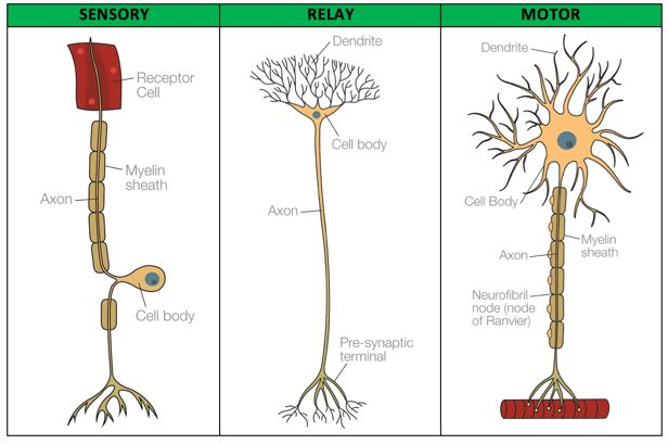
- Cell body- contains nucleus
- Dendrites- get information coming into the neurone
- Axon- carries information
- Dendrite- forms a synapse with the dendrite of the next neurone
- Myelin sheath- fatty sheath on axon that protects cell and speeds up transmission
- Only on sensory and motor neurones
- Receptor- receives information from the environment
- Only by sensory neurones
- Effector- causes a response like movement
- Only by motor neurones
- Relay neurons only ever in CNS, sensory and motor extend from PNS to CNS
Reflex arc
- Sensation detected at sensory receptors, e.g. pain
- Electrical signal goes along sensory neurone’s dendrite, axon and axon terminal
- Converts into a chemical signal, crosses synaptic cleft and is picked up by the relay neurone in the spine
- Reflex responses pass to motor neurone
- Signal reaches effector which causes muscle to move out of danger
Synaptic Transmission
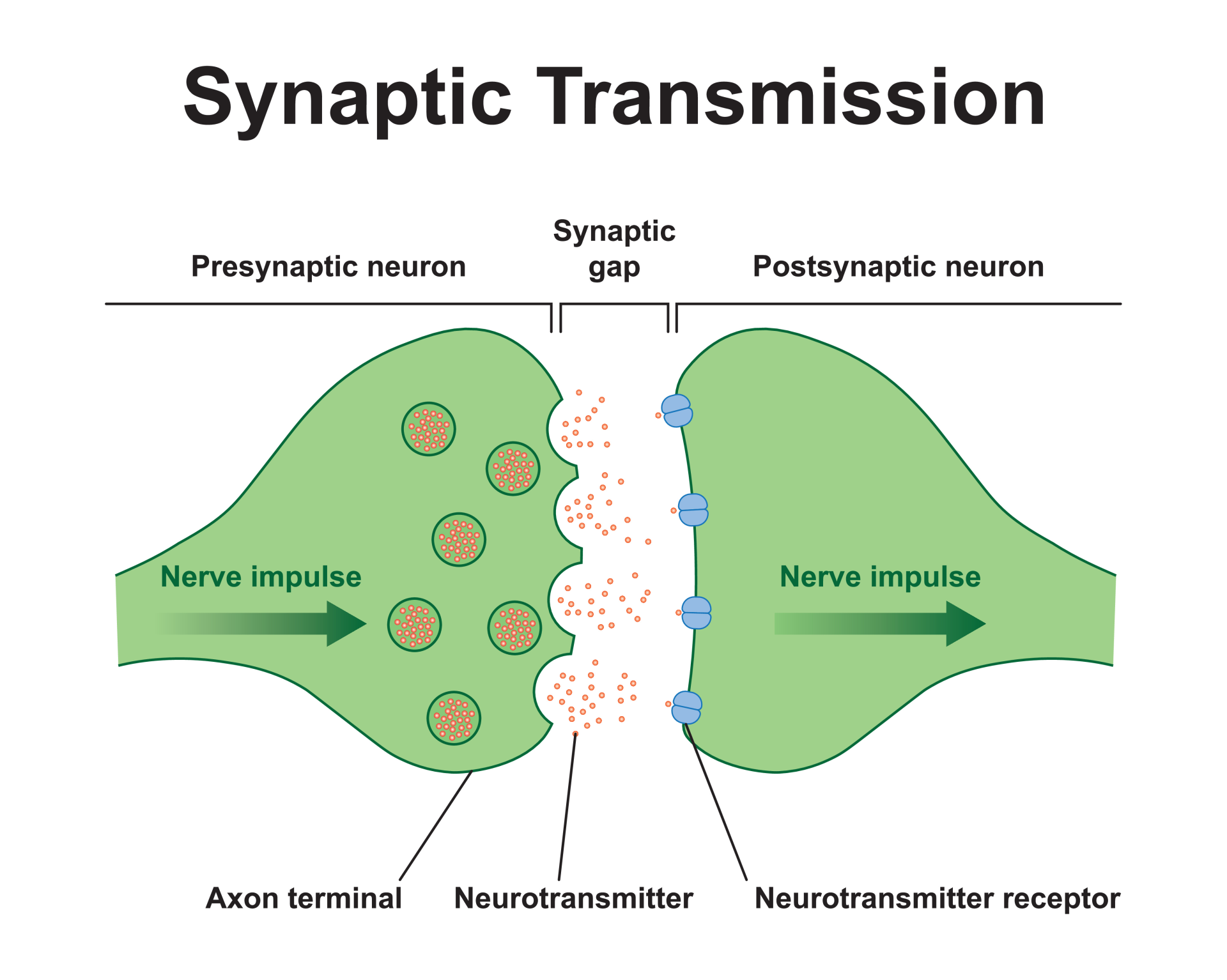
- Synapse- gap between two neurons (axon terminal to dendrite)
- Neurotransmitters- chemical messengers released by neurones, stimulate or inhibit the development of an action potential in the postsynaptic neurone
- Vesicles- contain neurotransmitters
- Excitatory neurotransmitters depolarise the post-synaptic neurone, make the electrical charge more positive and make the formation of a new action potential more likely
- Inhibitory neurotransmitters hyperpolarise the post-synaptic neurone, make the electrical charge more negative and make the formation of a new action potential less likely
- Summation- all influences of neurotransmitters on the post-synaptic neurones are summed
- Happens if multiple nerve impulses occur in the pre-synaptic neurone or if multiple synaptic transmissions happen at the same time
- Uni-directional- can only happen one way
- Drugs can interfere with this system
- Selective Serotonin Re-uptake Inhibitors (SSRIs) block the reuptake of serotonin and increase its level in the synapse
- Cocaine blocks reuptake of dopamine
- Action potential arrives at action terminal
- Vesicles fuse with the presynaptic membrane and neurotransmitters are released into the synaptic cleft
- Neurotransmitters diffuse across synaptic cleft and reach postsynaptic receptor sites
- If the threshold is reached, a new action potential is formed and travels down the post-synaptic neurone
- Neurotransmitters detach from the post-synaptic receptors and are reabsorbed back into the pre-synaptic neurone by re-uptake proteins to be used again (reuptake)
Endocrine System
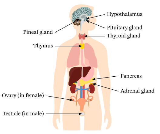
- Collection of glands around the body that regulate bodily and psychological functions by releasing hormones into the bloodstream
- Pituitary gland- ‘master gland’ at the bottom of the brain, controls other glands’ secretion of hormones
- ACTH activates other glands
- Can receive electrical signals
- Hypothalamus- above pituitary gland, receives electrical signals from somatic and autonomic nervous system and sends hormones to pituitary gland
- Links nervous system to endocrine system
- Releases CRH, detected by pituitary in fight or flight
- Pineal gland- in the brain, modulates sleep pattern, keeps body to a day/night circadian rhythm
- Releases melatonin
- Thyroid gland- in the front of the neck, influences metabolism
- Releases thyroxine
- Thymus gland- in the chest, only active until puberty, stimulates development of T cells that help immune system
- Releases thymosin
- Pancreas- just behind stomach, regulates blood sugar levels, problems lead to diabetes
- Releases insulin and glucagon
- Adrenal glands- one on top of each kidney, involved in fight or flight, heart rate, blood supply and sweating
- Adrenal medulla- inner part, releases adrenaline
- Adrenal cortex- outer part, releases cortisol
- Ovaries- either side of the uterus, female reproductive glands, develop secondary sexual characteristics at puberty
- Release oestrogen
- Testes- outside the body, male reproductive glands, develop secondary sexual characteristics at puberty
- Release testosterone
- Stimulus causes a gland to release a hormone into the bloodstream
- Hormones travel to target cells
- They bind to the receptors on target cells
Fight or Flight
- Evolutionary survival mechanism in response to a threat
- Involves endocrine system and sympathetic nervous system
- Somatic and autonomic nervous system → Hypothalamus → Pituitary gland → Adrenal gland
- Prepares mind and body for action- fighting for life or escaping the threat
- Body returns to homeostasis after the threat has passed
- Not designed for modern world, maladaptive in most situations
- Acute stress- most common form of stress in response to immediate pressures, can be exciting in small amounts and give focus and energy but exhausting if maintained
- Chronic stress- long term form of stress in response to prolonged emotional pressure, often occurs in situations the individual cannot control
- Effects of adrenaline and noradrenaline
- Pupil dilation
- Increased sweat
- Increased muscle tension
- Increased heart rate
- Increased breathing rate
- Decreased salivation
- Decreased digestion
- Psychological affects of increased anxiety, attention and alertness
- Constant triggering of fight or flight in chronic stress has long term effects on physical and mental health
- Increased risk of heart disease, obesity, IBS, general lowering of resistance to disease and depression
Fast response
- Hypothalamus sends electrical signals to SNS
- SNS activates adrenal medulla
- Adrenal medulla releases adrenaline
- Adrenaline binds to target cells and causes reactions
Slow response
- Hypothalamus sends hormones to pituitary gland
- Pituitary sends ACTH to adrenal cortex
- Adrenal cortex releases cortisol
- Cortisol binds to target cells
Rest and digest response
- Hypothalamus sends electrical signals to PNS
- Parasympathetic pathway causes opposite effects to fight or flight
:( Research has only used males, lacks population validity- some researchers suggest women have a tend and befriend response
:( Ignores individual differences in responses to danger, e.g. freeze response
Brain Localisation
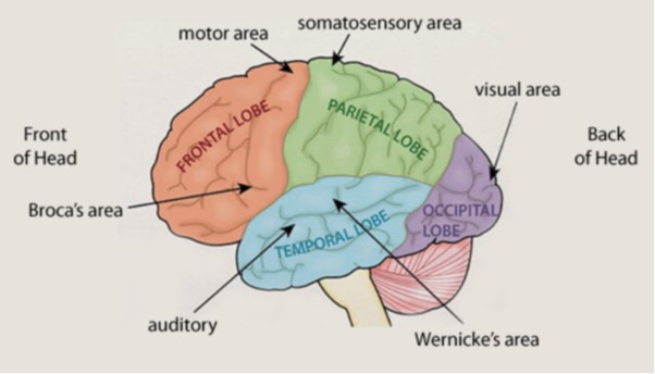
- Functions like movement, speech and memory are performed in distinct brain regions
- Opposite view is the brain acts holistically to perform functions
- Hemispheric lateralisation- each hemisphere is specialised to different functions
- Left hemisphere is specialised to language (Broca’s and Wernicke’s areas), right hemisphere is specialised to visual and spatial processing
- Contralateral- each hemisphere of the brain controls the opposite side of the body
- Cortex- grey matter on the surface layer of the brain, 2-4mm thick and folded for extra surface area for processing. White matter is myelinated axons with few cell bodies
- Motor cortex- Responsible for voluntary motor movements, both hemispheres, contralateral
- Damage causes loss of muscle function or paralysis on the opposite side of the body
- Separated from the somatosensory cortex by the central sulcus
- Somatosensory cortex- Responsible for receiving sense impressions from around the body, both hemispheres, contralateral
- Damage causes loss of sensation, ignoring areas of the body (neglect syndrome), losing ability to recognise objects by their feel (agnosia)
- Separated from the motor cortex by the central sulcus
- Visual cortex- Visual processing centre, both hemispheres, contralateral
- Damage causes partial or complete loss of vision (cortical blindness)
- Damage to one side can cause loss of vision in the opposite visual field
- Auditory cortex- Receives and processes sound info from ears, both hemispheres
- Damage causes cortical deafness
- Broca’s area- Responsible for speech production, left hemisphere (lateralised)
- Damage causes Broca’s aphasia- difficulty producing fluent speech, speech is slow and effortful, misses out words
- Discovered after case study of Tan- brain damage, could understand language but only say “tan”
- Wernicke’s area- Responsible for speech comprehension, left hemisphere (lateralised)
- Damage causes Wernicke’s aphasia- difficulty understanding speech or written language, and producing speech that sounds fluent but lacks meaning
- Discovered after case studies of people who could produce fluent sounding speech that made little sense and not understand others
- Damage to both Broca’s and Wernicke’s areas can cause global aphasia, inability to produce or understand speech
:) Post-mortem case studies (such as Tan)
:) fMRI scans show activation in regions associated when healthy participants perform language tasks
:( Recent MRI scan of Tan’s brain showed more damage than just in Broca’s area, so other areas could be responsible for speech production
:( Some functions like consciousness appear to not be localised at all, brain is highly connected and should be looked at holistically
:( Study found slight individual differences in active brain regions while reading, suggests individual differences in brain organisation
Brain Plasticity and Functional Recovery
- Plasticity- brain adapts due to environmental changes like direct brain damage, indirect damage like brain bleeding from a stroke, learning new skills or developmental changes
- Can adapt function and structure
- Functional recovery- healthy areas of the rain compensating for lost or damaged areas by performing their functions (functional reorganisation)
- Neuronal unmasking- silent synapse becomes active after a period of inactivity
- Rewiring- axons from regions that used to communicate with the damaged region form new connections with nearby regions
- Synapse strengthening- synapses in regions near the damaged region are reinforced to let nearby neurons take on the function of the damaged region
- Synaptic pruning- synapses that are used frequently become stronger over time and unused synaptic connections are lost, so the brain becomes more efficient
- More active in babies and children, as people develop connections are pruned
- Neuronal regeneration- growth of new neurons and connections
- Denervation supersensitivity- to compensate for the loss of axons in a pathway, remaining axons become more sensitive (more likely to fire)
- Can cause chronic sensitivity to pain
- Factors affecting functional recovery
- Age- younger someone is, more likely they are to recover- children are the best, then young adults
- Gender- women are more able to recover from brain damage
- Access to rehabilitative therapy
- Constraint induced therapy- stops patient from using coping strategies like body language for communication or undamaged limbs for tasks
- Makes them improve via functional reorganisation
Taxi Driver Study
- 16 taxi drivers who completed a test called The Knowledge (landmarks and routes in London) had MRI scans and were compared to 16 matched controls
- Taxi drivers had significantly higher posterior hippocampi
- Size of posterior hippocampus was positively correlated with time as a taxi driver
- Suggests physical structure of the brain is plastic and able to reconfigure itself to better adapt to psychological demands
:) Objective scientific method
:( Correlation, not causation
:) Case study of EB- had a hemispherectomy of the left side of his brain as an infant to remove a tumour, lost Broca’s and Wernicke’s areas. After 2 years he had recovered language ability, then developed normally with age except some dyslexia like symptoms and slight speech difficulties. fMRI scans as a teenager showed his right hemisphere functioned like a left hemisphere, suggests brain can adapt and recover after significant damage
:) Can help with rehabilitative therapy and help people return to work- benefits economy
:( Individual differences- meta-analysis showed higher IQ and better educational background are positively correlated with better outcomes after brain damage, suggests some people can recover better than others
Split-Brain Research
- Corpus callosum- connection between hemispheres, bundle of 200-300 million nerve fibres (white matter)
- If corpus callosum is cut, stops communication between each hemisphere
- Used to be a treatment for severe epilepsy
Sperry’s Split-Brain Studies
- Research study on 11 patients with a cut corpus callosum
- Participants were in front of a screen, covered one eye and were asked to look at a dot
- Words and images were flashed independently to right and left visual fields
- If word seen on RVF, they could say the word as info was in the LH where language centres are
- If word seen on LVF, they could draw the image or pick up the object as info was in the RH which has control of a hand
:) Helped understanding of consciousness and identity, suggests the brain is a combination of separate intelligent processes working together
:( Small sample of people who had epilepsy, had all undergone drug therapy and had varying amounts of connection cut- hard to generalise to wider population due to participant variables. Control group also didn’t have epilepsy
:( Experimental procedure is unlike how people would process info and act in everyday life- lacks mundane realism and external validity
Ways of Studying the Brain
- Spatial resolution- accuracy of position in space
- Temporal resolution- accuracy of position in time
Post Mortem
- Brains of dead people are treated and cut precisely
- Usually on unusual brains like those that suffered trauma or with people with mental illnesses
- Compared with neurotypical brains, physical differences could be linked to behavioural differences
- Post mortem of Tan was how Broca’s area was identified
:) High spatial resolution
:( Correlational data
:( Low temporal resolution
:( Confounding variables like medicines
fMRI
- Functional magnetic resonance imaging
- Uses magnets to detect blood flow in the brain
- Active parts of brain use more oxygen so blood flow is increased
:) High spatial resolution, can identify which brain regions are active while completing experimental conditions
:) Non-invasive and safe
:( Low temporal resolution- delay between activity and blood flow, many processes are too fast to study
:( Expensive to build and operate
:( Just because a region has increased blood flow during a task doesn’t mean there is a causal relationship between it and the task
EEG
- Electroencephalogram
- Electrodes are connected to scalp
- Output is a series of lines showing brain waves
- Amplitude showed intensity, frequency shows speed
:) Cheaper than fMRI, portable, can be used in experiments where participants move
:) High temporal resolution- matter of milliseconds
:( Low spatial resolution- cannot identify exact location, also only detects activity in cortex
ERP
- Event-related potential
- Uses same technique as EEG but presents a stimulus many times and creates an average brain wave
- Removes background noise unrelated to the stimulus
:) Allows researchers to isolate and study how individual cognitive processes happen in the brain
:) High temporal resolution
:( Low spatial resolution
Circadian Rhythms
- Biological rhythm that lasts 24 hours
- Sleep-wake cycle, release of hormones, varying body temperature and blood pressure
- Endogenous pacemakers- internal body clocks that keep biological processes to time
- Exogenous zeitgebers- external cues that alter body clocks to match the environment
Sleep-wake cycle
- Period of sleep and period of wakefulness every day
- 7am- wake up, 11am- most alert, afternoon- dip in alertness, 11pm- sleep, 2am- least alert
- Pineal gland releases melatonin, which activates sleep centres and inhibits wakefulness centres
- There must be an endogenous pacemaker that keeps the pattern
- Conflicts between EP and EZ like light and social cues in situations like let lag
- Suprachiasmatic nucleus (SCN)- EP for the sleep-wake cycle, also known as master clock
- Part of hypothalamus within the limbic system
- At the optic chiasm, point where optic nerves cross
- When it detects light it sends a signal to the pineal gland and stops melatonin production
- :) Study- swapped SCN of hamster groups, adopted sleep-wake cycle of other group
- Siffre tested the concept of a free-running sleep-wake cycle (not affected by EZs) by spending 6 months in a cave with no EZs. He maintained a regular cycle of around 25 hours
- Longer than normal 24-hour cycle
- Reported thinking less time had passed
- :( Low generalisability
:) Study support- when 27 office workers were exposed to strong blue light, they shifted circadian rhythms to match office lighting. Control group with normal lighting matched natural light of dawn
:( Artificial lights may affect measurements
Infradian Rhythms
- Biological rhythm that last more than 24 hours
- Seasonal Affective Disorder (SAD)- emotional mood lowers in winter and improves in summer
Menstrual Cycle
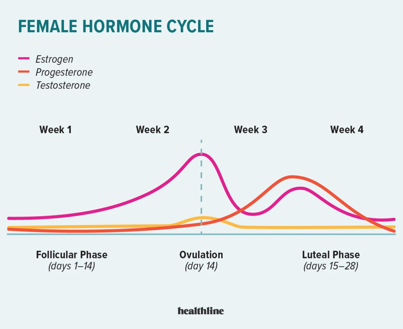
- 28 day cycle with a 6 day period of fertility
- Can range between 24-35 days
- Biological aspects- ovulation, thickening of womb lining, losing womb lining during menstruation
- Regulated by oestrogen pre-ovulation and progesterone post-ovulation
- Endogenous pacemakers that keep the biological processes to time
- Study- 135 women who lived in the same dorm at university, appeared to synchronise cycles with friendship groups
- Study- women wiped pads on their top lips taken from the armpits of other women at varying stages of menstrual cycle. Found cycle would change to match the donor depending on when in the cycle the pad was collected
- Suggests synchronisation due to pheromones (exogenous zeitgeber)
:) Evolutionarily advantageous- stops one male from impregnating a whole group, leads to genetic diversity
:( Many studies have failed to find results that support the theory of pheromone synchronisation, one study found cohabiting lesbian couples did not synchronise
:( As menstrual cycles can vary in length, women can appear to synchronise just due to variability in cycles
Ultradian Rhythms
- Biological rhythms that last less than 24 hours
Sleep-Wake Cycle
- EEG brainwave features
- Frequency- waves per second
- Amplitude- change in voltage
- Delta, theta, alpha, beta and gamma waves
- Theta and delta distinguish sleep stages
- Stages of sleep- cycle takes around 90 minutes
- N1- falling asleep sensation, easy to wake and body may move suddenly (hypnic jerks), may be a sensation of falling, mild auditory or visual hallucinations (hypnagogic hallucinations)
- EEG shows theta waves, low frequency but high amplitude in comparison to awake
- N2- deeper than N1, harder to wake, heart rate and body temp lower, eyes are still
- EEG shows theta waves with slow frequency but occasional sleep spindles
- N3- deepest sleep, very hard to wake, body at its most relaxed, heart rate lowest, where physical recovery of the body happens
- EEG shows delta waves with slow frequency and high amplitude
- REM- brain returns to active state passing back through N1 and N2, body is paralysed, rapid eye movement, where dreaming happens
- EEG shows patterns similar to being awake
- Body returns to N1 after REM, cycle is repeated 4-5 times each night
- Each cycle includes a larger proportion of REM and a shorter duration of N3
- Study- EEGs of 33 adults over a nights sleep, brain waves showed a cyclic pattern of activation, body relaxed during slow wave and high activation during REM
:) Development of technology and devices that help track and improve sleep, e.g. avoid waking in REM
:( Newborn babies spend 80% in REM and 20-25% in adults, suggests stages of sleep are not simple and adapt to developmental needs of the individual