Chapter 8: Transport in Mammals
- Transport system consists of:
- Blood System
- Lymphatic System
Blood:
Is a tissue fluid.
Structure and Composition of Blood:
- %%Plasma:%%
Is a pale yellowish liquid. It is 90% water and a complex mixture of various substances. These substances include:
- Soluble proteins- fibrinogen and prothrombin - play an important role in clotting blood.
- Dissolved mineral salts - (chlorides, bicarbonates, sulphates and phosphates) all occur as ions + calcium is also present - helps in clotting of blood.
- Food substances such as glucose, amino acids, fats and vitamins.
- Also contains; excretory products, hormones.
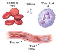
- %%Red Blood Cells (RBC’s)\ erythrocytes:%%
They are red, circular, flattened, biconcave disc making the centre of the cell thinner than its edges.
- Has no nucleus which helps it squeeze into capillaries easily.
- Produced in bone marrow.
- Lifespan of 3 to 4 months - when worn out destroyed in spleen and liver.
- Haemoglobin - give it red colour - enables RBC’s to transport oxygen from lungs to all cells of the body.
- %%White Blood Cells (WBC’s)\ leucocytes:%%
Are colourless and have no haemoglobin.
- Larger than RBC’s.
- Irregular in shape and contain nucleus - can diffuse in tissue.
- two main kinds - lymphocytes and phagocytes.
- Lymphocytes has large, rounded nucleus and a relatively small amount of non-granular cytoplasm. - only show limited movements.
- Phagocytes ingest foreign particles like bacteria.
- Role of WBC’s:
- fight foreign bodies.
- %%Blood platelets/ thrombocytes:%%
Are not true cells but are fragments of cytoplasm.
- Play a part in clotting of blood.
- Important:
- When oxygen combines with haemoglobin it forms oxyhaemoglobin - it gives it a bright red colour.
- Haemoglobin without oxygen is purplish red.
- It is this colour difference which accounts for the red colour of arteries and blue colour of veins. (discussed ahead).
Function of Blood:
- Is a transport medium for carrying substances.
- Protects the body against germs.
- %%Phagocytosis%% occur.
%%Phagocytosis%%: is the process of engulfing and ingesting foreign particles by WBC’s.
- ^^production of antibodies^^
- When disease-causing germs gan entry into the bloodstream, they produce poisonous substance called toxin. The blood then produces certain chemical substances called antibodies. They act as antioxidants which neutralize the poisonous effect of the toxins + they kill bacteria in bloodstream. Then this bacteria can be easily ingested by phagocytes.
- ^^clotting of blood^^
- Seals the wounds, preventing excessive loss of blood. This also stops foreign particles from entering the bloodstream.
- process of clotting
- An enzyme thrombokinase is released which converts prothrombin (inactive) to thrombin (active) This thrombin is also an enzyme it catalyses the reaction of fibrinogen to fibrin thread. These fibrin threads produce a clot.
Organ Transplant and Tissue rejection
- When a person’s tissue is ^^damaged it is replaced with a healthy one is called^^ %%Organ Transplant.%%
- Due to this a person’s lymphocytes respond negatively by producing antibodies to destroy the transplanted organ this is called tissue transplant.
- Prevention of tissue rejection:
- To reduce the risk of this both the recipient and donor should be genetically close.
- Immunosuppressant drugs can be taken.
- X-ray radiation of the bone marrow and lymphoid tissue may inhibit this issue.
- Circulatory system:
- Heart is a muscular organ which drives the blood around the body by pumping
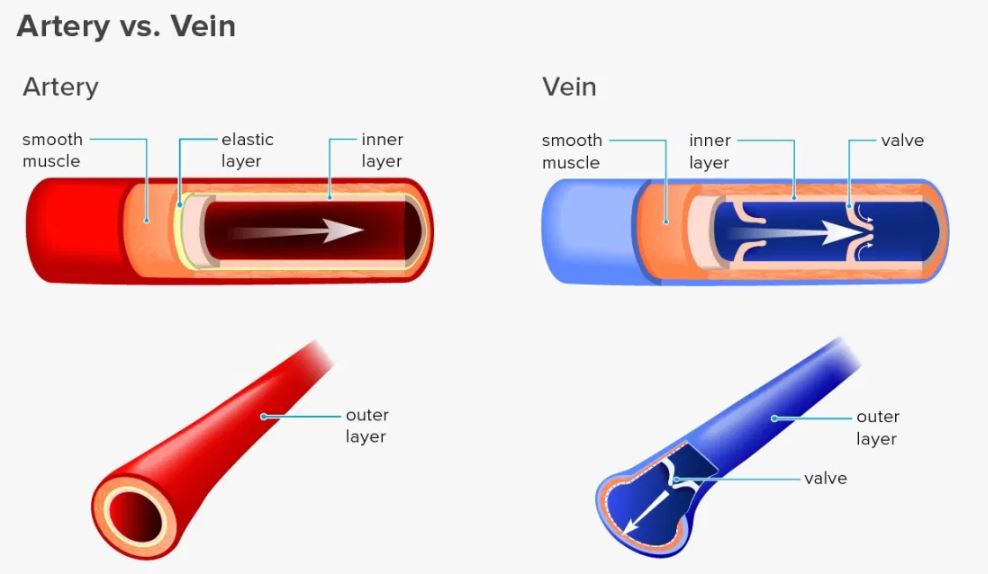
- Blood vessels which carry blood away from the heart are arteries. (you can remember this like ‘a’ from away ‘a’ from arteries) - they are further branched as arterioles - they are further divided as capillaries
- Veins are blood vessels which convey blood towards the heart
- Capillaries are microscopic thin walled blood vessels which carry blood from a small artery to a small vein
- Arteries
- Have thick, muscular and elastic walls. - this helps in maintaining high blood pressure and pushing blood along.
- When an artery constricts, its lumen narrows and less blood flows through it.
- When an artery dilates, its lumen becomes wider and more blood flows through.
- Has no valves.
- Veins:
- Has low pressure in comparison to arteries.
- Walls are not thick, elastic and muscular.
- Semilunar valve are folds of the inner walls, shaped like half moons.
- Movement of blood in veins is assisted by the action of skeletal muscles.
- Muscular exercises increases the pressure exerted on the veins and moves the blood quickly.
Double Circulation:
Blood passes ^^through the heart twice in one complete circuit.^^ Blood ^^flows from one the main circulation to the heart, then to the lungs and back to the heart again before it is pumped into the main circulation.^^
- Large veins carry deoxygenated blood from various parts of the to the heart. From the heart, the pulmonary arteries carry the blood to the lungs. Oxygenated blood is returned to the heart by the pulmonary veins. The circulation linking the lungs to the heart is known as the %%pulmonary circulation.%%
- Oxygenated blood leaves the left side of the heart and is distributed by arteries to all parts of the body (except the lungs). Veins carry the blood from all parts of the body back to the right side of the heart. This circulation is known as %%systemic circulation.%%
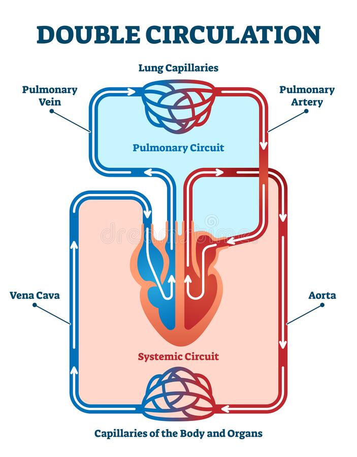
- Benefit of double circulation is; blood is pumped at a faster rate.
The Heart:
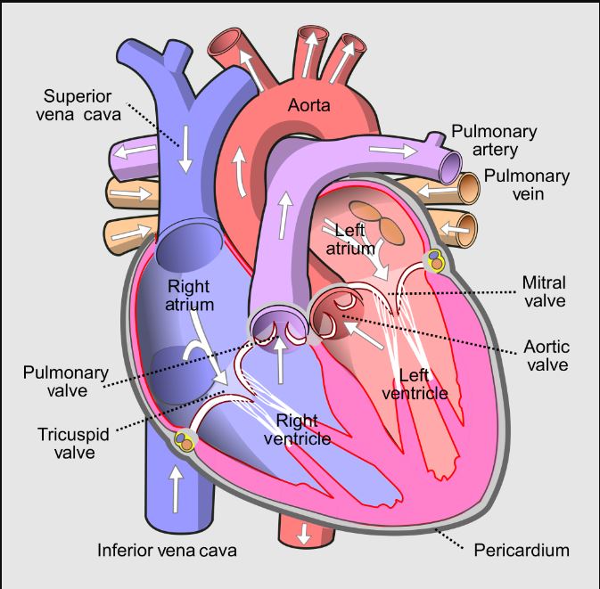
structure
- Whole heart is surrounded by __pericardium__
- Divided in four chambers:
- Upper two chambers; atria.
- Lower two chambers; ventricle.
- Right and left side separated by __septum __- helps in separating oxygenated and deoxygenated blood.
- Blood (from head, neck and arms) is returned to right atrium by large vein; __superior vena cava__. blood from other parts (excluding lungs) is brought by __inferior vena cava__.
- When the right atrium contracts, the blood flows into the right ventricle.
- __Tricuspid valve__ separates __right atrium and right ventricle.__
- When right ventricle contracts, tricuspid flaps to close the opening of atrium.
- Blood leaves the right ventricle by __pulmonary arch__ - it later divides into pulmonary arteries (to the lungs).
- Oxygenated blood from lungs is brought back with the help of __pulmonary veins__ which opens in left atrium.
- When __left atrium__ contracts, the blood enters the __left ventricle__.
- B/w left atrium and left ventricle is __bicuspid valve__.
- When __left ventricle__ contracts, blood leaves by way of aortic arch - blood is distributed to all parts except lungs - aortic arch divides into arteries.
Main arteries in the body:
- Pulmonary artery is the only artery which carries deoxygenated blood. (the rest contain oxygenated blood from the heart)
- From the aortic, the following arteries arise:
- Carotid arteries - supplies head and neck.
- Subclavian arteries - supplies arms and forelimbs.
- Hepatic artery - supplies liver.
- Renal artery - supplies to kidneys.
- Mesenteric arteries - supplies to intestines.
- Iliac arteries - supplies to hindlimb and legs.
Main veins in the body:
- Pulmonary vein is the only vein which carries oxygenated blood to the heart. (the rest carry deoxygenated).
- Jugular vein - brings from head and neck.
- Subclavian vein - brings from arms and forelimbs.
- Hepatic vein - brings from liver.
- Renal vein - brings from kidneys.
- Mesenteric vein - brings from intestines.
- Iliac vein - brings from hindlimb and legs.