skeletal system - dc anatomy & physiology
bones are organs
each bone is a complex living organ made up of many cells, protein fibers, and minerals
they come in many sizes and shapes
the largest bone is the femur and the smallest is the stapes
the humerus is pictured here
the skeletal system also includes…
joints
cartilages
ligaments* - bone to bone
tendon - muscle to bone
two basic types of bones
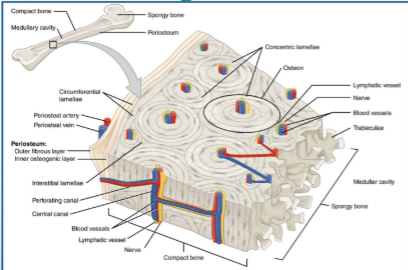
compact bone: dense and looks smooth and homogenous

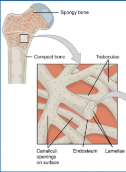
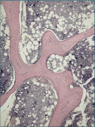
spongy bone: composed of small pieces of bone and lots of open space compact bone
in long bones, surrounds spongy bone at ends
along shaft surrounding medullary cavity
spongy bone
at ends or long bones
microscopic anatomy of spongy bone
the open spaces keep bones light
found in the ends of long bones
also fills short bones, flat bones and some parts of irregular bones
contains red marrow
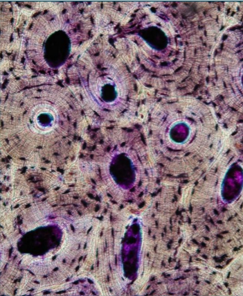
microscopic anatomy of compact bone
composed of a matrix of hard minera lsalts reinforced with tough collagen fibers
mature bone cells are called osteocytes
osteocytes are found in tiny cavities within the matrix called lacunae
gross anatomy of a long bone
diaphysis
shaft
composed of compact bone
epiphysis
end of the bone
composed of mostly spongy bone
other features of long bones
articular cartilages: cover epiphyses for smooth movement
epiphyseal line: marking left from growth at epiphyseal plate
periosteum: fibrous, connective tissue that covers the disaphysis
bone marrow
red marrow: in cavities of spongy bone in flat bonesa nd epiphyses of long bones, site of hematopoiesis
yellow marrow: fat storage in medullary cavity
anatomy of the skeletal system
two divisions of the skeleton
axial skeleton
z
axial skeleton
bones that form the longitudinal axis of the body make up the axial skeleton
verterbal column
vertebrae
sacrum
coccyx
bony thorax
ribs
sternum
skull
cranium
facial bones
vertebral column
composed of 33 bones before birth; some later fuse to form 26 separate bones (know numbers)
7 cervical vertebrae
7 vertebrae located in the neck
smallest and lightest vertebrae
12 thoracic vertebrae
articulate with ribs
larger than cervial vertebrae
long spinous process that hooks sharply downward
5 lumbar vertebrae
much larger than other vertebrae to support the weight of the upper body
sacrum: 5 fused
coccyx: 4 fused
atlas & axis
atlas: the first
bony thorax or rib cage
12 pairs of ribs articulate with the 12 throacic certebrae posterio
true ribs: pairs 1-7, articulate anteriorly directly to the sternum by cartilage
false ribs: pairs 8-12, articulate indirectly or not at all
last 2 apir do not connect at all and are floating ribs
sternum
manubrium
body
xiphoid process*
the skull
the skull is formed by two sets of bones
cranium
facial bones
all are joined by immovable joines except for the manible (jawbone)
hyoid bone
no direct articulation to another bone
provides attachment for these muscles:
floor of mouth
tongue
larynx
apiglottis
auditory ossicles
the samllest bones in the body are located in the middle ear
malleus
incus
stapes
appendicular skeleton
includes the limbs and the gidles which attach the limbs to axial skeleton
pectoral (shoulder)
collar bones
shoulder blades
upper limbs
arms
hands
pelvic hip
coxal bones
lower limbs
legs
feet
pectoral girdle
clavicle, scapula
upper limbs
humerus
ulna
radias
carpals
metcarpals
phalanges
pelvic girdle
ilium ischium
pubis
lower limbs
femur
patella
tibia
fibula
tarsals
metatarsals
phalanges
pectoral girdle
clavicle is commonly called the collar bone
the scapula is commonly called the shoulder blade
arm bones
humerus
upper arm
radius
lower arm; elbow to thumb side of the wrist
ulna
lower arm; elbow to pinkie side of the wrist
leg bones
femur: thigh bone
patella: knee cap
tibia: large bone in lower leg; sometimes called the shin bone
fibula: smaller bone in lower leg; forms the lateral ankle
shapes of bones
long
short
flat
irregular
sesamoid
bone markings
bumps, holes, and ridges where muscles, tendons, and ligaments are attached and where blood vessels and nerves pass through
two categories:
projections or processes
tuberosity
trochanter
tubercle
process
condyle
depressions, cavities, or openings
meatus
fossa
foramen
joints
also called articulatios, where two bones meet
holds bones together, but also gives mobility
three types
fibrous: no movement, (ex., skull)
cartilaginous: slightly moveable (ex., pubic symphysis, vertebrae)
synovial: bones separated by cavity filled with synovial fluid, allow the most movement
synovial joints
articular cartilage
fibrous articular capsule
joint cavity contains fluid
reinforced by ligaments
ball and socket joints
most moveable type of joint
found in the shoulder and hip
physiology of the skeletal system
support
the skeleton provides an internal framework that supports and anchors all soft organs
protection
some bones protect soft body organs
movement
skeletal muscles attach to bones by tendons
tendons use the bones as levers to move body parts
storage
fat storage in yellow marrow
the minerals calcium and phosphorous are stored in bone tissue
hematopoiesis: blood cell formation occurs in red marrow
developmental aspects of the skeletal system
fontanels
spaces between bones of the skull in an infant
“soft spots”
fully ossified by 2 years
allows for growth of the brain and skull
ossification: the formation of bone from cartilage
at birth, bones are part cartilage and part bone
the skeleton is fully ossified by age 2, except for epiphyseal (growth) plates
longitudinal growth
epiphyseal plates are fully ossified by the end of adolescence
bone formation and growth
ossification: bone formation
epiphyseal plates: provide for longitudinal growth; increase in length
appositional growth: increase in diameter
osteroblasts: bone-building cells
osteoclasts: bone-destroying cells
bone remodeling
breaking down and reforming of bone that occurs throughout life to maintain proportion and strength as well as healthy calcium levels
osteoporosis: weakening of the bone that occurs with aging
1 in 2 women and 1 in 4 men over age 50 will have an osteoporosis-related fracture
osteoporosis causes tissue loss as seen in the bottom image
top image shows healthy bone
hip fracture (femoral fracture)
occurs in the proximal end of the femur near the hip
the 1 year mortality rate after a hip fracture is over 30%
diseases and conditions of the skeletal system
arthritis: inflammation of the joints
rickets: lack of vitamin d, calcium, or phosphorus
bones fail to calcify; stay soft
usually in children ages 3-36 months
rare in developed countries
herniated disc
portruding discs of cartilage between the vertebrae
can irritate nearby nerves and result in pain, numbness, or weakness in an arm or leg
scoliosis: abnormal curvature of the spine
may be congenital or result from disease or trauma
skeletal system - dc anatomy & physiology
bones are organs
each bone is a complex living organ made up of many cells, protein fibers, and minerals
they come in many sizes and shapes
the largest bone is the femur and the smallest is the stapes
the humerus is pictured here
the skeletal system also includes…
joints
cartilages
ligaments* - bone to bone
tendon - muscle to bone
two basic types of bones
compact bone: dense and looks smooth and homogenous

spongy bone: composed of small pieces of bone and lots of open space compact bone
in long bones, surrounds spongy bone at ends
along shaft surrounding medullary cavity
spongy bone
at ends or long bones
microscopic anatomy of spongy bone
the open spaces keep bones light
found in the ends of long bones
also fills short bones, flat bones and some parts of irregular bones
contains red marrow
microscopic anatomy of compact bone
composed of a matrix of hard minera lsalts reinforced with tough collagen fibers
mature bone cells are called osteocytes
osteocytes are found in tiny cavities within the matrix called lacunae
gross anatomy of a long bone
diaphysis
shaft
composed of compact bone
epiphysis
end of the bone
composed of mostly spongy bone
other features of long bones
articular cartilages: cover epiphyses for smooth movement
epiphyseal line: marking left from growth at epiphyseal plate
periosteum: fibrous, connective tissue that covers the disaphysis
bone marrow
red marrow: in cavities of spongy bone in flat bonesa nd epiphyses of long bones, site of hematopoiesis
yellow marrow: fat storage in medullary cavity
anatomy of the skeletal system
two divisions of the skeleton
axial skeleton
z
axial skeleton
bones that form the longitudinal axis of the body make up the axial skeleton
verterbal column
vertebrae
sacrum
coccyx
bony thorax
ribs
sternum
skull
cranium
facial bones
vertebral column
composed of 33 bones before birth; some later fuse to form 26 separate bones (know numbers)
7 cervical vertebrae
7 vertebrae located in the neck
smallest and lightest vertebrae
12 thoracic vertebrae
articulate with ribs
larger than cervial vertebrae
long spinous process that hooks sharply downward
5 lumbar vertebrae
much larger than other vertebrae to support the weight of the upper body
sacrum: 5 fused
coccyx: 4 fused
atlas & axis
atlas: the first
bony thorax or rib cage
12 pairs of ribs articulate with the 12 throacic certebrae posterio
true ribs: pairs 1-7, articulate anteriorly directly to the sternum by cartilage
false ribs: pairs 8-12, articulate indirectly or not at all
last 2 apir do not connect at all and are floating ribs
sternum
manubrium
body
xiphoid process*
the skull
the skull is formed by two sets of bones
cranium
facial bones
all are joined by immovable joines except for the manible (jawbone)
hyoid bone
no direct articulation to another bone
provides attachment for these muscles:
floor of mouth
tongue
larynx
apiglottis
auditory ossicles
the samllest bones in the body are located in the middle ear
malleus
incus
stapes
appendicular skeleton
includes the limbs and the gidles which attach the limbs to axial skeleton
pectoral (shoulder)
collar bones
shoulder blades
upper limbs
arms
hands
pelvic hip
coxal bones
lower limbs
legs
feet
pectoral girdle
clavicle, scapula
upper limbs
humerus
ulna
radias
carpals
metcarpals
phalanges
pelvic girdle
ilium ischium
pubis
lower limbs
femur
patella
tibia
fibula
tarsals
metatarsals
phalanges
pectoral girdle
clavicle is commonly called the collar bone
the scapula is commonly called the shoulder blade
arm bones
humerus
upper arm
radius
lower arm; elbow to thumb side of the wrist
ulna
lower arm; elbow to pinkie side of the wrist
leg bones
femur: thigh bone
patella: knee cap
tibia: large bone in lower leg; sometimes called the shin bone
fibula: smaller bone in lower leg; forms the lateral ankle
shapes of bones
long
short
flat
irregular
sesamoid
bone markings
bumps, holes, and ridges where muscles, tendons, and ligaments are attached and where blood vessels and nerves pass through
two categories:
projections or processes
tuberosity
trochanter
tubercle
process
condyle
depressions, cavities, or openings
meatus
fossa
foramen
joints
also called articulatios, where two bones meet
holds bones together, but also gives mobility
three types
fibrous: no movement, (ex., skull)
cartilaginous: slightly moveable (ex., pubic symphysis, vertebrae)
synovial: bones separated by cavity filled with synovial fluid, allow the most movement
synovial joints
articular cartilage
fibrous articular capsule
joint cavity contains fluid
reinforced by ligaments
ball and socket joints
most moveable type of joint
found in the shoulder and hip
physiology of the skeletal system
support
the skeleton provides an internal framework that supports and anchors all soft organs
protection
some bones protect soft body organs
movement
skeletal muscles attach to bones by tendons
tendons use the bones as levers to move body parts
storage
fat storage in yellow marrow
the minerals calcium and phosphorous are stored in bone tissue
hematopoiesis: blood cell formation occurs in red marrow
developmental aspects of the skeletal system
fontanels
spaces between bones of the skull in an infant
“soft spots”
fully ossified by 2 years
allows for growth of the brain and skull
ossification: the formation of bone from cartilage
at birth, bones are part cartilage and part bone
the skeleton is fully ossified by age 2, except for epiphyseal (growth) plates
longitudinal growth
epiphyseal plates are fully ossified by the end of adolescence
bone formation and growth
ossification: bone formation
epiphyseal plates: provide for longitudinal growth; increase in length
appositional growth: increase in diameter
osteroblasts: bone-building cells
osteoclasts: bone-destroying cells
bone remodeling
breaking down and reforming of bone that occurs throughout life to maintain proportion and strength as well as healthy calcium levels
osteoporosis: weakening of the bone that occurs with aging
1 in 2 women and 1 in 4 men over age 50 will have an osteoporosis-related fracture
osteoporosis causes tissue loss as seen in the bottom image
top image shows healthy bone
hip fracture (femoral fracture)
occurs in the proximal end of the femur near the hip
the 1 year mortality rate after a hip fracture is over 30%
diseases and conditions of the skeletal system
arthritis: inflammation of the joints
rickets: lack of vitamin d, calcium, or phosphorus
bones fail to calcify; stay soft
usually in children ages 3-36 months
rare in developed countries
herniated disc
portruding discs of cartilage between the vertebrae
can irritate nearby nerves and result in pain, numbness, or weakness in an arm or leg
scoliosis: abnormal curvature of the spine
may be congenital or result from disease or trauma
 Knowt
Knowt
