Muscular System
Bold = Main, general points
Highlight = shortened definitions/main idea of bolded thing, for easy remembering
Functions:
Provides movement (locomotion of whole body, facial expressions, blood circulation, food passage) through contraction and relaxation of muscles.
Maintains posture by stabilizing the body and supporting its structure (works against gravity)
Produces heat as a byproduct of muscle activity/cellular respiration, contributing to body temperature regulation.
4 Characteristics of Muscle Tissue:
Excitability: The ability to respond to stimuli (ex. nerve impulses) from motor neuron/hormone
Contractility: Ability to shorten when stimulated (think like a bicep curl, your bicep contracts/gets shorter when pulling the weight closer)
Extensibility: The ability to stretch without being damaged, allowing muscles to increase in length even past OG shape (think extend)
Elasticity: Ability to return to its original length after being stretched
3 Types of Muscle Tissue:

Skeletal Muscle:
Connected to bones
Cylindrical
Striated (striped)
Multinucleated (multiple nuclei)
Voluntarily controlled
Contracts slowly or very quickly (most versatile in contraction speed)
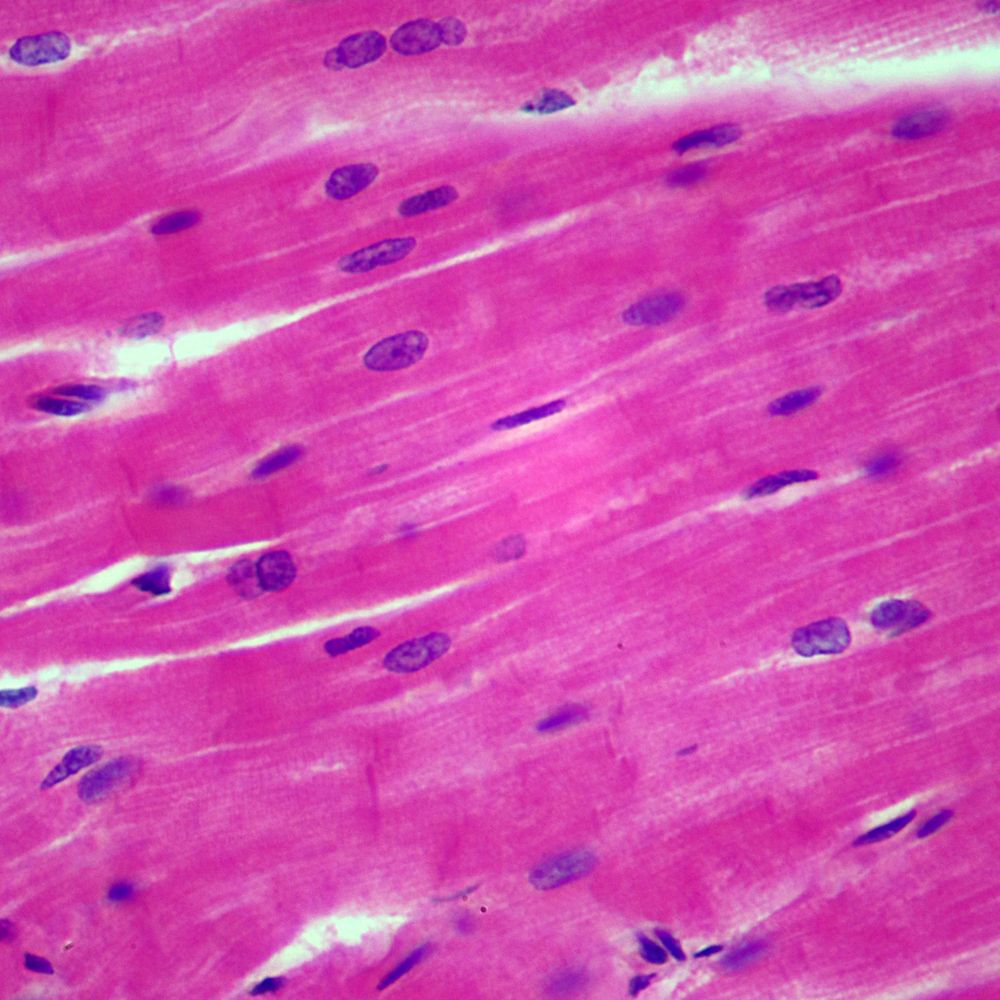
Cardiac Muscle:
In heart
Branched (somewhat connected to each other)
Striated (striped)
Uninucleated (ONE nucleus)
Involuntarily controlled
Slow and steady contractions EXCEPT during short periods of activity/exercise
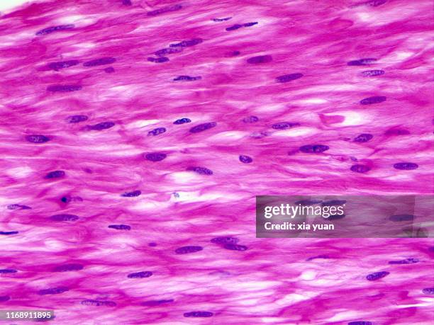
Smooth Muscle:
In walls of internal organs
Arranged in uniform layers
Nonstriated (NOT striped)
Uninucleated (ONE nucleus)
Involuntarily controlled
Slow contractions, sustained for long periods of time
Microscopic Structure of a Skeletal Muscle:
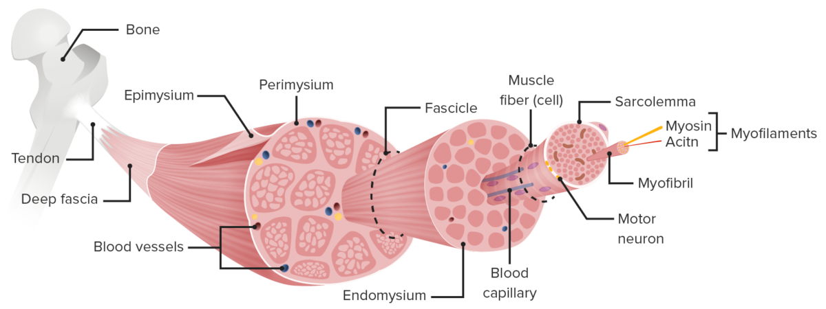
Largest - Smallest
Muscle -
Skeletal muscle is attached to bone by tendons
Made up of many bundles of fibers
Fascicle -
Bundles of fiber within muscle
Muscle Fiber -
Long, thin muscle cells
Each covered by sarcoplasmic reticulum, which transmit an impulse to the muscle fiber
Myofibril -
Thread-like organelles of muscle fibers
Structure in long, striated units = sarcomeres
Myofilaments -
Actin (thin) + Myosin (thick) make up sliding filament model of muscle (contraction)
Muscle Membranes:
Membranes allow for muscle fibers to slide and keeps them contained to prevent bursting during contractions
Epimysium - covers whole muscle
Perimysium - covers fascicle
Endomysium - covers individual muscle fiber
Muscle Contraction:
Arrangement of myofilaments -
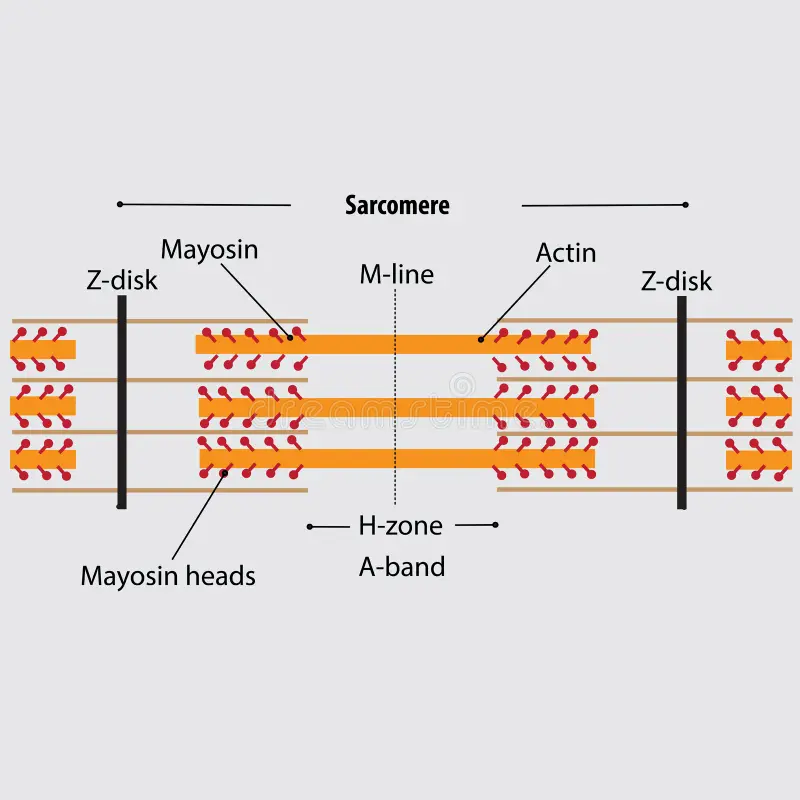
Actin + Myosin = Contraction
Attached to each other at Z line (Z disk on diagram woops)
Sarcomere = space between two Z lines
Actin and myosin interact to pull muscle fiber towards M-line, SHORTENS muscle fiber
Contraction:
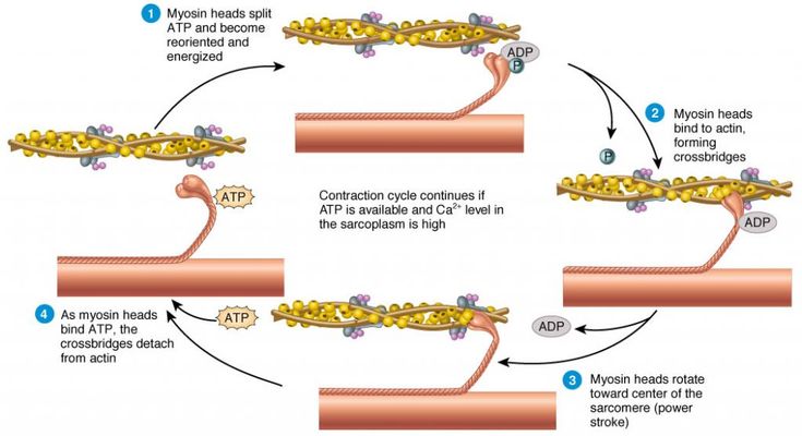
Nerve impulses are sent to muscle fibers to begin contraction
Myosin filaments have round extensions/ heads that attach to twisted actin filaments and pull on them, bringing the Z-lines closer, making the sarcomere shorter
Repeats until contraction is complete
ATP FUELS THIS PROCESS