y9 health
#ffc500 - yellow
UNIT 2: BODY SYSTEMS
SKELETAL SYSTEM:
The skeletal system consists of 270 bones at birth, and decreases to 206 by adulthood, due to some bones fusing together.
Types of bones to memorise:
Cranium - The skull
Mandible - The jaw bone
Clavicle - The collarbone
Pelvis - Near the hip area
Fibula - The smaller lower leg bone
Calcaneus - The balls of the feet
Cervical - The first seven vertebrae of the spinal cord, near the skull
Scapula - The ‘wing bones’ in the back
Humerus - The ‘funny bone’, the upper bone of the arm.
Ulna - The smaller bone in the lower arm
Femur - The largest bone in the body, located in the upper leg/thigh.
Tarsals - Bones that form the ankle and heel
Sacrum - Located above the coccyx (tailbone).
Coccyx - the tailbone
Radius - The larger bone of the lower arm.
Carpals - The wrist bones
Metacarpals - The bones that form your fingers
Patella - The kneecap
Metatarsals - The bones that are after the tarsals
Lumbar - 5 vertebrae between the rib cage and pelvis.
Phalanges (for the hands) - the bones that form your fingertips
Phalanges (for the feet) - bones that form your toes.
Sternum - The breast bone
Ribs - A cage that protects your lungs.
Tibia - The larger lower leg bone
Thoracic - 12 vertebrae that are after the cervical vertebrae.
Regions of vertebrae:
Cervical - 7 vertebrae
Thoracic = 12 vertebrae
Lumbar - 5 vertebrae
Sacrum - 5 fused
Coccyx - 4 fused (tailbone)
Tips for memorising bones:
Fibula - ‘fine’
Tibia - ‘thick’
Ulna - pinky side
Radius - thumb side
Tarsals - feet
Carpals - hands
Vertebrae:
The vertebrae are the bones that make up the spine. As you move down the body, the size of the vertebrae increases. The spinal cord runs through the hole in the vertebrae, and it relies on vertebrae for protection.
Functions of the skeletal system:
Allows movement - bones provide surfaces for muscles to attach, and they allow muscles to pull on them to produce more movement.
Shape and protection - bones give the correct shape to the body and protect organs.
Mineral storage - they store minerals such as calcium, iron, potassium and phosphorous. These will be released when the body needs them.
Production of blood cells - bone marrow (inside most bones) produce red and white blood cells.
Red blood cells - carry oxygen to muscles, are red because of a protein called haemoglobin, which contains iron, and the production of red blood cells are abundant during growth years, and decrease with age.
White blood cells - fight infection in the body, part of the immune system.
MUSCULAR SYSTEM:
The muscular system is an organ system that consists of approximately 600 muscles.
Types of muscles:
Trapezius - The muscle near the scapula, in the upper back.
Deltoid - The upper part of your arm, near your shoulders.
Pectoralis major - The chest muscles
Biceps brachii - The muscle below the deltoids
Triceps brachii - The muscle behind the biceps
Wrist flexors - Muscles in your front forearm
Rectus abdominis - The ab muscles
Obliques - The sides of your hips, on either side of the abs
Adductors - The muscles near your pelvic area, in the front.
Quadriceps - The frontal thigh muscles
Gastrocnemius - The muscle below the knee, on the back of the leg
Soleus - Muscles near your ankles
Tibialis anterior - Near your tibia
Erector spinae - The muscle in the middle lower back
Latissimus dorsi - The muscles on either side of the erector spinae
Gluteus maximus - The ‘butt’ muscles
Abductors - Muscles below the pelvic area, on the back side
Hamstrings - Thigh muscles, on the back of the leg
Functions of muscles:
Create movement - muscles pull on bones to produce movement
Posture - skeletal muscles are essential to keeping posture, and their flexibility and strength help keep posture
Heat production - When muscles contract, they create heat.
MUSCULOSKELETAL SYSTEM:
The musculoskeletal system relates to the bones, ligaments, tendons and skeletal muscles. The collaboration between the skeletal and muscular systems allow movement.
Ligaments and tendons:
Ligaments - Strong fibrous bonds that connect bone to bone. They provide stability at the joint where the muscles occur.
Tendons - Strong fibrous bonds that connect muscle to bone. When muscles contract, they pull on tendons, which pull on bones, which allows movement.
Flexion and extension:
Flexion - flexing a muscle
Extension - extending a muscle
Reciprocal inhibition: When muscles work in pairs. The agonist muscle (the one that contracts/shortens) contracts, while the other muscle (the antagonist muscle) relaxes/lengthens.
CARDIOVASCULAR SYSTEM:
The cardiovascular system, also known as the circulatory system, consists of the heart and blood vessels working together to transport gases and nutrients around the body.
Functions of the cardiovascular system:
Circulates blood around the body
Transports oxygen and nutrients to the cells
Transports waste away from the cells
Maintains stable body temperature
Types of blood vessels:
Arteries - have thick, elastic walls, and is responsible for transporting oxygenated blood to the muscles
Capillaries - have thin, non-elastic walls, and are responsible for the exchange of nutrients and waste between body cells. They transport both oxygenated and de-oxygenated blood.
Veins - have thin, not-so-elastic walls, carry de-oxygenated blood to the heart, and have valves inside, to prevent backflow. They rely on skeletal muscles to pump blood to the heart.
The heart:
The heart is a pump designed to push blood through the cardiovascular system. It is located slightly left to the chest, and is protected by the ribs. The adult heart is around the size of large fist.
In diagrams of the heart, the left and right sides are swapped, because you are looking at the front of it.
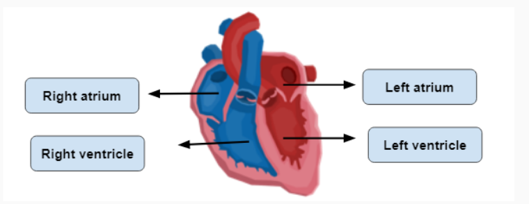
Atria are chambers that receive blood, while ventricles are chambers that pump blood.
The septum divides the heart into two halves. The right carries de-oxygenated blood, while the left carries oxygenated blood.
Pathway of blood: The veins first take blood into the right atrium, via the superior and inferior vena cava, and then into the right ventricle, which pumps blood to the lungs via pulmonary arteries. The blood travels via the lungs to alveoli, which are small air sacs at the end of bronchi. The alveoli are connected to capillaries, where gaseous exchange takes place. After that, the capillaries take blood to the heart via the pulmonary veins, to the left atrium, then the left ventricle, then out into the aorta and the rest of the body.
RESPIRATORY SYSTEM:
The respiratory system consists of the nose, mouth, throat, voicebox, windpipe and lungs, and is specialised for gas exchange.
Functions of the respiratory system:
Brings air from atmosphere to lungs
Transfers oxygen from air into the blood
Removes carbon dioxide from blood
Expels heat in exhaled air
Allow vocal chords to create speech
The lungs:
The lungs are the major organ in the respiratory system, located behind the ribs in the chest cavity. It allows oxygen inside the blood, and helps get rid of carbon dioxide.
Alveoli:
Alveoli are tiny air sacs in the lungs, and the site where oxygen and carbon dioxide are exchanged.
Diaphragm:
The diaphragm is a muscle in the respiratory system, and it helps us breathe in and out.
Breathing:
Is the process of taking air in and out of the lungs. It is broken into two phases; inspiration and expiration.
Concentration:
Breathing relies on tendency of air particles to move from areas of high concentration (high pressure) to areas of low concentration (low pressure).
Inspiration:
The process of inhalation, or air going into the lungs.
The diaphragm contracts, moving downwards.
The muscles between the ribs (intercostal muscles) contract, increasing the size of the chest cavity.
These steps causes pressure to decrease in the lungs.
Air is drawn into the lungs as gases move from areas of high concentration to low concentration.
Expiration:
The process of exhalation, or air getting expelled from the lungs.
The diaphragm relaxes, moving upwards.
The muscles between the ribs (intercostal muscles) relax, decreasing the size of the chest cavity.
These steps cause pressure to increase in the lungs.
Air is drawn out of the lungs as gases move from areas of high concentration to low concentration.
Gaseous exchange:
ALVEOLI AND CAPILLARIES
This happens between the alveoli and capillaries at the lungs. Carbon dioxide moves into the alveoli, and oxygen moves into the capillaries.
CAPILLARIES AROUND SKELETAL MUSCLES AND MUSCLE CELLS
Oxygenated blood becomes deoxygenated, as carbon dioxide moves to the capillaries, and oxygen moves to the muscles.
Menstruation:
Every woman has the ability to give birth
The menstrual cycle is the time from one period to the next.
There are four phases of the menstrual cycle
Menstruation - Your period starts, and you start to bleed, and is considered the beginning of the menstrual cycle.
It can last from 2-7 days
The uterus lining sheds over the days of your period.
Hormone levels are low, and you feel moody
Low-intensity movement is recommended.
You need to reduce salt intake, and caffeine
Increase consumption of good fatty acids
Drink more water.
Follicular - Starts on the last day of the period, and lasts to day 13 or 14, which ends in ovulation.
Around 11-20 ovarian follicles begin to develop, but only one matures into an ovum (egg).
The uterus lining (endometrium) begins to thicken.
Hormones are released, and you feel more energetic and happy.
The body is primed for maximal training, such as high-intensity training.
Appetite may be reduced
Intake more healthy carbs and fats
Intake iron-rich foods
Ovulation - Usually happens once a month, around two weeks before the next period (approx. days 15-19).
Can last from 16-32 hours.
The egg is released, and moves along fallopian tubes to the uterus.
Pregnancy can occur if a sperm cell reaches the uterus, and joins the egg.
People may feel more sensitive to emotional trigger, which can lead to the feeling of irritability or sadness.
The recommended movement is related to follicular phase.
Balanced hormone levels
Reduced appetite
Need carbs and fats, and foods that are high in fibre to reduce levels of estrogen.
Luteal - The body is working to support a positive pregnancy, and people may experience pre-menstrual syndrome.
Thickening of uterus lining
Slight increase in body temperature
Irritability, mood swings, anxiety, etc
Recommended exercise - low intensity to recover
Eat complex carbohydrates and high fiber foods to make you full for longer
Eat dark chocolate and fruits for satisfying sweet cravings.