Clinical Chem Acid-Base Balance & Electrolytes
Learning outcome
- Explain the role of buffer systems in maintaining acid-base balance and pH
- Explain the role of buffer systems in regulating pH of the intracellular fluid and extracellular fluid
- To understand the concept of fluid and electrolyte balance
- To understand homeostasis of selected electrolytes and related disorders.
Key Terms
Acid - proton (H+) donor
Strong acid - low affinity between acids & protons; highly dissociated in aqueous solution
Weak acid - high affinity between acids & protons; poorly dissociated in aqueous solution
Base - proton (H+) acceptor
Alkali - a base that is soluble in water & produces hydroxyl ion (OH-)
Concepts
Acid-base balance
- a state of equilibrium between acidity & alkalinity of the body fluids
- mechanisms of our body to maintain the body fluids close to neutral pH to ensure proper physiological functions
- measured using the pH scale
pH
- used to determine the acidity or alkalinity of a fluid
- measure the hydrogen ion (H+) concentration relative to that of a given standard solution
- negative logarithm of H+ concentration
pH = -log [H+]
- a scale from 0-14
- neutral (pH = 7.0)
- acidic (pH < 7.0)
- Basic/alkaline (pH > 7.0)
- very slight change in pH will have disastrous effects on cells & tissues
- acid-base balance is regulated within a narrow range for normal physiological functions
- normal blood pH: 7.35 - 7.45
- acidosis (acidemia)
- blood has low pH of less than 7.35
- overproduction of acid or excessive loss of bicarbonate or buildup of carbon dioxide
- metabolic or respiratory acidosis
- alkalosis (alkalemia)
- blood has high pH of greater than 7.45
- over-abundance of bicarbonate or a loss of acid or a low level of carbon dioxide
- metabolic or respiratory alkalosis
- Different mechanisms to regulate and maintain the blood pH within the fairly narrow optimum range
- Involve:
- lungs
- kidneys
- chemical buffer systems
Buffers
- solutions that can resist significant changes in pH
- maintain stable [H+] in biological systems
- consist of a conjugate acid-base pair
- present in both intracellular & extracellular fluids
- finite buffering capacity
- most common buffer systems:
- bicarbonate buffer system
- phosphate buffer system
- protein buffer system
- ]]Bicarbonate buffer system]]
- Major extracellular buffer system operates in both lungs and kidneys
- maintain pH homeostasis of the blood
- consists of carbonic acids (H2CO3) as the weak acids and its conjugate bases, bicarbonate ions
- H2CO3 is formed when dissolved carbon dioxide combines with the water in the bloodstream
- ↑ [H+] - HCO3- will accept H+ to form H2CO3
- ↓ [H+] - H2CO3 will donate H+ and turn in to HCO3-
- Compensation for the pH:
1. lungs → decrease the carbonic acids level through exhalation
→ adjust the respiration rate to decrease or increase the CO2 2. kidneys → reabsorbs bicarbonate ions or regenerate new bicarbonate ions
→ produce more acidic or more alkaline urine
- ]]Phosphate buffer system]]
important in buffering renal tubular fluid and intracellular fluid
comprised of hydrogen phosphate ions & dihydrogen phosphate ions
Hydrogen phosphate ion is freely filtered through glomerulus → high concentration intracellularly & in urine
critical renal & urinary buffer → allow secretion of H+ ions from the tubular cells in conjuction with the generation of HCO3-
↑ [H+] - HPO4(2-) will accept H+ to form H2PO4-
↓ [H+] - H2PO4- will donate H+ and turn in to HPO4(2-)
Catalysed by enzyme carbonic anyhydrase
- ]]Protein buffer system]]
- most abundant and important buffer system in the body fluids
- either intracellular or extracellular
- protein molecule carries both basic and acidic groups → acts as proton (H+) acceptor or donors
- Haemoglobin (Hb) is the major intracellular buffer system
Lungs
- HbO2 is formed from HHb by releasing H+ which will react with HCO3 and form H2CO3 + CO2 + H2)
- the CO2 is then eliminated by exhalation
Tissues
- CO2 produced by metabolism enters the blood, hydrated to form H2CO3
- the H2CO3 ionises to form H+ & HCO3-
- HbO2 accepts the H+ to form HHb
Disorders of Acid-Base Balance
- imbalances in acid-base equilibrium
- respiratory acid-base disorders
- caused by ventilatory dysfunction
- a change in the pCO2
- metabolic (non-respiratory) acid-base disorders
- a change in the bicarbonate level
- resulting from a change in renal or metabolic functions
Respiratory Acidosis
- decreased alveolar ventilation (hypoventilation) leads to a decrease in the elimination of CO2 from the lungs
- Possible causes:
- ineffective removal of CO2 from the blood in lung diseases
- trauma, infection or inflammation of central nervous system
- drugs (e.g., barbiturates, morphine) % alcohol
- congestive heart failure → decreased cardiac output
- Laboratory findings:
- pH < 7.35
- ↑ pCO2
- normal bicarbonate concentration
- acute respiratory acidosis: pH drops 0.1 unit for every 15 mmHg increase in pCO2
- chronic respiratory acidosis: pH drops 0.05 unit for every 15 mmHg increase in pCO2
- Compensatory mechanisms
- via the haemoglobin and protein buffer systems
- through metabolic processes in kidneys
- ↑ excretion of H+
- ↑ reabsorption of HCO3-
- ↑ formation of ammonia
- via respiratory organs (if functional)
- ↑ rate & depth of breathing
Respiratory Alkalosis
- Results from an increased rate or depth of breathing or both
- excessive elimination of CO2 by the lungs/deficit in pCO2
- Possible causes:
- hypoxemia- & hysteria-induced hyperventilation
- deugs, e.g., nicotine & salicylates
- pulmonary emboli & pneumonia
- gram-negative septicemia, meningitis or encephalitis
- Laboratory findings:
- ph > 7.45
- ↓ pCO2
- normal bicarbonate concentration
- Compensatory mechanisms
- haemoglobin & protein buffer systems
- kidneys excrete more HCO3 in urine
Metabolic Acidosis
- ↓ HCO3- level (< 24 mmol/L)
- Possible causes:
- direct administration or ingestion of acid-producing substances (e.g., ammonium chloride, calcium chloride, ethanol)
- production of organic acids (e.g., in diabetic ketoacidosis and lactic acidosis)
- reduced excretion of acids (e.g., renal tubular acidosis0
- excessive loss of HCO3- from diarrhea
- Laboratory findings:
- pH < 7.35
- ↓ pCO2
- ↓ bicarbonate concentration
- normal or increased anion gap
- Compensatory mechanisms
- via respiratory mechanisms (i.e., quick & shallow breathing)
- by kidneys (similar to those occur in respiratory acidosis)
Metabolic Alkalosis
- an excess or gain in HCO3-
- possible causes:
- increase in bases (i.e., massive blood transfusions, infusion of intravenous solution high in HCO3-, ingestion of large quantities of antacids)
- decreased excretion of bases (i.e., prolonged use of diuretics)
- loss of acidic fluids (i.e., prolonged vomiting, upper duodenal obstruction, cystic fibrosis)
- laboratory findings:
- pH > 7.45
- normal pCO2
- ↑ bicarbonate concentration
- compensatory mechanisms
- via respiratory system to retain CO2 (i.e., slower & depper breaths)
- by kidneys (excrete > HCO3- & form < NH3)
Fluid & Electrolytes
- Average water content: 40% - 75% of total body weight
- ~60% in men, ~55% in women
- Located in intracellular & extracellular compartments
- intracellular fluid (ICF)
- extracellular fluid (ECF)
- intravascular ECF (plasma)
- interstitial cell fluid
- Movement of water & distribution of water in different body fluid compartments are
- determined by osmolality & colloid osmotic pressure
- controlled by maintaining the concentration of electrolytes and proteins
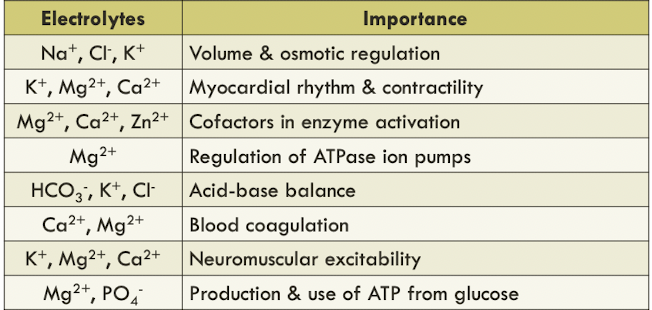
- Electrolytes are charged atoms or molecules found kn body fluids that are important for
- regulation of water distribution, osmotic pressure, cell permeability
- nerve transmissions to muscles
- oxidation-reduction reactions, maintenance of blood pH
- May be classified as:
- anions (negatively charged)
- cations (positively charged)
- Important physiologic electrolytes
- sodium (Na+), potassium (K+), chloride, (Cl-) & bicarbonate (HCO3-) occur primarily as free ions
- known as electrolyte profile
> 40% of calcium (Ca2+) & magnesium (Mg2+) are bound by proteins
- Electrolyte balance - the quantities of electrolytes gained is equal to those it loses
- Electrolyte imbalance is life-threatening
- Anion gap - the difference between the unmeasured anions and the unmeasured cations
- Calculation of anion gap
- determine certain types of electrolyte disorders
- as a marker of quality control of electrolyte testing
- a trend of increased or decreased anion gap in a run of patient specimens may indicate consistent testing errors in one or more electrolytes
Selected Electrolytes & Disorders
Sodium (Na+)
- Major cation in extracellular fluid (~90%)
- main source: sodium containing food additives, e.g., table salt, monosodium glutamate
- excess sodium is excreted in the urine or through sweating
Regulation
- depends on the intake & excretion of water, and renal regulation of Na+
- 3 primary processes:
- intake of water
- excretion of water
- excretion of Na+ through aldosterone, angiotensin II & atrial natriuretic peptide (ANP)
- 2 major homeostatic systems
- renin-angiotensis-aldosterone (RAA) system
- antidiuretic hormone (ADH)
- stimulated via:
- hypovolemia
- hypotension
- decreased renal perfusion
- hyperosmolality
Clinical Significance
- hyponatremia
- serum/plasma level of Na+ <135mmol/L
- caused by:
- increased Na+ loss (e.g., prolonged vomiting, diuretic use, severe burns)
- increased water retention (e.g., renal failure, hepatic cirrhosis, congestive heart failure)
- water imbalance (e.g., excess water intake)
- classification based on serum/plasma osmolality (ECF volume)
- low osmolality (e.g., ↑sodium loss, ↑water retention)
- normal osmolality (e.g., severe hyperkalemia, hyperproteinemoa)
- high osmolality (e.g., hyperglycaemia, mannitol infusion)
- acute hyponatremia (<48 hr); chronic hyponatremia (longer period)
- pseudohyponatremia
- an uncommon artifact results from in vitro hemolysis during blood sample processing in the laboratory
- a decrease of serum [Na+] but normal serum osmolality
- treatment aims to correct the underlying causes
- conventional treatment:
- fluid restriction
- hypertonic saline and/or other pharmacologic agents (i.e., AVP receptor antagonist)
- possible complications:
- osmotic demyelination syndrome
- cerebral edema
- Hypernatremia
- ↑serum level of Na+
- serum [Na+] > 160mmol/L has mortality rate of 60-75%
- caused by:
- excess loss of water relative to Na+ loss
- decreased water intake
- increased Na+ intake or retention (i.e., excess ingestion of salt)
- symptoms:
- altered mental status
- lethargy
- irritability & restlessness
- seizures
- muscle twitching & hyperreflexes
- fever
- nausea/vomiting
- difficult respiration & increased thirst
Chloride (Cl-)
- Major anion in extracellular fluid
- involved in maintaining osmolality, blood volume & electric neutrality
- filtered out by glomerulus & passively reabsorbed by the proximal tubules
- excess chloride is excreted in the urine and sweat
- Maintain electrical neutrality
- reabsorption of Na+ along with Cl- in proximal renal tubules
- Cl- acts as rate-limiting factor or
- through chloride shift
Clinical Significance
- Hypochloremia
- decreased level of Cl- in plasma
- due to prolonged vomiting, diabetic ketoacidosis, aldosterone deficiency, pyelonephritis
- conditions associated with high serum [HCO3-]
- Hyperchloremia
- increased level of Cl- in plasma
- caused by dehydration, renal tubule acidosis, prolonged diarrhea & diabetes insipidus
- excess loss of HCO3-