Neurons: resti ng potential, action potential and propagation (copy) (copy)
The Generic Cell Structure
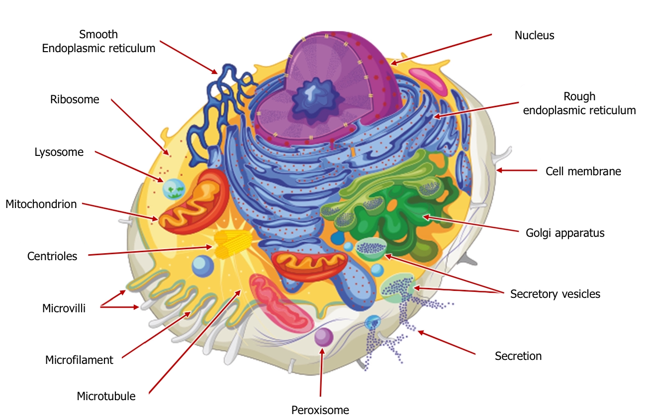
Components include:
Smooth Endoplasmic Reticulum: Synthesizes lipids and detoxifies certain chemicals.
Rough Endoplasmic Reticulum: Studded with ribosomes; involved in protein synthesis and transport.
Mitochondrion: Powerhouse of the cell; produces ATP through cellular respiration.
Centrioles: Involved in cell division by helping organize microtubules.
Microvilli: Increase surface area for absorption and interaction.
Microfilament: Part of the cytoskeleton, providing structure and support.
Ribosome: Site of protein synthesis.
Lysosome: Contains enzymes for digesting cellular waste and foreign material.
Cell Membrane: Semi-permeable barrier that controls the passage of substances into and out of the cell.
Golgi Apparatus: Modifies, sorts, and packages proteins for secretion or use within the cell.
Secretory Vesicles: Transport materials to be excreted from the cell.
Nucleus: Contains the cell's genetic material and regulates gene expression.
Peroxisome: Breaks down fatty acids and detoxifies harmful substances.
Microtubule: Component of the cytoskeleton; involved in maintaining cell shape and transport.
Glial Cells
Glial cells are non-neuronal cells that support and protect neurons, playing a critical role in maintaining homeostasis and forming myelin
Functions:
Insulation (myelination): Creates a myelin sheath around axons to enhance signal speed.
Protection: Protects neurons from injury and infection.
Nutrition and waste removal: Supplies nutrients to neurons and removes waste products.
Circulation of cerebrospinal fluid (CSF): Helps maintain the environment around the neurons:
Myelinating Cells: the type of glial cells that insulate neuronal axons, enhancing the speed of electrical signal conduction.
Schwann Cells: Located in Peripheral Nervous System (PNS); myelinate single neuronal axons (about 1mm each).
Oligodendrocytes: Located in Central Nervous System (CNS); myelinate multiple sections of different neuronal axons. Provideinsulation and support.
Support Cells: General term for cells that provide structural and functional support within the nervous system.
Microglia: Located in CNS; phagocytic cells that clear extra cellular debris and act as immune responders of glial cells.Have 9 sub-types and are extremely plastic. They divide and multiply at a fast rate.
Astrocytes: Star-shaped cells in CNS; Help provide (3D) structural support and are involved in the physical structuring of the brain. wrap around or surround the connections between neurons, called synapses. End feet contact blood vessels and are thought to be involved in regulation of blood flow.
Ependymal Cells: found in ventricles and central canal of spinal cord; produce and regulate CSF form the 'Ependyma' which is the tissue lining the ventricular system and central canal of the spinal cord., The top of the cells have cilia which beat in a coordinated pattern to circulate CSF. Extensions of the basal membrane attach to astrocytes..
The Neuron Structure
Neurons communicate via neurotransmitters at synapses, where electrical signals are converted to chemical signals and transmitted between cells.
Components of a neuron:
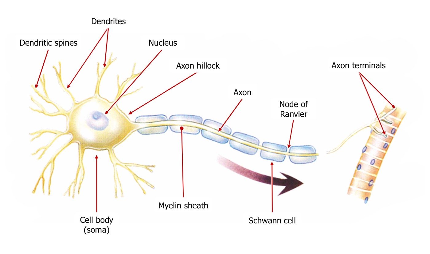
Axon terminals: Release neurotransmitters to communicate with other neurons.
Node of Ranvier: Gaps in the myelin sheath where ion exchange occurs.
Myelin sheath: Insulating layer surrounding the axon; increases transmission speed.
Schwann cell: Type of glial cell involved in myelination in the PNS.
Axon: Long projection that conducts electrical impulses away from the cell body.
Cell body (soma): Contains the nucleus and organelles of the neuron.
Axon hillock: Area where action potentials are initiated.
Nucleus: Houses genetic information and regulates neuron activity.
Dendrites: Branch-like structures that receive signals from other neurons.
Dendritic spines: Small protrusions on dendrites that form synapses with other neurons.
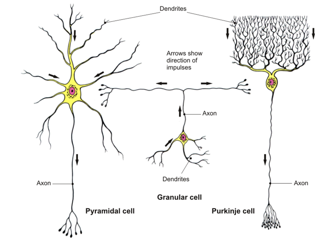
Classification by direction of impulses" in terms of neurons refers to how neurons are grouped based on the direction in which they carry electrical signals (impulses).
There are three main types of neurons based on this direction:
Sensory / afferent neurons: These carry impulses from sensory organs (like your eyes, skin, or ears) toward the brain and spinal cord (CNS).
Motor neurons: These carry impulses from the brain and spinal cord to muscles and glands. They tell your muscles to move or glands to release hormones.
Interneurons: These are found between sensory and motor neurons in the brain and spinal cord. They help process and relay signals between the other types of neurons.
Types of neurons
Bipolar Neurons: Typically found in sensory pathways, having two distinct processes—dendrite and axon. Found in special sense organs, sensory neuron
Unipolar Neurons: found in sensory neurones of peripheral nervous system characterized by a single process extending from the cell body, which then splits into two branches (one branch acts like a dendrite, and the other acts like an axon).
Multipolar Neurons: Have multiple processes; one axon and many dendrites, , most common neuron, involved in motor and interneuron functions.,
Cortical Cells - Detailed Description
the nerve cells (neurons) found in the cortex.
Examples include:
Granular cells: Small, densely packed neurons primarily found in the granular layer of the cerebellum. send signals to other types of cells in the cerebellum. Process info for movement.
Pyramidal cells: Large neurons with a pyramid-shaped cell body in the cortex cortex; send messages between different parts of the brain and control things like movement and thought.
Purkinje cells: Large neurons in the cerebellum with extensive dendritic trees, crucial for motor control.
Neuronal Cell Membrane
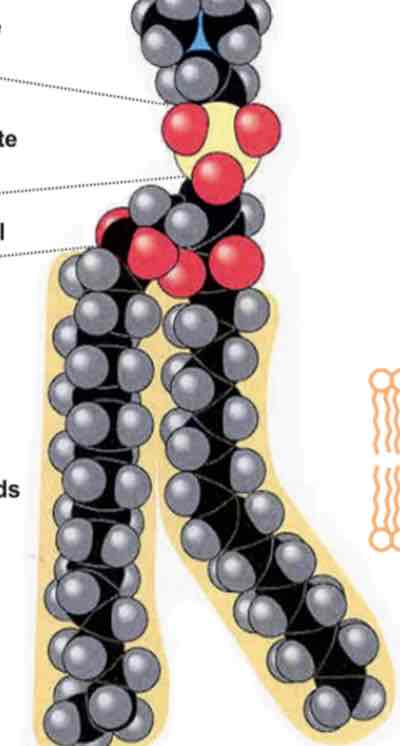
Composition includes phospholipids and proteins crucial for membrane function, forming a semi-permeable barrier vital for ion exchange.
Ions
Neutral particles: Have no net charge.
Anions: Carry a net negative charge.
Cations: Carry a net positive charge.
The Resting Potential
A neuron at rest has a voltage of approximately -70mV due to the uneven distribution of ions across its membrane, primarily maintained by ion pumps and channels
Active ion transport: 2 K+ ions are pumped by sodium potassium pumps into the cell while 3 Na+ ions are pumped out, maintaining concentration gradients essential for resting membrane potential.
Depolarization Process
Local currents modify the phospholipid bilayer, triggering the opening of ion channels, leading to K+ outflux and Na+ influx. This process ultimately initiates an action potential via voltage-gated ion channels.
The Action Potential
A rapid change in membrane potential, triggered when the neuron reaches a threshold level (-55mV). This involves depolarization (Na+ influx) followed by repolarization (K+ efflux).
Key voltages:
Resting: -70mV
Threshold for action potential: -55mV
Peak membrane potential: +40mV
Propagation of the Action Potential
Action potentials travel along axons via two mechanisms: continuous conduction in unmyelinated fibers and saltatory conduction in myelinated fibers, enhancing speed and efficiency
Myelinated Fibers (Aα): Conduct impulses at 80-120 m/s, showcasing rapid transmission.
Non-myelinated Fibers (C-fiber): Conduct impulses at a slower rate of 0.5-2.0 m/s, illustrating the effect of myelination on conduction speed.
Synaptic Transmission
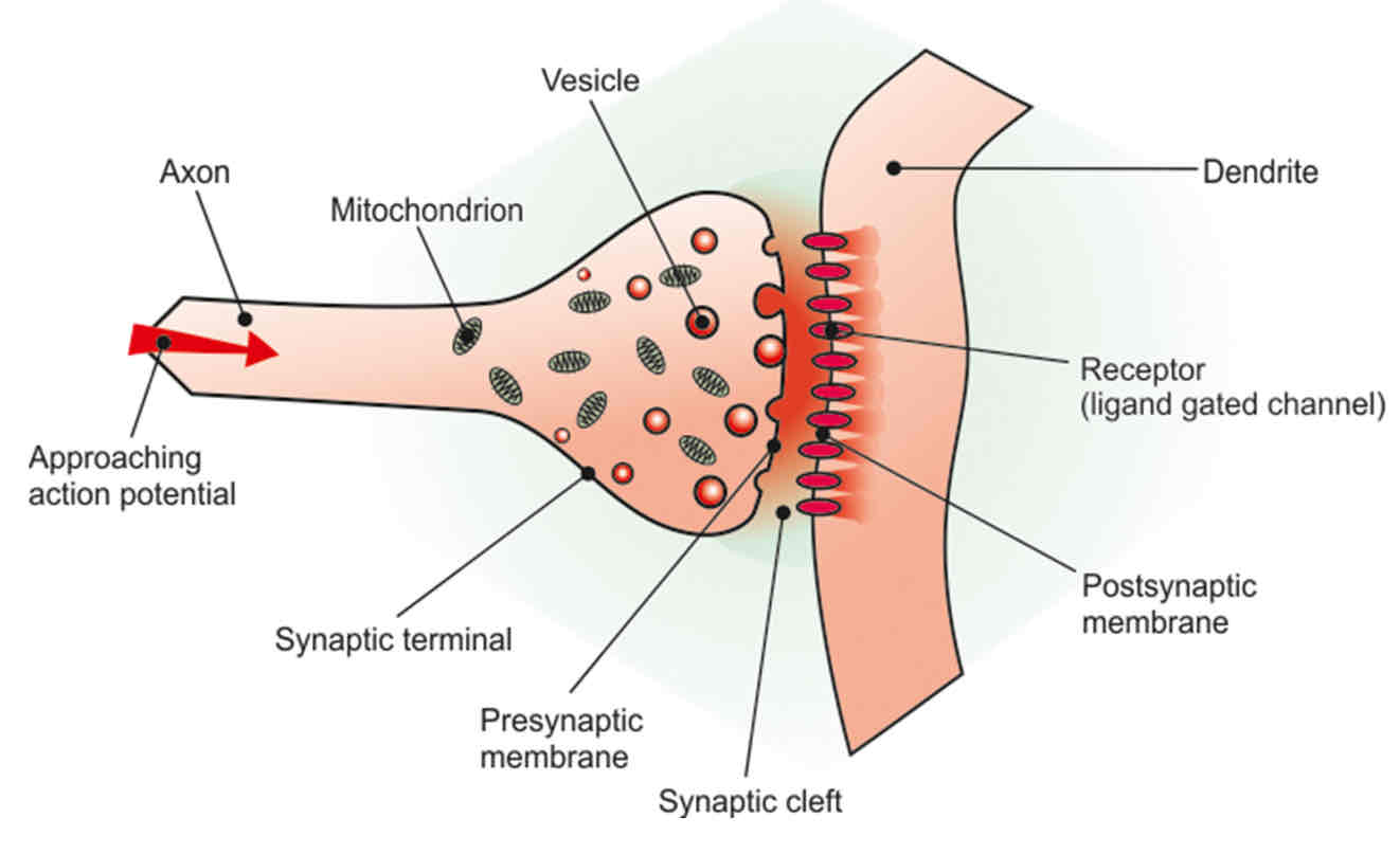
Key elements: Vesicles, axon, dendrite, receptors (ligand-gated channels), synaptic cleft, postsynaptic, and presynaptic membranes.
Ligand Gated Receptors
Functionality at the synaptic cleft: Neurotransmitter binds to the receptor on the postsynaptic membrane, initiating signal transduction and resulting in Na+ influx that may lead to depolarization.
.