MCAT Biology
Class 4 - 20/06/24:
Viruses, bacteria, reproduction of bacteria
Viruses: intracellular parasite
Virus structure: made up of a capsid(coat) with a nucleic acid genome inside(Can’t have both DNA and RNA)
Basic Steps: attachment(adsorption) - specific attachment but not infected yet; and injection - penetration - from bacterium to host
Lytic Cycle: transcribe and translate viral genome; replicate; lysis of host
Early genes - hydrolase and capsid
Hydrolase: destroy host cell genome
Replicate genome
Lysis of host and release of viral particles
Lysogenic Cycle: integrate viral genome into host then induce with normal host activity and excision and lytic cycle happens
Transduction - insertion of new DNA that was not present before
Productive Cycle: does not destroy the host cell
RNA Viral Genomes: can be both positive and negative types of RNA viruses
(+) RNA requires translation of RNA to protein - RNA dependant and RNA polymerases make the proteins
(-) RNA need a copy of RNA pol., and translate the now + RNA to proteins that negative
Prions - do not follow central dogma because they are self-replicating proteins
No DNA/RNA
no membranes
no organelles
very small
extremely stable
Prion categories = normal and mutagenic - mutant can lead to cell death
Mutant = Bad prions - come from a mutation in a prion, can be inherited, or by ingesting a bad prion → bad ones can make good ones bad too
Viroids: circular RNA, no capsid, must be co-infected, no protein code - block translation
two different mechanisms make viroids -
one by taking negative RNA, translating it to positive RNA to form many +RNA to form viroid copies
One by taking -RNA and wrap +RNA to form a viroid copy
Bacteria:
Can have three different shapes:
Round = coccus
Rod = bacilli
Spiral = spirillum
can have a flagella to move it or cilia
Bacteria have a cell wall and a cell membrane
gram + = stain dark and have a cell membrane covered by thick peptidoglycan(2 layers)- easier to get in
gram - = stain light and have an inner membrane covered by a cell wall covered by an outer cell membrane peptidoglycan(3 layers) - harder to get in
Temperature-dependent bacteria:
mesophiles → 30*C
Thermophiles → 100*C
psychrophiles → 0*C
Oxygen uses in Bacteria:
Obligate aerobe = use it and need it
Facultative anaerobe = can use it and survives
Tolerant anaerobe = doesn’t need it but can tolerate
Obligate anaerobe = can’t use it and can die due to O2
Energy/Nutrients of Bacteria
Photoauto = uses light and makes it on its own
Chemoauto = use chemicals by self
Photohetero = carnivorous plants
Chemohetero = need other energy sources
Reproduction - use of binary fission to duplicate identical copies
Binary Fission - growth follows an exponential growth pattern
Conjugation(genetic Diversity) - helps to provide genetic diversity, rather than increase population size
horizontal gene transfer - donor-to-recipient transfer with direct contact
F- is the donor(male) and F+ is the female recipient - gives an F plasmid, not a genome
Class 5 - 27/06/24:
Cell Biology, colligative properties, membranes, mitosis, cancer
Cell Biology and organelles → Eukaryotes
Nucleus and Nucleolus - DNA; makes ribosomes
Ribosomes - make Proteins(RNA to amino acids)
Rough ER - Makes post-translational protein modifications, proteins for the cell to use
Smooth ER - production/metabolism to fats and steroids
Golgi apparatus - prepares to ship/modify/sort proteins/lipids for in-cell and outside use
Lysosomes - break down foreign particles, eliminate toxins
Peroxisomes - eliminate H2O2, oxidative reactions of reactive O2 species
ALL transcription in the nucleus, and ALL translation in the cytosol
Secreted, transmembrane, lysosomal proteins are made in the Rough ER → resident proteins
Start in the nucleus(transcription, mRNA processes) → goes to the cytosol(begin all translation) → some proteins finish translation in the Rough ER → signal sequence tells the 3 proteins made by the Rough ER
Components of the cell membrane
Phospholipids - the membrane bilayer
Cholesterol - stabilizes membrane and keeps it fluid
Proteins
Carbohydrates
Colligative properties depend on the number to solute but not their identity
Freezing point, boiling point, vapour pressure, osmotic pressure
Electrolytes: free ions in a solution that come by dissolving ionic substances
ex. NaCl → Na + cl-
Van’t Hoff factor: the number of ions produced per molecule of an electrolyte when dissolved in water
ex. NaCl = 2 → 2 ions are produced per NaCl molecule
Freezing Point depression:
The freezing point of 1 KG H2O is OºC
FP depression Tf = -kf x i x m
kf water = 1.9℃
i = Van’t Hoff factor
m = # of moles
Vapour pressure depression:
need to raise the temperature to boil and evaporate molecules in a liquid
Boiling Point elevation:
BP Elevation Tb = kb x i x m
kb water = 0.5℃
Osmotic pressure elevation: we care about the number of particles(that change osmolarity)
Osmotic pressure = particle [C]
TT = i x M x R x T
M = molarity
R = gas constant
T = temperature
Diffusion: particles moving down a gradient → high [C] to low [C]
Osmosis: movement of water → water moves from high [C] to low [C]
Hypertonic = more particles than…
Hypotonic = fewer particles than…
Isotonic = the same amount of particles than…
Pressure is required to resist the movement of water by osmosis
osmotic pressure = particle [C]
ex. put a RBC(which is 0.9% NaCl into a beaker with 20% NaCl → water wants to leave the cell to equalize it, but the cell will shrivel → hypertonic
ex. put the same RBC into a beaker with 1% NaCl, very close to the RBC, some water leaves but not a lot → isotonic
Passive transport: no energy is needed and relies on the concentration gradient for movement
Simple diffusion and facilitated diffusion
Simple → works well for small hydrophobic molecules, ex. steroids, CO2, O2, lipids
Facilitated → still moves down a gradient and uses small hydrophilic molecules ex. glucose, amino acids, ions, H2O → need helper protein
Helper Proteins: pores, channels, porters
Pores: limits things in/out by size only
Channels: highly specific → Na/K channels
Porter: can undergo a conformational change to move molecules → The shapeshifters
Active transport: requires E and can move molecules without the need for concentration gradients
Primary: use ATP
Na/K pumps(every 2K for 3 Na)
K leak channels → can go by the concentration gradient
These maintain osmotic balance, establish e-gradient, set up gradient for secondary transport
Secondary: uses ATP indirectly and relies on the setup of the primary
G-Protein: adenylyl cyclase → makes cAMP → activates cAMP-dep kinases → phosphorylates enzymes and changes enzyme activity in cells
cAMP is a secondary messenger, signal amplification, fast and temporary
Phospholipase C → breaks phosphoinositol biphosphate → breaks into IP3 and DAG → DAG activates kinase and changes enzyme activity
Cytoskeleton:
Microtubules: made of a and b tubulin and are large in diameter and are used for mitotic spindle, intracellular transport, and cilia/flagella
Cilia/flagella → 9 microtubules surrounding 2 lone tubules
Microfilament: made of actin protein, smaller in diameter, and used for muscle contraction, pseudopod formation, cytokinesis
Intermediate filament: several different protein types, medium in diameter, and used in many structural roles
Cell Junctions:
desmosomes = general adhesive junctions
tight junctions: seal lumens and separate environments
Lumen also has gap junction → cell-to-cell communication
Cell Cycle: Interphase to mitosis(PMAT)
Sister chromatids: absolutely identical → same order, genes, alleles
Homologous chromosomes: same genes, same order, but can have different alleles
Interphase: cell growth and synthesis of DNA
G1 = cell growth, normal cell activities,
S = synthesis of DNA and DNA replication
G2 = growth and prepare for mitosis
Mitosis = PMAT → ends with two identical daughter cells similar to the parent
Prophase: condenses DNA, forms mitotic spindle, nuclear membrane breaks down
Metaphase: chromosomes align on the center
Anaphase: the sister chromatids separate and travel to the ends of the two poles and begin cytokinesis
Telophase: reverse prophase(DNA un-condenses and nuclear membrane forms again) - cytokinesis finishes
Cancer: mutation to DNA, starts from a single cell with mutations, goes through the cell cycle rapidly and out of control, spreads to other tissue → Metatsis
Two types of Cancer genes: oncogenes and tumour suppressors
Proto-oncogenes: genes are normally present in the cell and code for proteins that regulate the cell cycle →
Active types: fetal development, growth, and healing
Inactive types: when healing or growth is not required
Oncogenes: the mutated version of proto-oncogenes that are permanently on(always active and dividing)
Tumour suppressor genes: code for proteins that stop the cell cycle, and monitor the genome of cells in the cell cycle, if DNA damaged they initiate repair mechanisms, if DNA cannot be repaired then they initiate cell death(apoptosis)
If they lose their function to save or get rid of mutations, cancers can come from those mutations that were not ‘killed’ off by apoptosis
Class 6 - 01/07/24:
Genetics, Hardy-Weinberg, and Evolution
Meiosis - making of 4 cells that differ from the parent cell and each other → unique cells
Crossing over happens of DNA → leads to diversity
Non-disjunction - failure to separate DNA during meiosis
Genetics - the study of genes
Allele - the genes found on a chromosome
Trait - the characteristic that appears from the alleles
Polymorphic - several types of one trait
polygenic - several genes that determine a trait
Classical Dominance: homozygous dominant/recessive, heterozygous
Genotype: combination of alleles
Phenotype: physical characteristics
Incomplete: display a blend of the parental phenotypes
ex. red flower RR x white flower WW = pink flower RW
Co-dominance: both alleles are expressed independently and at the same time
Ex. Blood types → IA IB i
Epistasis: dominance between two different genes - one gene can mask or modify the expression of another gene, for example, Albinism
Test-cross: where one of an unknown genotype is crossed with another of a homozygous recessive genotype
Backcross - F1 x P
Mendel’s Laws
Law of segregation - alleles separate during gamete formation
Law of independent assortment - one allele is independent of another allele
Single-gene crosses - 4 types
Homozygote 1 x Homozygote 1
Homozygous dominant x homozygous recessive
heterozygote x homozygote dom/rec
heterozygoye x heterozygote
Rules of Probability
A AND B - multiply the probabilities
A OR B - add individual probabilities
Linked Genes: genes found close together on the chromosome
Dihybrid crosses = crosses between two traits
F1xF1 = 9:3:3:1 → unlinked
F1xHomozygous recessive Parent = 1:1:1:1 → unlinked
When the actual ratio differs from this, they will be linked genes as they don’t follow the expected ratios
Recombination: genes that do not assort independently
recombination frequency = # recombinants/total offspring x 100
Tells us the map units(mu) distance between genes on the chromosome
1 mu = 1 cM(Centimorgan)
Hardy Weinberg: tells us that allele frequencies within a population do not change from generation to generation
p + q = 1 → allele frequency where p = dominant allele and q = recessive allele
pp + 2pg + qq = 1 → genotype frequency where 2pq is the heterozygous allele
5 Conditions where Hardy-Weinberg hold:
No mutation
No natural selection
No migration
Total random mating
Large population size
Class 7 - 10/07/2024:
Nervous System, Auditory, and Optical systems
Neuron Structure
Specialized cells of the nervous system
Have a soma(central), dendrites, axon, axon terminus
Dendrites receive signals
Axon sends off signals
Axon has myelin covering it
Speeds and protects the axon
Types of Neurons:
Multipolar - connects and receives from many neurons
Bipolar - depends on the direction of the synapse - two-sided
Unipolar - soma is attached to one node only
Resting Potentials
at -70mV
sodium/potassium pump out one net positive ion, creating a Na/K gradient
many + ions lost via K leakage channels
The result is that the cell is more negative inside than outside
Action Potentials
When the cell reaches positive levels to send a signal across the neuron - All or None event
Depolarization - cell becomes more positive
Hyper polarization - when the cell becomes more negative
Repolarization - return to rest
Equilibrium potential - when there is no driving force on the ion, neither +/-
At -70mV → resting potential
At -55 → threshold - Na+ channels open
Depolarization upto +35mV - Na+ channels inactivate and K+ fully open
Hyperpolarization to -90mV - Na+ and K+ close
Repolarization to -70mV by Na/K pumps
This happens in a matter of 2-3 msec
Nerve Impulse: by a synapse from neuron-neuron or neuron-organ junction
Refractory Periods
Absolute: not able to fire a second action potential due to Na channels being inactive and the cell is too positive at the moment
Relative refractory period: there is a small chance of firing a second AP since Na channels are closed and the cell is too negative
Electrical Synapse - relatively rare but important in muscle cells
Require:
Physical connections - gap junctions
Always excitatory - AP in post-synapse
Bi-directional - either pre/post synapse
Unregulated
Chemical Synapse - more common - transport of neurotransmitters
One neuron can only make one type of NT but can respond to many
NT in the synaptic cleft can be re-uptaken or broken down
Response of the post-synapse depends on the receptors, not the NT
Need more than one vesicle of NT to make a change to post-synapse
EPSPs and IPSPs
EPSP = excitatory post-synaptic potential → Many accumulate to make an action potential - help reach the threshold
IPSP = inhibitory post-synaptic potential → Many accumulate to prevent an AP from reaching a threshold
EPSP and IPSP can cancel each other out
Summation
Spatial: The add-up of inputs from multiple sources
Temporal: the add-up of frequent impulses from a single source
General System Functions
Sensory Input - PNS
Info coming into the CNS
carried on the sensory neurons → afferent → towards CNS
Integration - CNS
decision making
interneurons - entirely contained within the CNS
Motor Output - PNS
commands sent out to the body
Carried on the motor neurons → efferent → exit CNS
Reflexes
rapid integration to avoid potential injury
Patellar tendon stretch reflex
CNS Anatomy
Telencephalon: cerebrum
Cerebral hemispheres - left and right - connected by the corpus callosum
Cerebral cortex - divided into 4 lobes
Frontal - complex processes and voluntary movement
Parietal - general sensations - touch, temperature, taste
Temporal lobe - sound and audition and olfactory, STM, language
Occipital lobe - Visual sensations
Diencephalon:
Epithalamus: Pineal gland and secretion of melatonin - links to the limbic system
Thalamus: sensory - all sensory (except olfactory)
Hypothalamus: sends hormones to the pituitary - primary link to endocrine - homeostasis and behaviour/emotions
Hindbrain:
cerebellum - movement and balance
Medulla - controls vital autonomic functions and relays info between other areas - respiratory centers located here
pons - the role is posture and balance
Spinal Cord: connects brain and body and is protected by the CSF
Limbic System: works for emotion and memory
White Matter vs Grey Matter:
White: myelinated axons - cell-to-cell communication
CNS to brain = Tract
CNS to cord = tract/column
PNS = nerve
Grey: non-myelinated axons - decision-making
CNS to deep brain = nucleus
CNS to brain surface = cortex(conscious mind)
CNS to cord = horn
PNS = ganglion
PNS
all nerves and sensory systems outside of the CNS
Somatic vs Autonomic
Somatic = voluntary control of the skeletal muscles
uses Ach only, excitatory only, single neuron effector
Autonomic = involuntary control of glands and smooth muscle
uses Ach and Norepinephrine, can be excitatory or inhibitory, a chain of two effectors
Parasympathetic vs. Sympathetic
Para = rest and digest
decrease HR, breathing, BP
Increase Digestion
release Ach to organs either inhibit(heart rate down) or excite(increase digest)
Symp = fight or flight
Fight, flight, fright, sex
increase body activity
decrease digestion
increase blood flow to skeletal muscles
release norepinephrine at the organ level
Sensory receptors - 5 Classes:
Mechanoreceptors: by physical shape changes, touch
Chemoreceptors: by chemicals, pH, O2, taste buds
Thermoreceptors: stimulated by temperature, hot or cold
Nociceptors: stimulated by pain, free nerve endings, chemicals, heat
Photoreceptors: by light, rods and cones
General Sensory Processing
Absolute threshold: the minimum stimulus required to trigger a receptor
Difference threshold: how much a stimulus must change to detect it
Sensory adaption: receptor stops responding to constant stimulus
pain receptors do not adapt
Bottom-up processing:
from the environment to the brain → sensory receptors take in the info, send it to the brain, and the brain uses the info
Top-down processing:
from inside to environment → The brain applies prior knowledge to identify the environment
Visual System
Cone cells: colour vision, stimulated in light only - three kinds: red, green, blue
Cone like a triangle; like a prism; prism makes rainbow - colour!
in the Fovea centralis only
Rod cells: susceptible to light and work in low light conditions
Concentrated in the retina
Auditory System
Sound waves → auricle → external auditory canal → tympanic membrane→ malleus → incus → stapes → oval window → perilymph → endolymph → basilar membrane - auditory hair cells → tectorial membrane → Neurotransmitters stimulate bipolar auditory neurons → brain → perception
Class 8 - 20/07/24:
Endocrine system, Cardiovascular system, Immune system
Endocrine system: hormones → through the bloodstream → no ducts
Exocrine system: hormones by way of ducts → into the intestinal lumen
Peptide hormones: made from amino acids, the receptor is on the cell surface, 2nd messengers, fast effects but temporary
Steroid hormones: made from cholesterol, intracellular binding, binds to DNA and modifies transcription, effects are slower but last longer
Mechanisms to control hormone release: neural, hormonal, humoral(in the blood)
Hypothalamus - pituitary: The hypothalamus controls neurally and humoral while the pituitary has divided control
Anterior pituitary: made of gland tissue, secretes six major hormones: FLAT PIG
FSH, LH, ACTH, TSH, Prolactin, Endorphins, growth hormone
Has hormones making cells and many veins
Posterior Pituitary: made of nervous tissue, stores and secretes two hormones - Oxytocin and ADH(vasopressin)
Many neurons and capillaries
Blood vessels: Veins and arteries
Veins: lower pressure, blood moves back to the heart - more rigid and made of collagen
Arteries: higher pressure, moves blood away from the heart - more elastic and can control dilation/constriction
Capillaries - smallest in size but largest SA → can exchange products like O2
The inner layer of blood vessels is endothelial cells
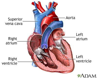
The heart - 4 chambers:

Blood carries from the aorta to the body → vena cava carries blood back to the heart from the body → into the right atrium → to the right ventricle → to the pulmonary artery to the lungs(deox) → blood comes back from the lungs into the pulmonary veins(oxy) → into left atrium → into left ventricle → to the aorta
Systole: ventricular contraction - empty
Diastole: ventricular filling
Lub Dub:
Lub: close AV valves and begin systole
Dub close SL valves and begin diastole
systole/diastole = Blood Pressure
BP is directly proportional to CO and peripheral resistance
CO = cardiac output = stoke volume x HR
volume pumped per minute x beats per minute
Stroke volume = change in blood volume, activity level, posture
Peripheral resistance = how hard it is to move blood through the vessels
Vasoconstriction and Vasodilation
Constrict = smaller diameter, lower flow, higher resistance, higher BP
Dilate = larger diameter, higher flow, lower resistance, lower BP
Tetany - tetanic contraction = involuntary muscle contraction from overstimulated neurons and low Ca levels
Cardiac cell potential: slow opening of CA+ channels, fast depolarization to 20+, then potassium channels open and repolarization back - unstable resting potential due to NA leakages
Cardiac Conduction: SA node → AV node → HIS Bundle → Purkinje Fibers
Blood composition: 54% plasma, 45% RBC, 1% leukocytes/WBC
Oxygen Dissociation Curve: oxygen is 3% dissolved in plasma and 97% dissolved in Hb → The higher the O2 levels, the higher the Hb saturation, and the more it is exchanged to tissues
Immunity:
Antigen: a foreign protein that can trigger an immune response
Antibody: a specific marker for anti-gen
Pathogen: disease-causing organism
B cells - humoral immunity - make antibodies
Produce and secrete antibodies into the blood - when stimulated, will clone into thousands of B cells to enhance antibody production - rearrange antibody genes(DNA) to generate antibody diversity
T cells - kill virus-infected cells, and tumour cells, and control the immune response(helper T cells)
T = thymus(develop in childhood)
MHC 1 → found on all cells, allows the display of cell contents
MHC 2 → macrophages and B cells, allows cells to display eaten stuff on the cell surface
Classes of Antibodies:
IgM = blood and B cell surface - initial immune response
IgG = blood - ongoing immune response
IgD = B cell surface - with IgM, antigen on B cells
IgA = secretions(saliva, mucus, tears, breast milk, etc) - protect newborns
IgE = blood - allergic reactions
Autoimmunity: uneliminated B and T cells(that did not undergo apoptosis) that get released into the body and go by attacking normal body proteins leading to an autoimmune reaction and other inflammation responses.
Class 9 - 27/07/24:
Excretory organs:
Colon - eliminates solid waste - material not absorbed into the blood
Liver - eliminates hydrophobic water - material too hydrophobic to be dissolved into the plasma
Kidney: eliminates hydrophilic waste - material eaten and absorbed into the blood and dissolved into the plasma
Kidney -> ureter -> bladder -> internal sphincter -> external sphincter -> urethra
The kidney has the ureter that connects to the renal pelvis, to the medulla and medullary pyramids, to the nephrons -> outer side of the kidney is the cortex
3 processes to produce urine:
filtration(moving a substance across a membrane using pressure
Reabsorption(move a substance from the filtrate to the blood(glucose, amino acids, water) -> glomerular filtration
Secretion: move a substance from the blood to the filtrate(drugs, toxins, creatine)
Nephron Structure:
Afferent arterioles come into the glomerulus leading to the PCT(mostly reabsorption and secretion), then to the loop of Henle - descending is permeable to H2O and the ascending is permeable to salt - DCT is specialized for absorption and reabsorption then the collecting duct(regulated H2O reabsorption)
Urine and Blood flow are opposites
Renin-Angiotensin system:
Angiotensinogen -> angiotensin 1 -> angiotensin 2 -> increases release of aldosterone and leads to systemic vasoconstriction
Juxtaglomerular apparatus(JGA): contact point between afferent arteriole and distal convoluted tubule
Afferent = baroreceptor
Distal = chemoreceptor
ANP - blood pressure regulation:
High BP -> arteria of heart stretch -> right atrium releases atrial natriuretic peptide(ANP) -> vasodilation and inhibits renin release
Accessory Digestive Organs: Liver and gallbladder
Liver = produces bile(amphipathic) → attack lipids(fats)
Gallbladder → stores the bile
Pancreas: Endocrine role = insulin and glucagon and Exocrine role = digestive enzymes, proteases, lipases, amylases, nucleases, and bicarbonate(pH control)
Alimentary Canal: Contains mucosa(epithelium), submucosa(connective tissue), serosa, and circular and longitudinal muscle
peristalsis → movement in one direction only
Regions of the alimentary canal:
Mouth = grind food and begin starch digestion by amylase and lingual lipase(fats) and use of lysozyme to kill bacteria
Esophagus = tube from mouth to stomach by the movement of peristalsis and starts off as voluntary(skeletal) to involuntary(smooth) → also has the cardiac sphincter. that prevents the movement of acid reflux
Stomach = storage tank for food and grinds food down more by gastric glands - parietal cells make HCL and Cheif cells make pepsinogen to make pepsin(by HCl) - the G cells in the stomach secrete gastrin and activate gastric glands(negative feedback loop)
Small Intestine: all digestion and absorption happens here in three regions - duodenum to jejunum to ileum - food brushes up against the microvilli to absorb nutrients - enterokinase(activates trypsin for pancreatic enzymes) and brush border enzymes(to break up peptides) as well as enterogastrone(reduce stomach mobility and empty food), CCK(bile release), and secretin(if pH too low, release bicarbonate by pancreas)
Large Intestine: absorbs water and feces only - has bacteria, internal and external anal sphincters - bacteria break up remaining stuff and make/absorbs vitamins A, K, D, C
Class 10 - 01/08/24:
Musculoskeletal System and Respiratory System
Skeletal muscle overview: voluntary function, on the bones, multinucleate and striated appearance
Hierarchy:
protein filament - actin and myosin → they do not shorten
Sarcomere - a unit of contraction → Shortening happens here → Depolarization
1 sarcomere is one Z line to the next Z line
Actin, thin filaments are held by the Z-line
The myosin, thick filaments, is centred in each sarcomere but doesn’t reach the Z-line
The I band (isotropic) are the regions with full actin or ½ actin
The A band(anisotropic) are the regions where there are both actin and myosin - it is both dark and light - ends at the ends of the myosin
H zone is the light zone where there is only myosin and no overlap with actin
The M line is the middle of the sarcomere
Myofibril
Covered by sarcoplasmic reticulum(holds Ca)
T-tubules - the plasma membrane goes in deep to help action potential travel to the interior of the cell
Muscle cell - myofiber
Fascicle
Whole muscle
Sliding Filament Theory:
Myosin binds to actin (cross bridge formation)→ needs calcium
myosin pulls actin towards the center of the sarcomere - power stroke → myosin returns to low-E state
Myosin release actin → needs ATP but doesn’t break it down
Myosin resets to high-E → ATP hydrolysis
When you run out of ATP, you can’t relax
Excitation-contraction coupling:
Excitation - depolarize, open voltage-gated Ca channels
Troponin binds Ca and changes shape, lifts tropomyosin off myosin binding sites, and myosin binds to actin
Contraction occurs
Motor Neuron - a neuron and all the muscle cells it controls
Contraction of the motor unit is all or none
Contraction of the whole muscle is graded
Large vs. Small:
Large is 1000s of m/n
Small is 10-20 m/n
Gross motor control - a few large motor units
Fine motor control - many many small motor units
Muscle Energy:
Fastest source of E = Creatine (substrate-level phosphorylation)→ reversible process
Medium source of E = Glycolysis(fermentation) - 2 ATP per glucose and lactic acid
Slowest source of E = aerobic respiration → 30 ATP, H2O, CO2 → store O2(myoglobin)
Oxygen Debt - extra O2 needed after exercise
replenish O2 stores in myoglobin
convert lactic acid into something useful → back into pyruvate
How to repay O2 debt? Bohr effect → pH and temp changes after O2 is used, then Hb is changed - rather than holding lots of O2, gives more of it back to tissues
Skeletal Muscle Types:

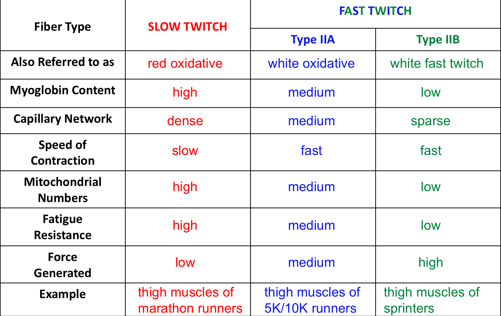
Slow Twitch- more myoglobin, more blood vessels, slow contraction, higher mitochondria, higher fatigue resistance, the low force generated
Fast Twitch IIA: medium level of myoglobin and blood vessels, faster contraction, medium mitochondria levels, medium resistance to fatigue, medium force generated
Fast Twitch 11B: low myoglobin levels, lesser capillary network, fast contraction and higher force, lower mitochondria, lower fatigue resistance
Fast twitch makes more glycolytic enzymes - more glycolysis
Cardiac and Smooth Muscle
Cardiac = auto, involuntary, only in the heart(vessels have smooth muscle) - uninucleate - striated appearance → The filaments overlap(the difference is that some of the calcium for contraction comes from the extracellular environment)
Smooth muscle = involuntary, neural, mechanical, hormonally stimulated; located in the walls of hollow organs; uninucleated; non-striated but has bundles of actin and myosin and still needs calcium
4 different tissues:
Muscle
Neural
Epithelial
The first three are mostly cells(living)
Connective - mostly non-living
Connective tissue: cells in a matrix
matrix is made of fibres and glop(ground substance)
Fibers are made of collagen and elastic fibres
glop is the glue that holds everything
Liquid or solid → Blood plasma or bone → in between is cartilage
osteoblasts - form new bone - can still divide and make the matrix
osteoclasts - break down bone
-cyte cells: the mature cells - don’t divide
Bone helps:
Support and movement
Store minerals - calcium and phosphate
Protection
blood cell formation
Osteoporosis - bone creation is slower than its removal → Weak and brittle bones
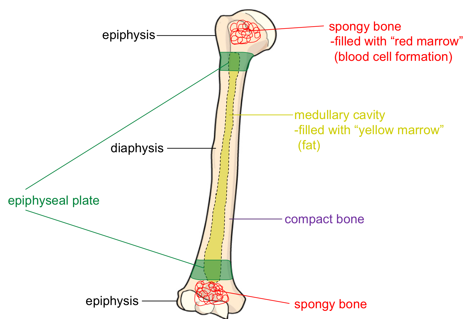
Long Bone Anatomy:
Shaft = Diaphysis
Ends = Epiphysis - holds spongy bone that makes RBC
Core = medullary cavity = yellow bone marrow(fat)
Surrounding the core is a compact bone
Epiphyseal plate = growth plate
ossification greater than cell division

Compact bone:
Osteons → have a central canal → central canal holds blood vessels → central canal made of rings that hold osteocytes
Bone Turnover:
PTH and Calcitonin
PTH - increases blood calcium by dissolving bones, increases Ca absorption in intestines and kidneys
Calcitonin - builds bone back, and decreases intestinal and kidney absorption
Vitamin D(calcitriol) - increases PTH effects and absorption in kidneys
osteoclasts - dissolve bone - eat bone, not like bone cells
Respiratory System:
Gas exchange and pH regulation
Ventilation = move in the air and out
Respiration = gas exchange (External and Internal)
Conduction zone = ventilation only
Air drawn in by the nose → nasal cavity → warmed up and filtered here → tissue is respiratory epithelium(mucus cells and cilia) → air to pharynx(naso, oro and laryngo pharynx) → air to larynx and travels down to trachea → separates to R/L primary bronchi → travels to secondary Bronchi in lobes → travels to tertiary bronchi → travels to terminal bronchioles to reach the respiratory bronchioles
The larynx is all cartilage → keeps airways open and separates air and food(epiglottis) and helps produce sound
Trachea: a muscle lined with cartilage rings and connective tissue membrane → The muscle can contract and pull the rings together → increases the speed of airflow(ex. coughing)
Primary bronchi → cartilage rings → cilia cells
Secondary bronchi → oddly shaped cartilage rings, some smooth muscles → short cells, no cilia
Tertiary bronchi → all smooth muscle, no cartilage → no cilia, short cells
Respiratory zone = Gas exchange
Air travels from respiratory bronchioles to alveolar ducts → enters alveolar sac and gas exchanges occur in the alveoli by the capillary network surrounding the sac → O2 is released into the blood and CO2 is picked up → CO2 travels back outside the lungs
Alveolar cells:
Type 1 = walls of alveoli
Type 2 = secrete surfactant → makes breathing easier and reduces tension/friction
The Lungs: there are two of them → The right lobe has 2 parts and the left lobe has 3
lungs stick to the chest cavity due to surface tension and slight negative pleural pressure(Inhale) → pressure wants to go positive, hence, becomes like environment(exhale)
Inspiration: Active → contraction of the diaphragm
Relaxed expiration: Passive → Diaphragm contracts
Forced expiration: Active → Abdominal muscles contract

Skin: has 3 layers
Epidermis: epithelial tissue
Dermis: connective tissue
Hypodermis: fat
Thermoregulation:
Cold = no sweat, shivering, vasoconstriction
Hot = sweat, no shivers, vasodilation
Class 11 - 15/08/24:
Reproductive System
Primary Sex Organs - testes and ovaries → make gametes(sperm or egg)
accessory sex organs - any organ or structure that has a role in development and reproduction that is not directly involved in the production of gametes
Male System:
Scrotum = suspends testes outside the body
Sperm viability needs high testosterone levels and slightly lower cooler levels than body temp
The testes: make sperm and testosterone(androgen) - higher in males
testes filled with seminiferous tubules with sustentacular cells(Sertoli cells) that sustain sperm development - secrete nutrients and ABP(androgen binding protein) → spermatogonia - cells that WILL become sperm(not yet)
The hormone FSH in males stimulates Sertoli cells and spermatogonia
Interstitial cells make testosterone - Leydig cells(surround seminiferous tubules) - stimulated by LH
Spermatogenesis - from puberty to death
Spermatogonia - can divide into many more spermatogonia(mitosis)
Then, activation to the Primary Spermatocyte(not yet meiosis)
Secondary spermatocytes after meiosis 1(haploid now and onward)
Spermatid forms after meiosis 2
Goes off to finish up and makes sperm
1 Spermatogonium makes 4 spermatids
Epididymis(6M long) - Sperm storage and secrete nutrients and give sperm their swimming ability(2-3 weeks to travel through here)
Vas deferens - long muscular duct and peristalsis for ejaculation and crosses body wall and enters body cavity(vasectomy happens here and can be undone(sometimes))
Urethra(shared pathway) - carries urine and semen(the fluid component, not sperm)
Accessory glands - sperm prefer basic environment - seminal vesicles, prostate, bulbourethral glands(alkaline mucus because urethra is also acidic(due to urine))
The penis: functions to inject sperm into the female tract
Sexual function:
Arousal by parasympathetic control, reception by dilating erectile arteries, and lubrication by activation of bulbourethral glands
Orgasm is sympathetic control → emission by mixing sperm and semen in the urethra(contract vas deferens and accessory glands) → ejaculation by reflexive contractions
Resolution → sympathetic control and constrict erectile arteries
Sex Development:
Male = Wolffian, XY, testes, testosterone, Wolffian ducts(WOLFGANG(by SKZ(boy group, get it?)), woof woof)
Female = Mullerian, XX, testes, Mullerian inhibiting factor(MIF) - making Mullerian ducts
Female External and Internal Genitalia
External = Labia, skin folds that enclose openings
Vaginal and urethral opening
greater vestibular glands: secrete alkaline mucus on arousal
Mammary glands: to produce milk for infants
Vagina = birth canal - muscular, stretchy, and acidic(to fight bacteria)
Cervix = opening to uteru, when non-fertile → closed and sticky and acidic; when fertile → open, dilated, thin, watery, alkaline mucus
Uterus = pregnancy develops here
Endometrium - layer which sheds every month and fertilizes the egg
Myometrium - the muscle part of the uterus → cells here can divide
Uterine tubes(fallopian) = connect the uterus and ovary - path for an egg to the uterus → covered with ciliated cells to push the egg down(fertilization happens here) → then the clump of cells move to the uterus(usually) → ectopic pregnancies - in the fallopian tube → tubal ligation happens here too(tie the tubes, usually can’t be reversed)
The Ovary and Oogenesis: The ovary make eggs(ova) and estrogen and progesterone
Oogenesis: not continuous unlike the male system
Prenatal → Oogonia undergo mitosis and make more oogonia(200k-400K) and made into primary oocytes by activation(females don’t make more eggs after birth) - > Stop
Then from puberty(up to menopause), take 5-6 primary oocytes and make secondary oocytes and polar body by meiosis 1 → usually 1 successful → the secondary oocyte is the one ovulated
If the oocyte is fertilized, undergoes meiosis 2, makes ovum and secondary polar body
Eggs are the only cells that can be seen with the naked eye(microscope)
From one oogonium, we get 1 ovum(and 2 polar bodies( the first polar body can divide, making a total of 3 polar bodies).
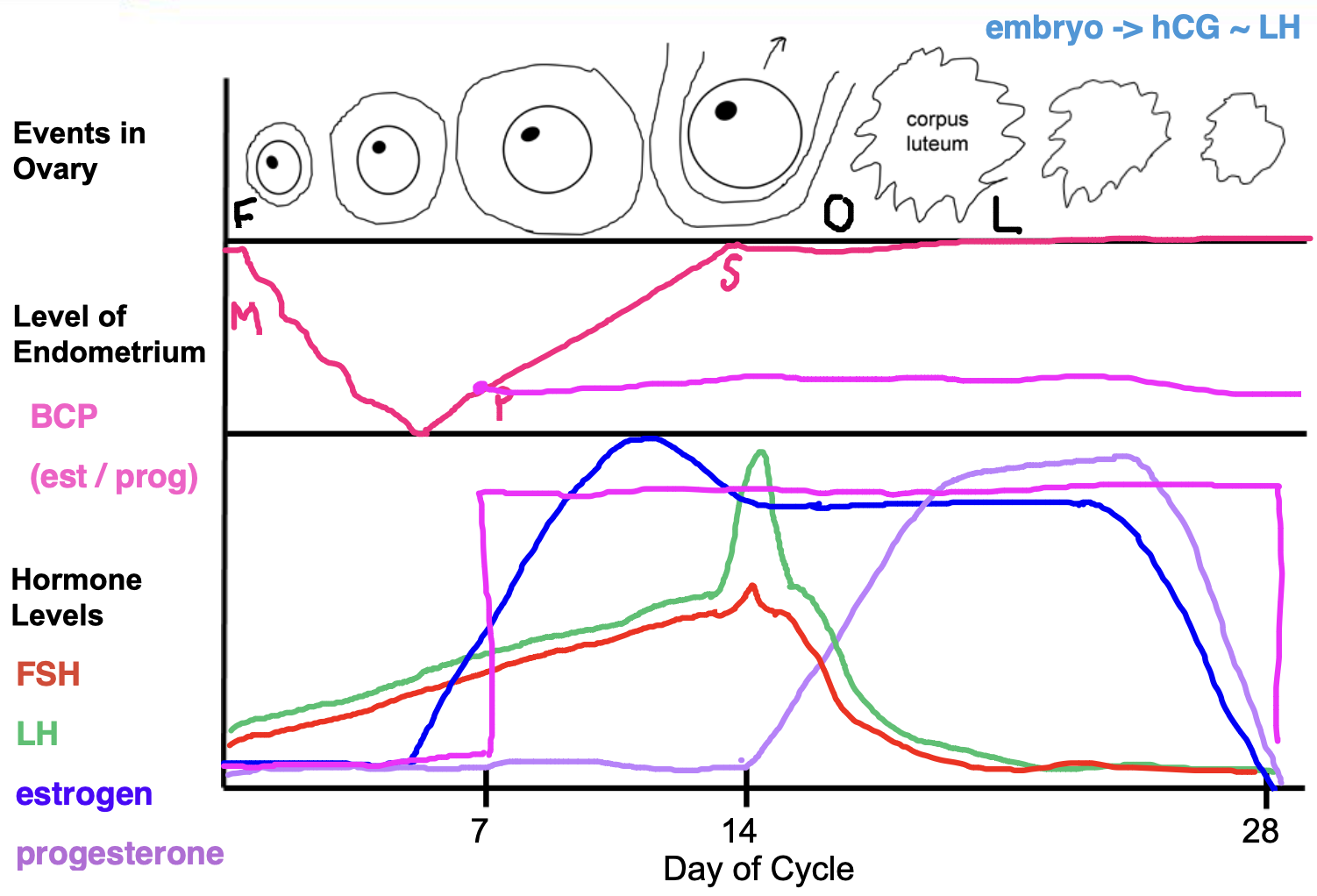
Ovarian Cycle:
hypothalamus → GNRH → anterior pituitary → LH and FSH → progesterone and estrogen(-feedback) → uterus
Ovarian Cycle - 1-13 days of follicular phase → build a follicle(oocyte and support cells(granulosa cells, fluid)); triggered by FSH to build it and secretes estrogen
Ovulation(day 14) - oocyte and some cells(corpus luteum left behind) released from the ovary and triggered by a surge of LH
Luteal Phase(15-28 days) - form corpus luteum from follicle remains, triggered by LH surge, and secretes estrogen and progesterone
Uterine cycle:
Estrogen Establishes Endometrium
Progestogen Protects Endometrium
menstruation(1-5 days) - shed off old endometrium(low Es and Pro)
Proliferative stage(6-14 days) - rebuild endometrium - low pro and rising est
Secretory phase(15-28) - enhance endometrium, stable est and high prog

Fertilization = when a sperm implants into the oocyte → only 1 can get through it → egg can block polyspermy(only letting 1 in) → egg depolarizes to repel other sperm or slow block that causes the egg surface to harden
Cleavage → dikaryon(one cell, 2 nuclei) → need to finish meiosis first and kick out second polar body → makes zygote → 24 hours turns into 2 cells stage → then morula which is a ball of cells into a blastula(hollow ball of cells) → a larger cell cluster that implants into the uterus in 4-5 days
The outer shell of the blastula is trophoblast that secretes hCG to maintain the corpus luteum → will become the placenta in 3 months and takes over hormone secretion and hCG falls - pregnancy tests for hCG level but only works before the first trimester —> the inner part of the cell is the embryo and turns into the umbilical cord and amniotic sack
The stem cells → can become many other cells and make more stem cells → found in blastocysts → embryonic stem cells
Embryonic stem cells → Determination(determines its path to which cell it wants to be) → Differentiation (physical change into a new cell type) → Pluripotent cells that can become any type of cell in the body but not the placenta
Stem cells in other parts of the body(adult cells) → ex. blood cell type in blood → but these cells can only become one type of cell(blood cells only and restricted)
Totipotent cells from zygote→ can be any body cell or the placenta
Embryonic Stages:
First 8 weeks → fertilization through 8 weeks → medically pregnant is 10 weeks here
Gastrulation → about 4 weeks to form 3 primary germ layers → endoderm(inner linings), mesoderm(organs, blood vessels, non-gland organs), ectoderm(nervous system, nails, skin)
Neurulation and organogenesis 4- 8 weeks → form all other body organs and structures → nothing new formed after this
Functional beating heart at 3 weeks!
Week 12 - ID the sex of the baby
Week 16 - Baby movements
week 24 - eyelids infuse and sound and light
Week 28 - testes descend in males
Week 33 - lung release surfactant
Week 38 - full term
Labor - 3 triggers
The placenta deteriorates, the uterus stretches, baby’s head stretches the cervix(ideal position)
Baby’s head stimulus hypothalamus releases oxytocin from post. pituitary, uterus contracts majorly → + feedback
Baby’s gain 1 pound a week during the last weeks of pregnancy
Baby changes:
close lung bypass - a hole between the Right and Left aorta and vessel connects the pulmonary artery to the aorta
Liver bypass
close umbilical vessel → close the arteries first after being born and veins close second
Baby stops making fetal hemoglobin → regular hemoglobin(mother) and baby’s hemoglobin has a higher affinity to O2 than mother’s → high affinity is then removed to go to regular hemoglobin
Mom’s Changes → more hormonal in nature → deliver placenta → drop in estrogen and progesterone → now prolactin is secreted(increase) to make milk, baby nurses, and makes more milk(NOT + Feedback). Baby nursing also causes oxytocin to rise
 Knowt
Knowt
