CHAPTER 10 Nervous System
Introduction
The nervous system is one of the most complex of all human body systems. More than 100 billion nerve cells operate constantly all over the body to coordinate the activities we do consciously and voluntarily, as well as those that occur unconsciously or involuntarily. We speak, move muscles, hear, taste, see, and think. Our glands secrete hormones, and we respond to danger, pain, temperature, and touch. All of these functions comprise only a small number of the many activities controlled by the nervous system.
Hệ thần kinh là một trong những hệ thống phức tạp nhất trong tất cả các hệ thống cơ thể con người. Hơn 100 tỷ tế bào thần kinh hoạt động liên tục trên khắp cơ thể để điều phối các hoạt động mà chúng ta thực hiện một cách có ý thức và tự nguyện, cũng như những hoạt động diễn ra vô thức hoặc không tự nguyện. Chúng ta nói, di chuyển cơ bắp, nghe, nếm, nhìn và suy nghĩ. Các tuyến của chúng ta tiết ra hormone, và chúng ta phản ứng với nguy hiểm, đau đớn, nhiệt độ và cảm giác chạm. Tất cả những chức năng này chỉ chiếm một số ít trong nhiều hoạt động do hệ thần kinh kiểm soát.
Fibers exiting from microscopic nerve cells (neurons) are collected into macroscopic bundles called nerves, which carry electrical messages all over the body. External stimuli, as well as internal chemicals such as acetylcholine, activate the cell membranes of nerve cells, which results in electrical discharges of these cells. These electrical discharges, nervous impulses, may then traverse the length of the associated nerves. External receptors (sense organs) as well as internal receptors in muscles and blood vessels receive these impulses and may in turn transmit impulses to the complex network of nerve cells in the brain and spinal cord. Within this central part of the nervous system, impulses are recognized, interpreted, and finally relayed to other nerve cells that extend out to all parts of the body, such as muscles, glands, and internal organs.
Các sợi thần kinh thoát ra từ các tế bào thần kinh vi mô (neurons) được thu thập thành các bó lớn gọi là dây thần kinh, mang thông điệp điện khắp cơ thể. Các kích thích bên ngoài, cũng như các hóa chất nội sinh như acetylcholine, kích hoạt màng tế bào của các tế bào thần kinh, dẫn đến các phóng điện của các tế bào này. Những phóng điện này, hay xung thần kinh, có thể đi qua chiều dài của các dây thần kinh liên quan. Các thụ thể bên ngoài (cơ quan cảm giác) cũng như các thụ thể bên trong cơ bắp và mạch máu nhận các xung thần kinh này và có thể truyền các xung thần kinh tới mạng lưới phức tạp của các tế bào thần kinh trong não và tủy sống. Trong phần trung tâm này của hệ thần kinh, các xung thần kinh được nhận diện, diễn giải và cuối cùng được chuyển tiếp tới các tế bào thần kinh khác mở rộng ra tất cả các phần của cơ thể như cơ bắp, tuyến và các cơ quan nội tạng.
General Structure of the Nervous System
(Cấu trúc chung của hệ thần kinh)
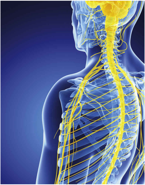
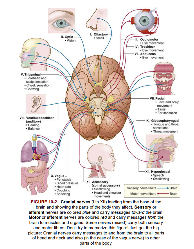
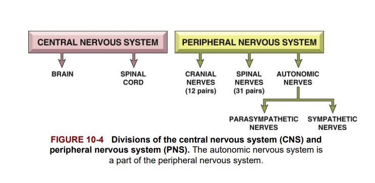
The nervous system is classified into two major divisions: the central nervous system (CNS) and the peripheral nervous system (PNS). The central nervous system consists of the brain and spinal cord. The peripheral nervous system consists of cranial nerves and spinal nerves, plexuses, and peripheral nerves throughout the body (Figure 10-1). Cranial nerves carry impulses between the brain and the head and neck. The one exception is the tenth cranial nerve, called the vagus nerve. It carries messages to and from the neck, chest, and abdomen. Figure 10-2 shows cranial nerves, their functions, and the parts of the body that they carry messages to and from. Spinal nerves carry messages between the spinal cord and the chest, abdomen, and extremities.
Hệ thần kinh được phân loại thành hai bộ phận chính: hệ thần kinh trung ương (CNS) và hệ thần kinh ngoại biên (PNS). Hệ thần kinh trung ương bao gồm não và tủy sống. Hệ thần kinh ngoại biên bao gồm các dây thần kinh sọ và dây thần kinh tủy sống, đám rối và các dây thần kinh ngoại biên trên khắp cơ thể. Dây thần kinh sọ mang các xung thần kinh giữa não và đầu và cổ. Ngoại lệ duy nhất là dây thần kinh sọ thứ mười, gọi là dây thần kinh vagus. Nó mang thông điệp đến và từ cổ, ngực và bụng. Dây thần kinh tủy sống mang thông điệp giữa tủy sống và ngực, bụng và chi.
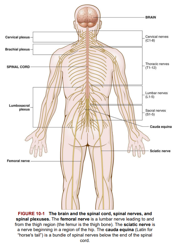
 A plexus is a large network of nerves in the peripheral nervous system. The cervical, brachial (brachi/o means arm), and lumbosacral plexuses are examples that include cervical, lumbar, and sacral nerves. Figure 10-1 illustrates the relationship of the brain and spinal cord to the spinal nerves and plexuses.
Một đám rối là một mạng lưới lớn các dây thần kinh trong hệ thần kinh ngoại biên. Các đám rối cổ, đám rối cánh tay, và đám rối thắt lưng cùng là những ví dụ bao gồm các dây thần kinh cổ, thắt lưng và cùng.
Plexus
There are other plexuses in the body—networks of intersecting blood vessels (vascular) and lymphatic vessels.
• Lymphatic plexus is an interconnecting network of lymph vessels.
• Rectal plexus is a plexus of veins in the rectal region.
• Vertebral plexus is a plexus of veins related to the backbone.
Đám rối
Có các đám rối khác trong cơ thể - mạng lưới của các mạch máu (mạch máu) và mạch bạch huyết:
Đám rối bạch huyết là một mạng lưới các mạch bạch huyết liên kết.
Đám rối trực tràng là một đám rối của các tĩnh mạch ở vùng trực tràng.
Đám rối đốt sống là một đám rối của các tĩnh mạch liên quan đến xương sống.
The spinal and cranial nerves are composed of nerves that help the body respond to changes in the outside world. They include sense receptors for sight (eye), hearing and balance (ear), smell (olfactory), and touch (skin sensation) and sensory (afferent) nerves that carry messages related to changes in the environment toward the spinal cord and brain. In addition, motor (efferent) nerves travel from the spinal cord and brain to muscles of the body, telling them how to respond. For example, when you touch a hot stove, temperature and pain receptors in the skin stimulate afferent nerves, which carry messages toward the spinal cord and brain. Instantaneously, the message is conveyed to efferent nerve cells in the spinal cord, which then activate voluntary muscles to pull your hand away from the stove.
Các dây thần kinh tủy sống và dây thần kinh sọ được cấu tạo từ các dây thần kinh giúp cơ thể phản ứng với những thay đổi từ thế giới bên ngoài. Chúng bao gồm các thụ thể cảm giác cho thị giác (mắt), thính giác và cân bằng (tai), khứu giác (mũi), và cảm giác (da) và các dây thần kinh cảm giác (afferent) mang thông điệp liên quan đến những thay đổi trong môi trường hướng về tủy sống và não. Ngoài ra, các dây thần kinh vận động (efferent) di chuyển từ tủy sống và não đến cơ bắp của cơ thể, cho chúng biết cách phản ứng.
In addition to the spinal and cranial nerves (whose functions are mainly voluntary and involved with sensations of smell, taste, sight, hearing, and muscle movements), the peripheral nervous system also contains a large group of nerves that function involuntarily or automatically, without conscious control. These peripheral nerves belong to the autonomic nervous system. This system of nerve fibers carries impulses to glands, heart, blood vessels, involuntary muscles found in the walls of tubes like the intestines, and hollow organs like the stomach and urinary bladder.
Hệ thần kinh ngoại biên cũng chứa một nhóm lớn các dây thần kinh hoạt động một cách tự động, không có sự kiểm soát của ý thức. Những dây thần kinh ngoại biên này thuộc hệ thần kinh tự trị, mang các xung thần kinh đến các tuyến, tim, mạch máu, cơ bắp không tự chủ nằm trong các ống như ruột, và các cơ quan rỗng như dạ dày và bàng quang.
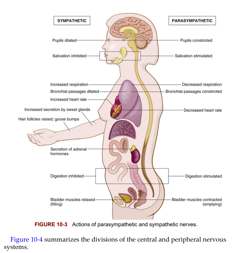
Some autonomic nerves are sympathetic nerves and others are parasympathetic nerves. The sympathetic nerves stimulate the body in times of stress and crisis. They increase heart rate and forcefulness, dilate (relax) airways so more oxygen can enter, and increase blood pressure. In addition, sympathetic neurons stimulate the adrenal glands to secrete epinephrine (adrenaline), while also inhibiting intestinal contractions to slow digestion. The parasympathetic nerves normally act as a balance for the sympathetic nerves. Parasympathetic nerves slow down heart rate, lower blood pressure, and stimulate intestinal contractions to clear the rectum. Figure 10-3 shows the differences in actions between the sympathetic and parasympathetic nerves.
Một số dây thần kinh tự trị là dây thần kinh giao cảm, và những dây thần kinh khác là dây thần kinh đối giao cảm. Dây thần kinh giao cảm kích thích cơ thể trong thời gian căng thẳng và khủng hoảng, tăng nhịp tim và sức mạnh, giãn các đường thở để tăng lượng oxy, và tăng huyết áp. Ngoài ra, các neuron giao cảm kích thích tuyến thượng thận tiết ra epinephrine (adrenaline), đồng thời ức chế các cơn co thắt ruột để làm chậm quá trình tiêu hóa. Dây thần kinh đối giao cảm thường hoạt động như một sự cân bằng cho các dây thần kinh giao cảm, làm chậm nhịp tim, giảm huyết áp và kích thích các cơn co thắt ruột để làm sạch trực tràng.
Neurons, Nerves, and Glial Cells
A neuron is an individual nerve cell, a microscopic structure. Impulses pass along the parts of a nerve cell in a definite manner and direction. The parts of a neuron are pictured in Figure 10-5; label it as you study the following.
Một neuron là một tế bào thần kinh đơn lẻ, một cấu trúc vi mô. Các xung thần kinh truyền qua các phần của một tế bào thần kinh theo một hướng nhất định. Các phần của một neuron bao gồm:
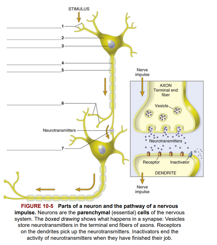 A stimulus begins an impulse in the branching fibers of the neuron, which are called dendrites [1]. A change in the electrical charge of the dendrite membranes is thus begun, and the nervous impulse moves along the dendrites like the movement of falling dominoes. The impulse, traveling in only one direction, next reaches the cell body [2], which contains the cell nucleus [3]. Small collections of nerve cell bodies outside the brain and spinal cord are called ganglia (singular: ganglion). Extending from the cell body is the axon [4], which carries the impulse away from the cell body. Axons can be covered with a fay tissue called myelin. The purpose of this myelin sheath [5] is to insulate the axon and speed transmission of the electrical impulse. Demyelination is loss of the myelin insulating the nerve fiber and is characteristic of multiple sclerosis, an acquired illness affecting the CNS.
Một kích thích bắt đầu một xung thần kinh trong các sợi nhánh của neuron, gọi là dendrites [1]. Sự thay đổi trong điện tích của màng dendrite bắt đầu, và xung thần kinh di chuyển dọc theo dendrite như sự đổ của các quân domino. Xung thần kinh, di chuyển theo một hướng duy nhất, tiếp theo sẽ đến thân tế bào [2], chứa nhân tế bào [3]. Các cụm nhỏ của thân tế bào thần kinh bên ngoài não và tủy sống được gọi là hạch (số ít: ganglion). Kéo dài từ thân tế bào là axon [4], mang xung thần kinh ra khỏi thân tế bào. Axon có thể được bao phủ bởi một lớp mô mỡ gọi là myelin. Mục đích của lớp myelin [5] là cách điện axon và tăng tốc độ truyền xung điện. Demyelination là sự mất myelin bao quanh sợi thần kinh và là đặc trưng của bệnh đa xơ cứng, một bệnh mắc phải ảnh hưởng đến hệ thần kinh trung ương.
The myelin sheath gives a white appearance to the nerve fiber—hence the term white matter, as in parts of the spinal cord and the white matter of the brain and most peripheral nerves. The gray matter of the brain and spinal cord is composed of the cell bodies of neurons that appear gray because they are not covered by a myelin sheath.
Lớp myelin tạo ra một vẻ ngoài trắng cho sợi thần kinh - do đó thuật ngữ chất trắng, như trong các phần của tủy sống và chất trắng của não và hầu hết các dây thần kinh ngoại biên. Chất xám của não và tủy sống được tạo thành từ thân tế bào của các neuron xuất hiện màu xám vì chúng không được bao phủ bởi lớp myelin.
The nervous impulse passes through the axon to leave the cell via the terminal end fibers [6] of the neuron. The space where the nervous impulse jumps from one neuron to another is called the synapse [7]. The transfer of the impulse across the synapse depends on the release of a chemical substance, called a neurotransmitter, by the neuron that brings the impulse to the synapse. See the boxed diagram in Figure 10-5. Tiny sacs (vesicles) containing the neurotransmitter are located at the ends of neurons, and they release the neurotransmitter into the synapse. Acetylcholine, norepinephrine, epinephrine (adrenaline), dopamine, serotonin, and endorphins are examples of neurotransmitters.
Xung thần kinh đi qua axon để rời khỏi tế bào thông qua các sợi đầu cuối [6] của neuron. Khoảng trống nơi xung thần kinh nhảy từ một neuron sang neuron khác được gọi là synapse [7]. Sự truyền xung thần kinh qua synapse phụ thuộc vào việc giải phóng một chất hóa học, gọi là chất dẫn truyền thần kinh, bởi neuron mang xung thần kinh đến synapse. Các túi nhỏ (vesicles) chứa chất dẫn truyền thần kinh được đặt ở cuối các neuron, và chúng giải phóng chất dẫn truyền thần kinh vào synapse. Acetylcholine, norepinephrine, epinephrine (adrenaline), dopamine, serotonin, và endorphins là các ví dụ về chất dẫn truyền thần kinh.
Whereas a neuron is a microscopic structure within the nervous system, a nerve is macroscopic, able to be seen with the naked eye. A nerve consists of many axons that travel together like strands of rope. Peripheral nerves that carry impulses to the brain and spinal cord from stimulus receptors like the skin, eye, ear, and nose are afferent or sensory nerves; those that carry impulses from the CNS to organs that produce responses, such as muscles and glands, are efferent or motor nerves.
Trong khi neuron là một cấu trúc vi mô trong hệ thần kinh, một dây thần kinh là một cấu trúc vĩ mô, có thể nhìn thấy bằng mắt thường. Một dây thần kinh bao gồm nhiều axon di chuyển cùng nhau như các sợi dây thừng. Các dây thần kinh ngoại biên mang xung thần kinh đến não và tủy sống từ các thụ thể kích thích như da, mắt, tai và mũi là dây thần kinh cảm giác hoặc afferent; những dây thần kinh mang xung thần kinh từ hệ thần kinh trung ương đến các cơ quan tạo ra phản ứng, như cơ và tuyến, là dây thần kinh vận động hoặc efferent.
Neurons and nerves are the parenchyma of the nervous system. Parenchyma is the essential distinguishing tissue of an organ. In the brain and spinal cord, neurons, which conduct electrical impulses, are the parenchymal tissue. Stroma of an organ is the connective and supportive tissue of an organ. The stromal tissue of the central nervous system consists of the glial (neuroglial) cells, which make up its supportive framework and help it ward off infection. Glial cells do not transmit impulses. They are far more numerous than neurons and can reproduce.
Neurons và dây thần kinh là parenchyma của hệ thần kinh. Parenchyma là mô phân biệt thiết yếu của một cơ quan. Trong não và tủy sống, các neuron dẫn truyền xung điện là mô parenchymal. Stroma của một cơ quan là mô liên kết và hỗ trợ của cơ quan. Mô stroma của hệ thần kinh trung ương bao gồm các tế bào glial (neuroglial), tạo nên khung hỗ trợ và giúp nó chống lại nhiễm trùng. Các tế bào glial không truyền xung thần kinh. Chúng nhiều hơn nhiều so với các neuron và có thể sinh sản.
There are four types of supporting or glial cells (see Figure 10-6). Astrocytes (astroglial cells) are star-like in appearance (astr/o means star) and transport water and salts between capillaries and neurons. Microglial cells are small cells with many branching processes (dendrites). As phagocytes, they protect neurons in response to inflammation.
Có bốn loại tế bào hỗ trợ (tế bào glial) (xem Hình 10-6). Tế bào astrocytes (tế bào astroglial) có hình dạng giống ngôi sao (astr/o nghĩa là ngôi sao) và vận chuyển nước và muối giữa mao mạch và neuron. Tế bào microglial là các tế bào nhỏ với nhiều nhánh (dendrite). Là các thực bào, chúng bảo vệ neuron trong phản ứng viêm.
Oligodendroglial cells (oligodendrocytes) have few (olig/o means few or scanty) dendrites. These cells form the myelin sheath in the CNS. By contrast, ependymal cells (Greek ependyma means upper garment) line membranes within the brain and spinal cord where CSF is produced and circulates.
Tế bào oligodendroglial (oligodendrocytes) có ít nhánh (olig/o nghĩa là ít hoặc thưa thớt). Các tế bào này hình thành lớp myelin trong hệ thần kinh trung ương (CNS). Ngược lại, tế bào ependymal (tiếng Hy Lạp ependyma nghĩa là áo ngoài) lót các màng trong não và tủy sống nơi sản xuất và tuần hoàn dịch não tủy (CSF).
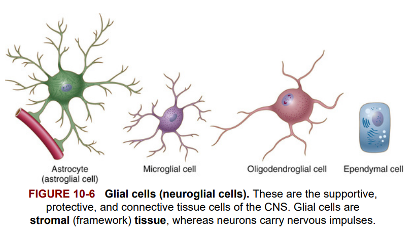 Glial cells, particularly the astrocytes, are associated with blood vessels and regulate the passage of potentially harmful substances from the blood into the nerve cells of the brain. This protective barrier between the blood and brain cells is called the blood-brain barrier (BBB). This barrier consists of special lining (endothelial) cells, which along with astrocytes separate capillaries from nerve cells. Delivery of chemotherapeutic drugs to treat brain tumors is thus difficult, because the BBB blocks drug access to brain tissues. Figure 10-6 illustrates glial cells.
Các tế bào glial, đặc biệt là astrocytes, có liên quan đến các mạch máu và điều chỉnh sự di chuyển của các chất có thể gây hại từ máu vào các tế bào thần kinh trong não. Hàng rào bảo vệ này giữa máu và các tế bào não được gọi là hàng rào máu-não (BBB). Hàng rào này bao gồm các tế bào lót đặc biệt (endothelial), cùng với astrocytes, tách mao mạch khỏi các tế bào thần kinh. Việc cung cấp các thuốc hóa trị để điều trị khối u não do đó gặp khó khăn, vì BBB ngăn chặn sự tiếp cận của thuốc vào các mô não. Hình 10-6 minh họa các tế bào glial.
The Brain (Não Bộ)
The brain controls body activities. In the human adult, it weighs about 3 pounds and has many different parts, all of which control different aspects of body functions.
Não điều khiển các hoạt động của cơ thể. Ở người trưởng thành, nó nặng khoảng 3 pound và có nhiều phần khác nhau, tất cả đều điều khiển các khía cạnh khác nhau của chức năng cơ thể.
The largest part of the brain is the “thinking” area, or cerebrum. On the surface of the cerebrum, nerve cells lie in sheets, which make up the cerebral cortex. These sheets, arranged in folds called gyri, are separated from each other by grooves known as sulci. The brain is divided in half, a right side and a left side, which are called cerebral hemispheres. Each hemisphere is subdivided into four major lobes named for the cranial (skull) bones that overlie them. Figure 10-7 shows these lobes—frontal, parietal, occipital, and temporal—as well as gyri and sulci.
Phần lớn nhất của não là khu vực "tư duy," hay đại não (cerebrum). Trên bề mặt của đại não, các tế bào thần kinh nằm thành các tấm, tạo thành vỏ não (cerebral cortex). Các tấm này, được sắp xếp thành các nếp gấp gọi là gyri, được tách ra bởi các rãnh gọi là sulci. Não được chia thành hai nửa, một bên phải và một bên trái, gọi là bán cầu đại não (cerebral hemispheres). Mỗi bán cầu được chia thành bốn thùy chính, được đặt tên theo các xương sọ (cranial) nằm trên chúng. Hình 10-7 cho thấy các thùy này—trán (frontal), đỉnh (parietal), chẩm (occipital), và thái dương (temporal)—cũng như các gyri và sulci.
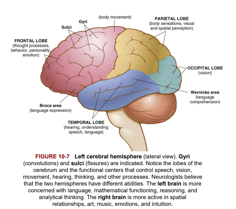 The cerebrum has many functions. It is responsible for thought, judgment, memory, association, and discrimination. In addition, sensory impulses are received through afferent cranial nerves, and when registered in the cortex, they are the basis for perception. Nerve impulses from the cerebrum extend to muscles and glands producing movement as well as internal changes in the body. Figure 10-7 shows the location of some of the centers in the cerebral cortex that control speech, vision, smell, movement, hearing, and thought processes.
Đại não có nhiều chức năng. Nó chịu trách nhiệm cho suy nghĩ, phán đoán, trí nhớ, liên kết và phân biệt. Ngoài ra, các xung cảm giác được nhận thông qua các dây thần kinh sọ hướng tâm và khi được đăng ký trong vỏ não, chúng là cơ sở cho nhận thức. Các xung thần kinh từ đại não mở rộng đến các cơ và tuyến, tạo ra chuyển động cũng như các thay đổi nội bộ trong cơ thể. Hình 10-7 cho thấy vị trí của một số trung tâm trong vỏ não điều khiển lời nói, thị giác, khứu giác, chuyển động, thính giác và các quá trình tư duy.
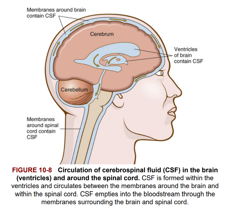
In the middle of the cerebrum are spaces, or canals, called ventricles (pictured in Figure 10-8). They contain a watery fluid that flows throughout the brain and around the spinal cord. This fluid is cerebrospinal fluid (CSF), and it protects the brain and spinal cord from shock by acting like a cushion. CSF usually is clear and colorless and contains lymphocytes, sugar, and proteins. Spinal fluid can be withdrawn for diagnosis or relief of pressure on the brain; this is called a lumbar puncture (LP). For this procedure, a hollow needle is inserted into the lumbar region of the spinal column below the region where the nervous tissue of the spinal cord ends, and CSF is withdrawn.
Ở giữa đại não có các không gian, hoặc kênh, gọi là các não thất (ventricles) (được minh họa trong Hình 10-8). Chúng chứa một chất lỏng dạng nước chảy khắp não và xung quanh tủy sống. Chất lỏng này là dịch não tủy (CSF), và nó bảo vệ não và tủy sống khỏi chấn động bằng cách hoạt động như một tấm đệm. Dịch não tủy thường trong suốt và không màu, chứa các tế bào lympho, đường và protein. Dịch tủy sống có thể được lấy ra để chẩn đoán hoặc giảm áp lực lên não; điều này được gọi là chọc dò tủy sống (LP). Để thực hiện thủ thuật này, một kim rỗng được chèn vào vùng thắt lưng của cột sống dưới vùng mà mô thần kinh của tủy sống kết thúc, và CSF được rút ra.
 Two other important parts of the brain are the thalamus and the hypothalamus (Figure 10-9). The thalamus acts like a triage center. It decides what is important and what is not, selectively processing and relaying sensory information to the cerebral cortex. The thalamus also plays a major role in maintaining levels of awareness and consciousness. The hypothalamus (below the thalamus) contains neurons that control body temperature, sleep, appetite, sexual desire, and emotions such as fear and pleasure. The hypothalamus also regulates the release of hormones from the pituitary gland at the base of the brain and integrates the activities of the sympathetic and parasympathetic nervous systems.
Hai phần quan trọng khác của não là đồi thị và vùng dưới đồi (Hình 10-9). Đồi thị hoạt động như một trung tâm phân loại. Nó quyết định điều gì là quan trọng và điều gì không, chọn lọc xử lý và chuyển tiếp thông tin cảm giác đến vỏ não. Đồi thị cũng đóng vai trò quan trọng trong việc duy trì mức độ nhận thức và ý thức. Vùng dưới đồi (nằm dưới đồi thị) chứa các nơ-ron kiểm soát nhiệt độ cơ thể, giấc ngủ, sự thèm ăn, ham muốn tình dục và các cảm xúc như sợ hãi và khoái cảm. Vùng dưới đồi cũng điều chỉnh sự giải phóng hormone từ tuyến yên ở đáy não và tích hợp các hoạt động của hệ thần kinh giao cảm và đối giao cảm.
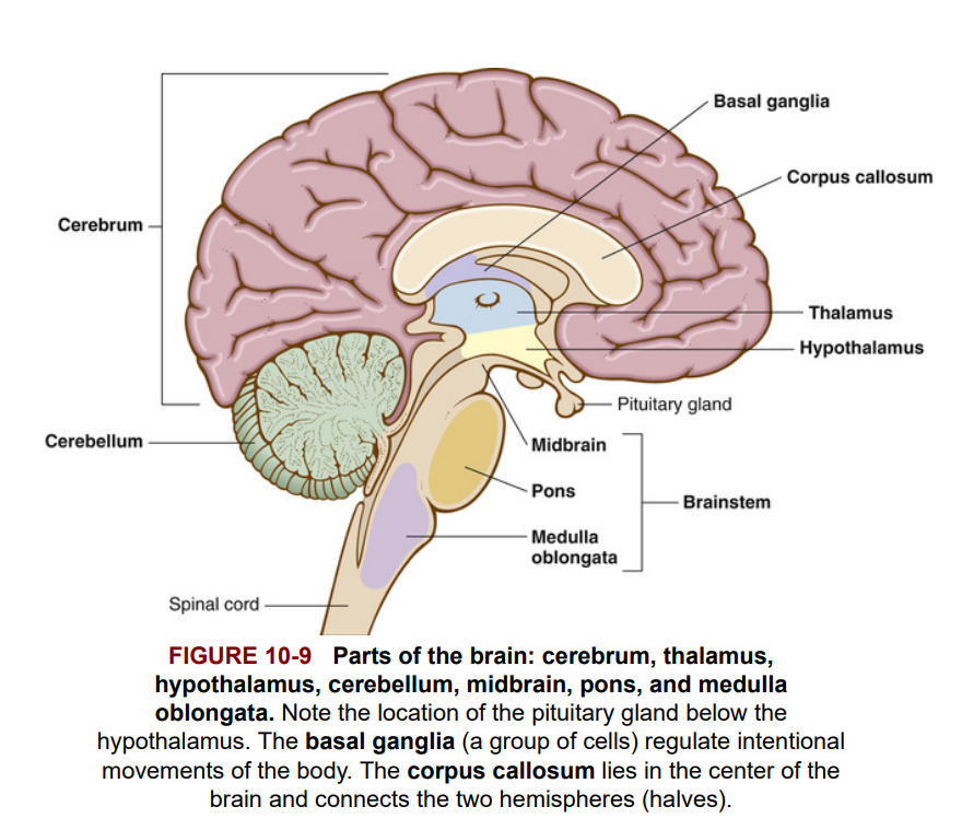 The following structures within the brain lie in the back and below the cerebrum and connect the cerebrum with the spinal cord: cerebellum, midbrain, pons, and medulla oblongata. The midbrain, pons and medulla are part of the brainstem. See Figure 10-9.
Các cấu trúc sau đây trong não nằm ở phía sau và dưới đại não, và kết nối đại não với tủy sống: tiểu não, não giữa, cầu não, và hành tủy. Não giữa, cầu não và hành tủy là một phần của thân não. Xem Hình 10-9.
The cerebellum functions to coordinate voluntary movements and to maintain balance and posture.
Tiểu não có chức năng điều phối các chuyển động tự nguyện và duy trì sự cân bằng và tư thế.
The midbrain is the uppermost portion of the brainstem. It contains pathways connecting the cerebrum with lower portions of the brain and structures involved with seeing and hearing.
Não giữa là phần trên cùng của thân não. Nó chứa các con đường kết nối đại não với các phần thấp hơn của não và các cấu trúc liên quan đến thị giác và thính giác.
The pons is a part of the brainstem that literally means bridge. It contains nerve fiber tracts that connect the cerebellum and cerebrum with the rest of the brain. Nerves affecting the face and eye movement are located here.
Cầu não là một phần của thân não mà nghĩa đen là cầu nối. Nó chứa các đường sợi thần kinh kết nối tiểu não và đại não với phần còn lại của não. Các dây thần kinh ảnh hưởng đến chuyển động của khuôn mặt và mắt nằm ở đây.
The medulla oblongata, also in the brainstem, connects the spinal cord with the rest of the brain. Nerve tracts cross from right to left and left to right in the medulla oblongata. For example, nerve cells that control movement of the left side of the body are found in the right half of the cerebrum. These cells send out axons that cross over (decussate) to the opposite side of the brain in the medulla oblongata and then travel down the spinal cord.
Hành tủy, cũng nằm trong thân não, kết nối tủy sống với phần còn lại của não. Các đường dẫn thần kinh bắt chéo từ phải sang trái và từ trái sang phải trong hành tủy. Ví dụ, các tế bào thần kinh điều khiển chuyển động của bên trái cơ thể nằm ở nửa phải của đại não. Những tế bào này phát ra các sợi trục bắt chéo sang phía đối diện của não trong hành tủy và sau đó di chuyển xuống tủy sống.
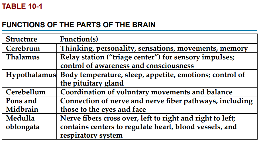
In addition, the medulla oblongata contains three important vital centers that regulate internal activities of the body:
1. Respiratory center—controls muscles of respiration in response to chemicals or other stimuli
2. Cardiac center—slows the heart rate when the heart is beating too rapidly
3. Vasomotor center—affects (constricts or dilates) the muscles in the walls of blood vessels, thus influencing blood pressure
Ngoài ra, hành tủy chứa ba trung tâm quan trọng điều chỉnh các hoạt động nội bộ của cơ thể:
Trung tâm hô hấp—kiểm soát các cơ hô hấp để đáp ứng với hóa chất hoặc các kích thích khác.
Trung tâm tim mạch—làm chậm nhịp tim khi tim đập quá nhanh.
Trung tâm vận mạch—ảnh hưởng đến (co lại hoặc giãn ra) các cơ trong thành mạch máu, do đó ảnh hưởng đến huyết áp.
Figure 10-9 shows the locations of the thalamus, hypothalamus, cerebellum, pons, and medulla oblongata. Table 10-1 reviews the functions of these parts of the brain.
Hình 10-9 cho thấy vị trí của đồi thị, vùng dưới đồi, tiểu não, cầu não và hành tủy. Bảng 10-1 tổng kết các chức năng của các phần này của não.
The Spinal Cord and Meninges
(Tủy sống và Màng não)
Spinal Cord (Tủy sống)
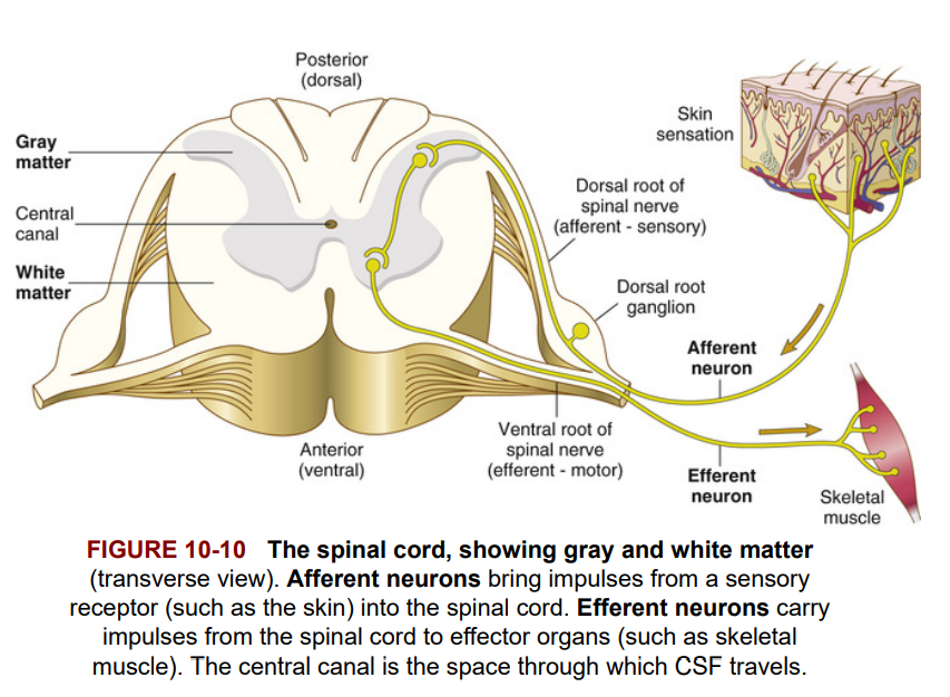
The spinal cord is a column of nervous tissue extending from the medulla oblongata to the second lumbar vertebra within the vertebral column. Below the end of the spinal cord is the cauda equina (Latin for “horse's tail”), a fan of nerve fibers (see Figure 10-1, page 323). The spinal cord carries all the nerves to and from the limbs and lower part of the body, and it is the pathway for impulses going to and from the brain. A cross-sectional view of the spinal cord (Figure 10-10) reveals an inner region of gray matter (containing cell bodies and dendrites) and an outer region of white matter (containing the nerve fiber tracts with myelin sheaths) conducting impulses to and from the brain.
Tủy sống là một cột mô thần kinh kéo dài từ hành tủy đến đốt sống thắt lưng thứ hai trong cột sống. Phía dưới phần cuối của tủy sống là chùm đuôi ngựa (tiếng Latin gọi là “đuôi ngựa”), một chùm các sợi thần kinh (xem Hình 10-1, trang 323). Tủy sống mang tất cả các dây thần kinh đến và từ các chi và phần dưới của cơ thể, và nó là con đường cho các xung động đi đến và đi từ não. Một cái nhìn cắt ngang của tủy sống (Hình 10-10) cho thấy một vùng bên trong của chất xám (chứa các thân tế bào và sợi nhánh) và một vùng bên ngoài của chất trắng (chứa các dải sợi thần kinh có bao myelin) dẫn truyền các xung động đến và từ não.
Meninges (Màng não)
The meninges are three layers of connective tissue membranes that surround the brain and spinal cord. Label Figure 10-11 as you study the following description of the meninges.
Màng não là ba lớp màng mô liên kết bao quanh não và tủy sống. Hãy đánh dấu Hình 10-11 khi bạn nghiên cứu mô tả sau đây về màng não.
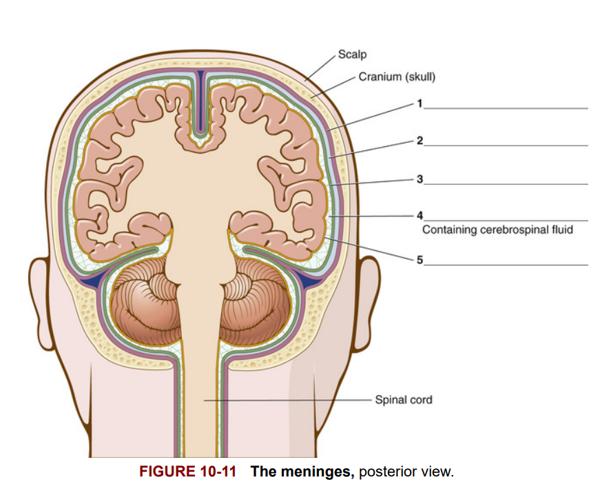
The outermost membrane of the meninges is the dura mater [1]. This thick, tough membrane contains channels (dural sinuses) that contain blood. The subdural space [2] is below the dural membrane. The second layer surrounding the brain and spinal cord is the arachnoid membrane [3]. The arachnoid (spider-like) membrane is loosely attached to the other meninges by web-like fibers, so there is a space for fluid between the fibers and the third membrane. This is the subarachnoid space [4], containing CSF. The third layer of the meninges, closest to the brain and spinal cord, is the pia mater [5]. It contains delicate (Latin pia) connective tissue with a rich supply of blood vessels. Most physicians refer to the pia and arachnoid membranes together as the pia-arachnoid.
Màng ngoài cùng của màng não là màng cứng [1]. Màng dày và chắc này chứa các kênh (xoang cứng) chứa máu. Khoang dưới màng cứng [2] nằm dưới màng cứng. Lớp thứ hai bao quanh não và tủy sống là màng nhện [3]. Màng nhện (giống như mạng nhện) được gắn lỏng lẻo với các màng khác bằng các sợi giống như mạng nhện, do đó có một khoảng không gian chứa dịch giữa các sợi và màng thứ ba. Đây là khoang dưới nhện [4], chứa dịch não tủy (CSF). Lớp thứ ba của màng não, gần nhất với não và tủy sống, là màng mềm [5]. Nó chứa mô liên kết mềm mại (tiếng Latin là pia) với nguồn cung cấp máu phong phú. Hầu hết các bác sĩ thường gọi màng mềm và màng nhện cùng nhau là màng mềm-nhện.
Vocabulary
acetylcholine
Neurotransmitter chemical released at the ends of nerve cells. (Chất truyền dẫn thần kinh được phát ra tại các đầu của tế bào thần kinh.)
afferent nerve
Carries messages toward the brain and spinal cord (sensory nerve). Afferent comes from af- (a form of ad-, meaning toward) and -ferent (meaning carrying). (Dây thần kinh đưa tin nhắn về hướng não và tủy sống (dây thần kinh cảm giác). "Afferent" xuất phát từ af- (một dạng của ad-, có nghĩa là về phía) và -ferent (có nghĩa là mang).)
arachnoid membrane
Middle layer of the three membranes (meninges) that surround the brain and spinal cord. The Greek arachne means spider. (Lớp màng giữa trong ba lớp màng (màng não) bao quanh não và tủy sống. "Arachnoid" có nguồn gốc từ tiếng Hy Lạp arachne nghĩa là nhện.)
astrocyte
Type of glial (neuroglial) cell that transports water and salts from capillaries in the nervous system.
autonomic nervous system
Nerves that control involuntary body functions of muscles, glands, and internal organs.
axon
Microscopic fiber that is part of a neuron and carries nervous impulse along a nerve cell.
blood-brain barrier
Protective separation between the blood and brain cells. This makes it difficult for substances (such as anticancer drugs) to penetrate capillary walls and enter the brain.
brainstem
Posterior portion of the brain that connects the cerebrum with the spinal cord; includes the midbrain, pons, and medulla oblongata.
cauda equina
Collection of spinal nerves below the end of the spinal cord.
cell body
Part of a nerve cell that contains the nucleus.
central nervous system (CNS)
Brain and spinal cord.
cerebellum
Posterior part of the brain that coordinates muscle movements and maintains balance.
cerebral cortex
Outer region of the cerebrum, containing sheets of nerve cells; gray matter of the brain.
cerebrospinal fluid (CSF)
Circulates throughout the brain and spinal cord.
cerebrum
Largest part of the brain; responsible for voluntary muscular activity, vision, speech, taste, hearing, thought, and memory.
cranial nerves
Nerves carry messages to and from the brain to all parts of head and neck and also (in the case of the vagus nerve) to other parts of the body. There are 12 pairs of cranial nerves.
dendrite
Microscopic branching fiber of a nerve cell (neuron) that is the first part to receive the nervous impulse.
dura mater
Thick, outermost layer of the meninges surrounding and protecting the brain and spinal cord. Latin for “hard mother.”
efferent nerve
Carries messages away from the brain and spinal cord; motor nerve. Efferent comes from ef- (meaning away from) and -ferent (meaning to carry).
ependymal cell
Glial cell that lines membranes within the brain and spinal cord and helps form cerebrospinal fluid.
ganglion (plural: ganglia)
Collection of nerve cell bodies in the peripheral nervous system.
glial cell (neuroglial cell)
Supportive and connective nerve cell that does not carry nervous impulses. Examples are astrocytes, microglial cells, ependymal cells, and oligodendrocytes. Glial cells can reproduce themselves, as opposed to neurons.
gyrus (plural: gyri)
Sheet of nerve cells that produces a rounded ridge on the surface of the cerebral cortex; convolution.
hypothalamus
Portion of the brain beneath the thalamus; controls sleep, appetite, body temperature, and secretions from the pituitary gland.
medulla oblongata
Part of the brain just above the spinal cord; controls breathing, heartbeat, and the size of blood vessels; nerve fibers cross over here.
meninges
Three protective membranes that surround the brain and spinal cord.
microglial cell
Phagocytic glial cell that removes waste products from the central nervous system.
midbrain
Uppermost portion of the brainstem.
motor nerve
Carries messages away from the brain and spinal cord to muscles and organs; efferent nerve.
myelin sheath
Covering of white fay tissue that surrounds and insulates the axon of a nerve cell. Myelin speeds impulse conduction along axons.
nerve
Macroscopic cord-like collection of fibers (axons) that carry electrical impulses.
neuron
Nerve cell that is necessary for impulses to be carried throughout the nervous system; parenchyma of the nervous system.
neurotransmitter
Chemical messenger released at the end of a nerve cell. It stimulates or inhibits another cell, which can be a nerve cell, muscle cell, or gland cell. Examples of neurotransmitters are acetylcholine, norepinephrine, dopamine, and serotonin.
oligodendroglial cell
Glial cell that forms the myelin sheath covering axons. Also called oligodendrocyte.
parasympathetic nerves
Involuntary autonomic nerves that regulate normal body functions such as heart rate, breathing, and muscles of the gastrointestinal tract.
parenchyma
Essential, distinguishing tissue of any organ or system. The parenchyma of the nervous system includes the neurons and nerves that carry nervous impulses. Parenchymal cells of the liver are hepatocytes, and parenchymal tissue of the kidney includes the nephrons, where urine is formed. Note the pronunciation: păr-ĔNkĭ-mă.
peripheral nervous system
Nerves outside the brain and spinal cord: cranial, spinal, and autonomic nerves.
pia mater
Thin, delicate inner membrane of the meninges.
plexus (plural: plexuses)
Large, interlacing network of nerves. Examples are lumbosacral, cervical, and brachial (brachi/o means arm) plexuses. The term originated from the Indo-European plek, meaning to weave together.
pons
Part of the brain anterior to the cerebellum and between the medulla and the rest of the midbrain. It is a bridge connecting various parts of the brain. In Latin, pons means bridge.
sciatic nerve
Nerve extending from the base of the spine down the thigh, lower leg, and foot. Sciatica is pain or inflammation along the course of the nerve.
sensory nerve
Carries messages toward the brain and spinal cord from a receptor; afferent nerve.
spinal nerves
Pairs of nerves, arising one on each side of the spinal column. They transmit messages to and from the spinal cord.
stimulus (plural: stimuli)
Agent of change in the internal or external environment that evokes a response. It may be light, sound, touch, pressure, or pain.
stroma
Connective and supporting tissue of an organ. Glial cells make up the stromal tissue of the brain.
sulcus (plural: sulci)
Depression or groove in the surface of the cerebral cortex; fissure.
sympathetic nerves
Autonomic nerves that influence bodily functions involuntarily in times of stress.
synapse
Space through which a nervous impulse travels between nerve cells or between nerve and muscle or glandular cells. From the Greek synapsis, a point of contact.
thalamus
Main relay center of the brain. It conducts impulses between the spinal cord and the cerebrum; incoming sensory messages are relayed through the thalamus to appropriate centers in the cerebrum. Latin thalamus means room. The Romans, who named this structure, thought this part of the brain was hollow, like a little room.
vagus nerve
Tenth cranial nerve (cranial nerve X). Its branches reach to the larynx, trachea, bronchi, lungs, aorta, esophagus, and stomach. Latin vagus means wandering. Unlike the other cranial nerves, the vagus leaves the head and “wanders” into the abdominal and thoracic cavities.
ventricles of the brain
Canals in the brain that contain cerebrospinal fluid. Ventricles are also found in the heart—they are the two lower chambers of the heart.