Human bio things to work on
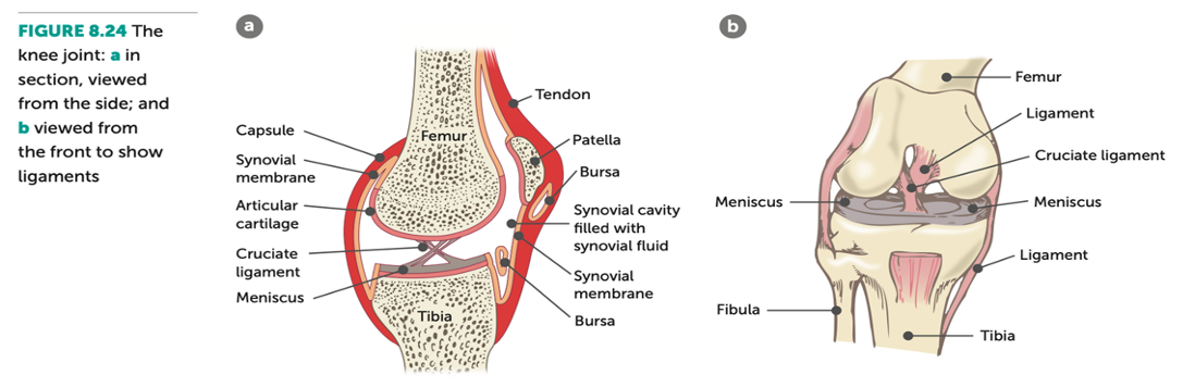
Articular discs – menisci
• Occur in some synovial joints. In the knee, these are menisci (singular is meniscus).
• Description: fibrocartilage extending inward from articular capsule, dividing synovial cavity in two.
• Function: provide cushioning in joint.
Bursae
• Description: small sacs of synovial fluid in some synovial joints.
• Function: positioned to help reduce friction between:
⚬ Bones and ligaments/tendons
⚬ Bone and skin
Accessory ligaments
• Description: made up strong fibrous connective tissue.
• Function: hold bones together many joints & resist extreme movements that may damage joint.
• E.g. anterior cruciate ligament (acl).

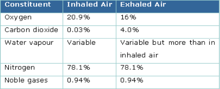
• Decrease of nearly 5% of oxygen in expired compared to inspired. This decrease due to some oxygen in inspired diffusing into blood across alveoli.
• Increase of nearly 4% of carbon dioxide in expired compared to inspired. This increase due to some CO2 in blood diffusing into air in alveoli.
• Noble gases - These gases are not very soluble in water and do not diffuse into the blood, so they remain at the same percentages in inhaled and exhaled air.
• The amount of water vapour in inspired air is variable as it depends on atmospheric conditions, particularly humidity. Expired air tends to have more water vapour as the water in the lungs can evaporate and add to the air we breathe out.

• Valves in the heart ensure that blood can only flow in one direction.
• Between atria and ventricles are the atrioventricular valves (AV valves).
• These are flaps of thin tissue with the edges held by tendons called chordae tendineae, attached to the heart on papillary muscles.
⚬ Tricuspid valve = between the right atrium & right ventricle.
⚬ Mitral (bicuspid) valve = between the left atrium & left ventricle.
• When the ventricles contract, blood catches behind the flaps and they billow out like a parachute, sealing the opening between the A and V.
• Blood must then leave through arteries, not flow back into the atria

• Where arteries leave the heart there are a second set of valves, stopping blood from flowing back into the ventricles when they relax - semilunar valves.
• Each semilunar valve has 3 cusps.
⚬ Pulmonary valve = between the right ventricle & pulmonary artery.
⚬ Aortic valve = between the left ventricle & aorta.
• When blood flows into the artery the cusps are pressed flat against the artery wall, allowing the blood to flow in.
• When the blood tries to flow back into the ventricle, the cusps fill out and seal off the artery.
• The closing of the valves in the heart gives the characteristic ‘lub dub’ sound

Carbon dioxide in blood
• Carbon dioxide is a waste product of cellular respiration.
• This gas diffuses out of the tissue cells into the tissue fluid and then into the blood in the tissue bed.
• Transport of carbon dioxide in the blood:
• 8% will be dissolved in the plasma.
• 22% will combine with haemoglobin in red blood cells forming carbaminohaemoglobin.
• 70% will undergo a chemical reaction and then be transported as bicarbonate ions in the plasma.
Carbon dioxide in haemoglobin
• Carbon dioxide diffuses into the red blood cell and combines with the globin part of a haemoglobin molecule to form carbaminohaemoglobin.
• Note: it doesn’t compete with oxygen transport (binds to the heme part).
Carbon dioxide in plasma
• Bicarbonate ions in the plasma:
⚬ Carbon dioxide diffuses into a red blood cell.
⚬ The carbon dioxide reacts with the water in the cytoplasm of this cell to form carbonic acid.
⚬ The dissolved carbonic acid ionises into hydrogen ions and bicarbonate ions.
• Bicarbonate ions in the plasma:
• Although this reaction also occurs in plasma, it is thousands of times faster in RBCs because they (and not the plasma) contain an enzyme that reversibly catalyses the conversion of carbon dioxide and water to carbonic acid.
• Once generated, bicarbonate ions move quickly from the RBCs into the plasma.
• In the lungs, the process is reversed
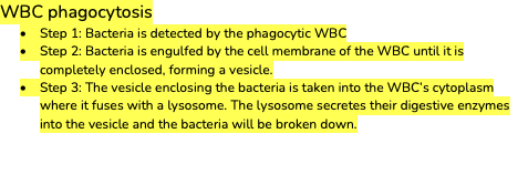
WBC phagocytosis
· Step 1: Bacteria is detected by the phagocytic WBC
· Step 2: Bacteria is engulfed by the cell membrane of the WBC until it is completely enclosed, forming a vesicle.
· Step 3: The vesicle enclosing the bacteria is taken into the WBC’s cytoplasm where it fuses with a lysosome. The lysosome secretes their digestive enzymes into the vesicle and the bacteria will be broken down.

• Whole blood transfusion:
⚬ Severe traumatic haemorrhage (severe blood loss).
• Red blood cell transfusion:
⚬ Spinning blood at very high speed in a centrifuge.
⚬ Treat low haemoglobin or red cell count, anaemia, heart disease.
• Platelet transfusion:
⚬ Treat low platelet count (thrombocytopaenia).
⚬ Caused by chemotherapy, bone marrow transplantation, major surgery, liver disease, severe trauma, some medications.
• Plasma transfusion:
⚬ Fresh frozen plasma (all blood cells are taken out).
⚬ Replace missing or low levels of blood proteins.
⚬ Liver disease, heart surgery, severe blood lost, severe burns.
• Cryoprecipitate:
⚬ Contains clotting factors (factor VIII, Factor XIII and von Willebrand factor) and fibrinogen.
⚬ Thawing fresh frozen plasma and then collecting the cold-insoluble proteins (precipitate); refrozen for storage.
⚬ Low levels of clotting proteins for conditions like Haemophilia A (lack factor VIII) and von Willebrand disease (lack this factor).

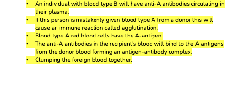
• An individual with blood type B will have anti-A antibodies circulating in their plasma.
• If this person is mistakenly given blood type A from a donor this will cause an immune reaction called agglutination.
• Blood type A red blood cells have the A-antigen.
• The anti-A antibodies in the recipient's blood will bind to the A antigens from the donor blood forming an antigen-antibody complex.
• Clumping the foreign blood together.
Fibrous:
• Fibrous = structural classification
• Fixed/immovable = functional classification
• Description:
⚬ Bones are held in place by fibrous connective tissue
⚬ No joint cavity is present
⚬ Mainly found in the axial skeleton = more stable
• Examples:
⚬ Skull, between teeth & jaw, between radius & ulna, between tibia & fibula
Cartilaginous:
• Cartilaginous = structural classification
• Slightly movable = functional classification
• Description:
⚬ Bones are held in place by fibrocartilage (compressible & resilient), allowing slight movement
⚬ No joint cavity is present
⚬ Mainly found in the axial skeleton = more stable
• Examples:
⚬ Pubic symphysis (junction of two pelvic bones), between vertebrae, between ribs & sternum
 | |||
 | |||
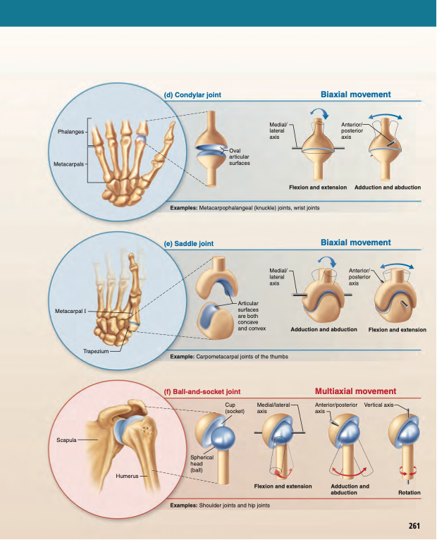
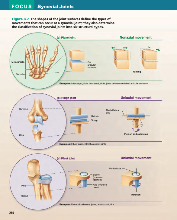
Synovial:
• Synovial = structural classification
• Freely movable = functional classification
• Description:
⚬ Contains a fluid-filled synovial cavity, enclosed in a fibrous capsule, between the articulating bone surfaces, allowing free movement in multiple directions but limited by ligaments, muscles, tendons & adjoining bones
⚬ Most commonly injured joint
• Examples:
⚬ Shoulder, elbow, wrist, fingers, hip, knee, ankle, toes

Bundles of muscle fibres
• Muscle cells are held together in bundles (fasicles).
• A sheath of connective tissue (perimysium) surrounds each bundle and allows bundles to slide over one and other.
• These sheaths taper to form a tendon, joining the muscle to bone.
• The connective tissue allows adjacent bundles to slide easily over one another as they contract.
• Muscle bundles contain many parallel muscle cells.
• Each cell is an elongated cylinder with many nuclei.
• Around the cell is a thin plasma membrane called the sarcolemma which contains cytoplasm, called the sarcoplasm.
Myofibrils
• Within the sarcoplasm of each muscle fibre/cell there are thread like structures, myofibrils that run the length of the fibre.
• Myofibrils are made from smaller myofilaments which are made of protein. This is what contracts.
• There are two types of myofilaments:
• Myosin - thick filaments
• Actin - thin filaments
• The arrangement of these filaments give skeletal muscle the banded appearance


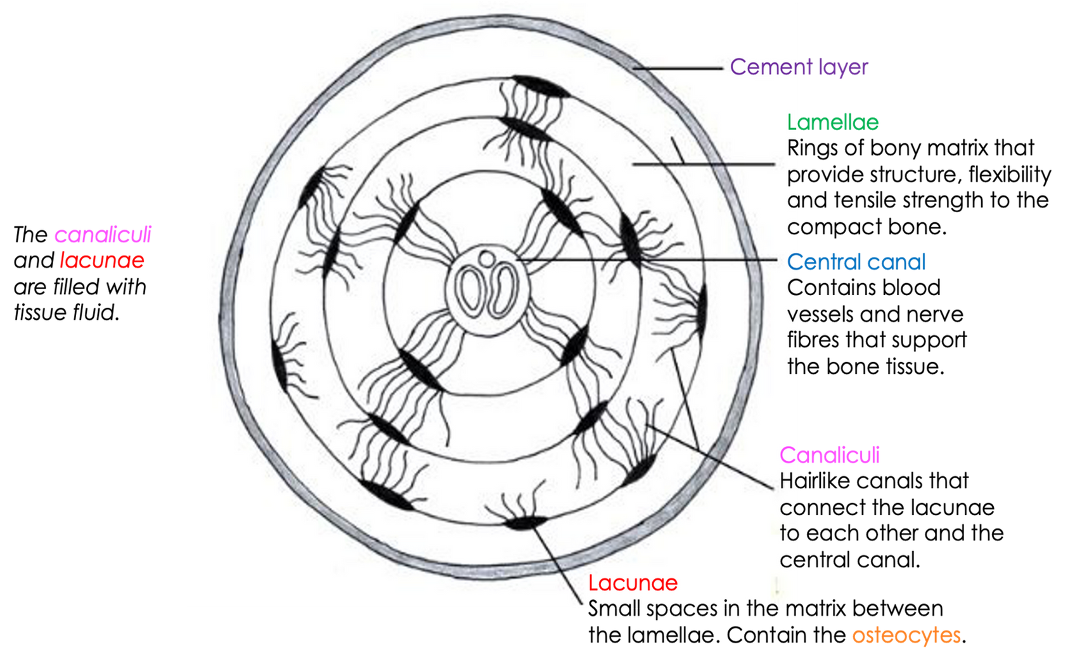

Spongy (cancellous bone)
• No osteons present.
• Instead, it is arranged in irregular, thin, bony plates called trabeculae.
• Bone cells (osteocytes) are in the lacunae (spaces in the trabeculae) but lamellae are irregularly arranged (not in concentric layers).
• Interconnected by canaliculi.
• Nutrients reach the osteocytes of spongy bone by diffusing through the canaliculi from capillaries in the endosteum surrounding the trabeculae.

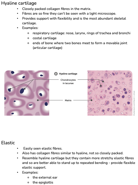
Hyaline cartilage
• Closely packed collagen fibres in the matrix.
• Fibres are so fine they can’t be seen with a light microscope.
• Provides support with flexibility and is the most abundant skeletal cartilage.
• Examples:
⚬ respiratory cartilage: nose, larynx, rings of trachea and bronchi
⚬ costal cartilage
⚬ ends of bone where two bones meet to form a movable joint (articular cartilage)
Elastic
• Easily seen elastic fibres.
• Also has collagen fibres similar to hyaline, not so closely packed.
• Resemble hyaline cartilage but they contain more stretchy elastic fibres and so are better able to stand up to repeated bending - provide flexible elastic support.
• Examples:
⚬ the external ear
⚬ the epiglottis
Fibrocartilage
• Coarse appearance from parallel bundles of thick collagen fibres.
• Not as compact as hyaline, therefore can be compressed.
• Highly compressible and has great tensile strength, great for weight-bearing areas.
• Occur in sites that are subjected to both pressure and stretch, for example:
⚬ the menisci in the knee,
⚬ the discs between the vertebrate
⚬ the tissue joining the two sides of the pelvis (pubic symphysis).
Muscle tone
• Muscle tone = maintaining partial contraction of skeletal muscles.
• Partial contraction of skeletal muscle to tighten muscles but not enough to produce movement.
• Muscle tone helps hold many body parts in position.
• Posture = the way a person holds their body when standing or sitting & relies on muscle tone.