Chapter 8: Anthropology and Odontology
8.1: Introduction
- Anthropology – the study of humans, their cultures, and their biology.
- Forensic Anthropology – the application of the study of humans to situations of modern legal or public concern. It takes the form of collecting and analyzing human skeletal remains to help identify victims and reconstruct the events surrounding their deaths.
Main Disciplines within Anthropology
- Paleontology – The biological study of past human populations.
- Anthropology – The biological study of current human populations.
- Archaeology – The study of past human cultures.
- Ethnology – The study of current human cultures.
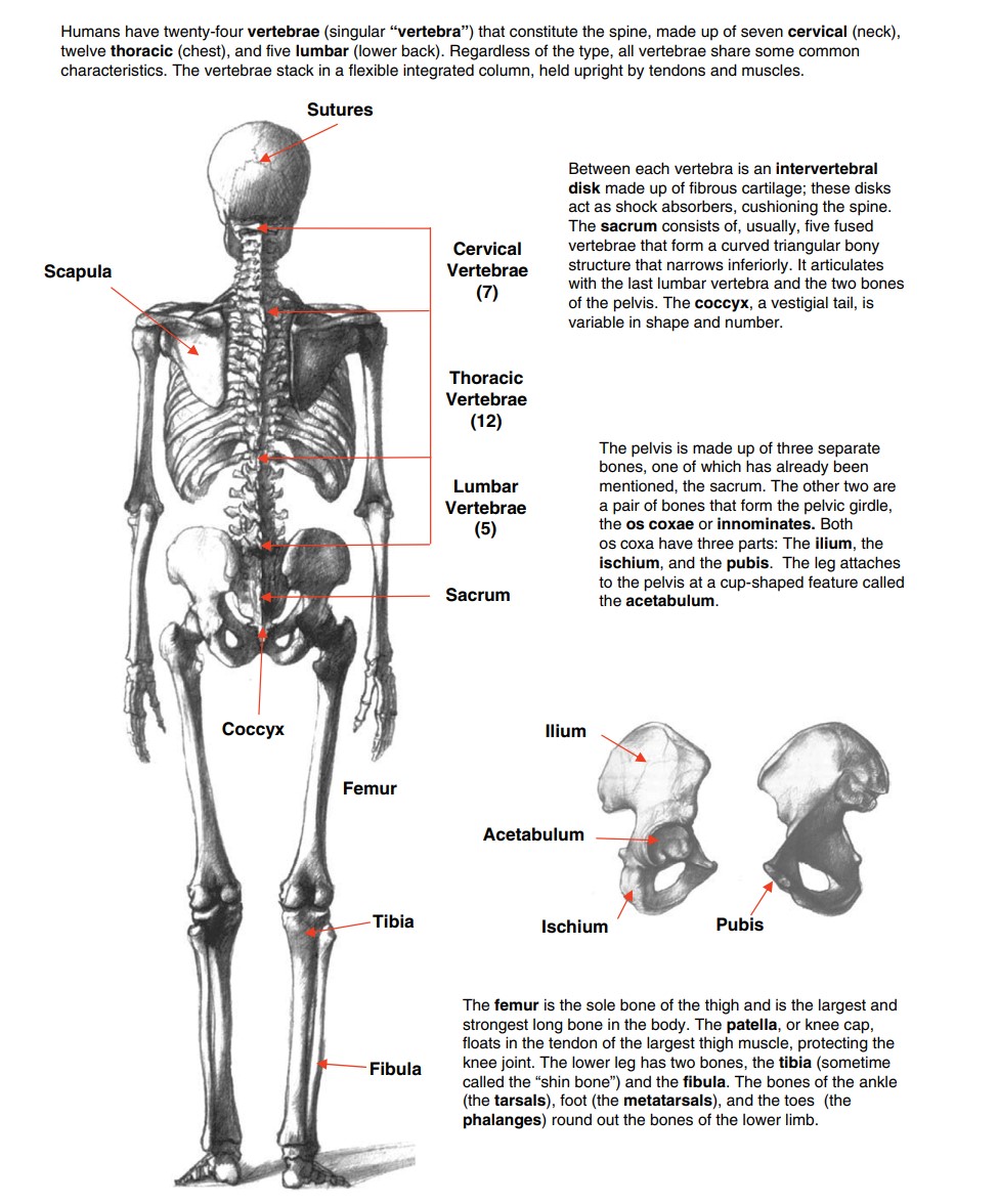
8.2: The Human Skeleton
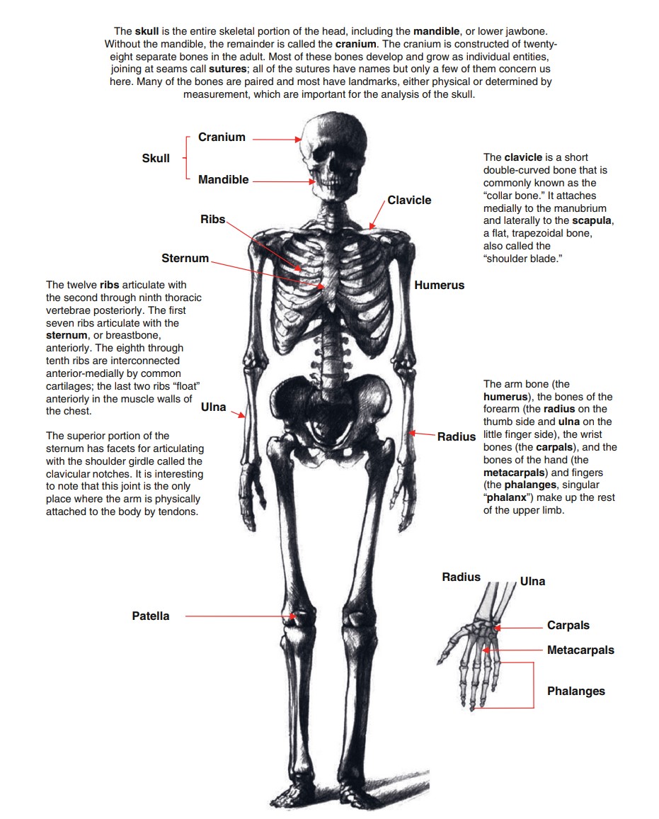
Bones perform four main functions for the body:
- The skeleton provides the infrastructure for attachment and support of the softer tissues in our bodies.
- These attachments allow the bones to act as levers, providing motion, powered by muscles, at the joints. The structure and arrangement of our bones set the range of motion for our limbs and bodies.
- The hard bones protect our soft organs from physical damage; this is especially true of the brain and the heart and the lungs.
- The bones are centers of growth from infancy to early adulthood; they also continue important physiological functions throughout our lives by housing the tissue that makes red blood cells.
Bone Organization and Growth
Bone growth and maintenance are complex processes that continue throughout our lives. Our skeletons must grow, mature, and repair at the macro and microscopic levels even as we use them.
Two types of bone growth characterize the human skeleton: endochondral and intramembranous. Endochondral bone growth starts with a “model” of a bone consisting of cartilage and centers of ossification.
The developing shaft of the bone is called the diaphysis and the ends are called epiphysis.
In intramembranous bone growth, instead of a cartilage model, the ossification occurs within a membrane and this occurs in many bones of the skull.
The osteoblasts, which form the osseous tissue, become encapsulated in lacunae but maintain contact with the vascular system via microscopic canaliculi. When they become encapsulated, they are referred to as osteocytes.
Between neighboring Haversian systems are non-concentric lamellae, devoid of Haversian canals, termed interstitial lamellae.
Vascular canals called Volkmann’s canals, traverse the long axis of the bone; they are always at right angles to Haversian canals. Their function is to link vascular canals of adjacent Haversian systems with each other and with the periosteal and endosteal blood vessels of the bone.
The outer perimeter of a long bone, beneath the osteogenic connective tissue (called periosteum), is composed of circumferential lamellae, which also lack Haversian canals.
This thick-walled hollow shaft of compact bone (the diaphysis) contains bone marrow.
- At the distal ends of long bones, where Haversian systems are not found, the bone appears spongy and is therefore called cancellous, or spongy, bone.
- The spongy appearance is misleading because careful examination of the architecture reveals a highly organized trabecular system providing maximal structural support with a minimum density of bony tissue.
The cavities of the epiphyseal spongy bone are in contact with the bone marrow core of the diaphysis except during the growth of long bones in young animals.
Interposed between the epiphysis and the diaphysis is the cartilaginous epiphyseal plate. The epiphyseal plate is joined to the diaphysis by columns of cancellous bone; this region is known as the metaphysis.
When bone is formed in and replaces a cartilaginous “model,” the process is termed endochondral ossification.
Some parts of the skull develop from osteogenic mesenchymal connective tissue, however, without a cartilaginous “model” having been formed first. This is termed intramembranous ossification and these bones are called membrane bones.
In both instances, three types of cells are associated with bone formation, growth, and maintenance:
- The osteoblasts produce osseous tissue (bone), become embedded in the matrix they manufacture, and are then renamed osteocytes, to reflect their change of status.
- They remain viable because they have access to the vascular supply via microscopic canaliculi through which cellular processes extend to receive nutrients and oxygen.
- Osteoclasts actively reabsorb and remodel bone as required for growth; these are giant, multinuclear, phagocytic, and osteolytic cells.
Cortical Bone – the outer layer of hard, smooth compact bone.
Trabecular bone – the inner layer is an infrastructure of sponge-like bone.
In the very center of long bones is the medullary cavity, which contains marrow — a fatty material that also houses blood-generating tissues.
Interstitial Bone – bone that lies between recently reworked bone.
8.3: Collecting Human Remains
- Forensic anthropologists rarely find skeletal remains that are aboveground.
- It is often a hiker, hunter, or some other civilian in a remote or uninhabited area who stumbles across the bones at a crime scene.
- Because the “evidence” has been found by untrained persons, securing the scene is the most effective way of initiating evidence protection.
- If the remains are scattered, each bone fragment should be flagged or marked.
- Context is even more important with skeletal remains and the individual bones should not be disturbed until the entire scene has been photographed and documented.
- All the bones on the surface, even animal bones, should be collected.
- Buried remains require more time and skill to retrieve.
- Archaeological techniques are employed to excavate buried skeletal materials and should be performed only by trained personnel under the supervision of an experienced archaeological excavator.
- A grid is set up with one point set as a datum, or reference point, from which all measurements originate.
- Soil and materials are removed by a thin layer at a time (usually 2–5cm) slowly exposing the buried items.
8.4: Analysis of Skeletal Materials
- Once the remains are determined to be human, then a biological profile can be developed for the individual(s) represented.
- A biological profile consists of assessing the sex, age at death, racial affinity, height, and any other aspects that would describe the individual-class-level information. It is the first step toward identifying whom the remains represent.
- The criteria that help determine the biological profile are either qualitative, that is, morphological (the presence or absence of a trait, or the shape or size of a landmark) or quantitative.
- Quantitative physical anthropology is dominated by statistical analysis and sometimes these analyzes, such as principal component analysis, are quite complex involving many measurements, samples, and relationships.
What is the sex?
The largest number of and most accurate traits for determining sex reside in the pelvis.
The major reason that male and female skeletal anatomy differs so much in the pelvic region is that only females carry and bear babies; human pelvic anatomy reflects this functional difference.
The male pelvis tends to be larger and more robust, whereas the female pelvis is broader and can exhibit pregnancy-specific traits.
A useful trait for distinguishing between the male and female pelvis is the sciatic notch, located on the inferior lateral border of the ileum.
- The sciatic notch is wide (an angle of about 60°) in females and narrow in males (about 30°).
Phenice Method – developed by Dr. Terrell Phenice in 1969, which uses three characteristics: the ventral arc, the subpubic concavity, and the ischiopubic ramus.
- Ventral Arc – a ridge on the anterior surface of the pubic bone that is present in females but absent in males.
- Subpubic Concavity – a depression on the medial border of the ischiopubic ramus, just inferior to the pubic symphysis.
- Concavity – wider and deeper in females and is only slight, if at all present, in males.
- Ischiopubic ramus – flatter and thinner in males, whereas in females it is wide and may even have a ridge on it.
Sex can be estimated from the cranium.
- The specific areas of interest are the brow ridges, mastoid processes, occipital area at the rear of the skull, upper palate, and the general architecture of the skull.
- The skull is one of the most, if not the most, studied, measured, and examined part of the skeleton. This metric enthusiasm extends to the determination of sex.
Thirty-four standard measurements are the minimum for the inclusion of a skull into the National Forensic DataBase.
- These measurements are taken with specialized rulers, called calipers, that are either spreading calipers or sliding calipers.
- Complicated statistical techniques are used to sort out the measurements, relate them to each other, and then compare them against an appropriate reference population.
- FOR DISC – a software that provides an easy way to analyze and compare data from skeletons.
Postcranial bones can also provide information about a person’s sex, but most of this information is based on size and therefore is quantitative.
How old was the person?
The range of human ages has been broken into various classes with associated years:
- Fetal (before birth),
- Infant (0–3),
- Child (3–12),
- Adolescent (12–20),
- Young adult (20–35),
- Adult (35–50), and
- Old adult (50+).
Bones can indicate the stage of development attained by the appearance and fusion of the various epiphyses throughout the body.
- Nonunited epiphyses are easy to observe because the diaphyseal surface is characteristically rough and irregular in appearance.
- Epiphyseal appearance and union occur over years and is a process, not an event; the degree of the union must be carefully assessed because this could indicate which extreme of an age range is being observed.
@@The three main stages of the union are:@@
- The epiphysis is open;
- The epiphysis is united but the junction is still visible; and
- The epiphysis is completely fused.
Pubic Symphysis – the junction of the two pubic bones lying roughly 4–5 in below the navel. This junction is bridged by cartilage that acts as a cushion between the two bones.
- Symphysis – a false joint.
Another area of morphological change with advancing adulthood is the sternal end of the fourth left rib. As the cartilage between the sternum and the ribs ages, it begins to ossify at a known and predictable rate.
Another method of estimating age at death is the examination of the changes in the auricular surface, where the ilium attaches to the sacrum.
The bone never rests. It is constantly remodeling in response to the stresses placed upon it. This remodeling can be seen in the microscopic structure of bone. In approximately the same way as a wall would be rebuilt, bone first needs to be torn down before it can be built up. This constant erosion and renewal leave permanent markers in bone: Once we die, these changes cease.
Ancestry
Human physical variation is often a subtle thing and people are sensitive to the labels other people place on them. While it is true that no pure ethnic groups exist, we identify people based partly on what we perceive their “race” to be.
Ancestry can be estimated by morphological or quantitative analysis and both of these methods are centered on the skull.
- Features of the skull, such as the general shape of the eye orbits, nasal aperture, dentition, and surrounding bone and the face, can offer indications of ancestry.
- Other features are more distinct, such as the scooped-out appearance of the lingual (tongue) side of the upper central incisors often found in individuals of Asian ancestry (so-called “shovel-shaped” incisors).
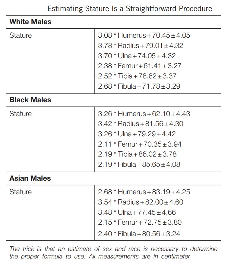
Stature
Our living stature directly relates to the length of our long bones, especially those of our lower limbs. Calculating stature from long-bone lengths is relatively simple and even partial bones can yield useful results. The only difficulty is that sex and ancestry must be known to correctly estimate height because humans vary within and between these categories.
Facial Reproductions
- An artist recreates the likeness of a person either by sculpting the soft tissues with clay in three dimensions or by drawing.
- Facial reconstructions require a high degree of artistic skill, a good knowledge of human anatomy and variation, and an appreciation of the human face. These likenesses are not used for identification purposes but are meant to stir the public’s recognition of otherwise unidentifiable remains.
8.5: Odontology
- Dental identification can be conducted through the comparison of dental remains to either antemortem or postmortem records.
- The most frequently performed examination is comparing the dentition of a deceased person to those of a person represented by an antemortem to determine if they are the same individual.
- The forensic dentist produces the postmortem record by careful charting and written descriptions of the dental structures and by taking radiographs.
- Once the postmortem record is complete, a comparison between it and any antemortem records can be conducted. The comparison is methodical and systematic: Each tooth and structure are examined and compared. Fillings, caps, and restorations play the largest role in the identification process.
Dental Anatomy
The anatomy of the mouth is important to forensic science for several reasons.
- Teeth are made of enamel — the hardest substance that the body produces, and teeth can survive severe conditions and still be viable for analysis.
- The teeth are the only part of the skeletal anatomy that directly interacts with the environment and, therefore, can reflect conditions the person experiences during life.
- Teeth and their related structures have the potential to be used in the identification of the deceased.
Teeth
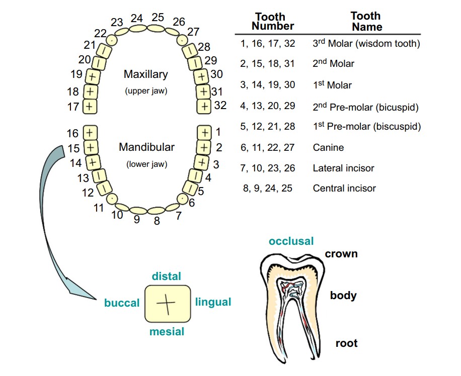
Tooth Development
- Teeth grow from the chewing surface, or cusps, downward to the roots. This continual process is usually broken up into phases that relate to the amount of tooth development.
- Humans have two sets of teeth, one when we are children, called “baby” teeth but more properly termed “deciduous” teeth, and one when we are adults, our permanent teeth.
- Different teeth develop at different rates, with incisors developing faster than molars.
- Teeth erupt through the gums when they are about one-half to three-fourths developed.
- Notable landmarks in tooth eruption are the first deciduous incisor at about 9 months, the first permanent molar at about 6 years, the first permanent incisor at about 7 years, and the third permanent molar at some time between 15 and 21 years.
Identification
- The goal of a forensic anthropological examination is individualizing a set of human remains, often referred to as a “ positive identification.”
- Because most people regularly visit their dentists, dental records and X-rays are the most common form of antemortem record that leads to a positive identification. A structure in the frontal bone, the frontal sinus, is considered to be unique to a reasonable degree of scientific certainty. Surgeries healed fractures, and disease may all be documented radiographically and also can lead to positive comparisons.
- Identification through the comparison of ante- and postmortem X-rays is considered the best method for skeletal remains.
\n