Lesson 2: Path of Blood (Arteries, Capillaries and Veins)

Arteries/ Arterioles
The middle layer is thicker than in veins
They have a solid structure
Arteries carry blood away from the heart
They have a very high-pressure
No oxygen except (pulmonary artery)
Very little diffusion of substances
Very muscular layer
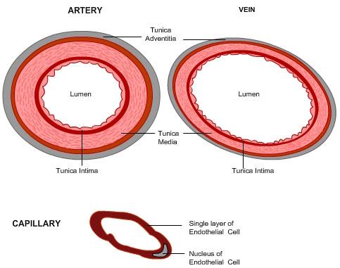
Capillaries
A single layer of cells forms the wall (endothelial cells)
Smallest blood vessel with a fine network
Blood flows from arteries to veins by travelling through many capillaries
Lowest pressure
Gradient (O2 is higher at the arteriole side and lowest at the venule side, CO2 is the opposite)
Nutrients: Into AND out of
O2: out of blood into the cells
CO2: out of cells into the blood
Single-cell thick only one cell can pass at a time
Veins/ Venules
The middle layer is thinner than in the arteries
It has an oval shape that can be collapsible
Veins carry blood towards the heart
Has the lower pressure
Lots of oxygen - pulmonary vein
Very little diffusion happens
Veins have one-way valves, that prevent blood from moving back into your system. (when valves fail it can lead to varicose veins)
Why is blood considered a tissue?
Blood is considered a tissue because it is a collection of similar specialized cells that serve particular functions.
What is blood plasma and its Functions?
Plasma is the fluid portion of one's blood, which makes up 55% of the blood volume. Plasma consists of water, dissolved gases, proteins, sugars, vitamins, minerals and waste products. Plasma is a clear, yellowish fluid composed of around 92% water and 7% dissolved blood proteins. The remaining 1% consists of organic and inorganic substances like sodium, potassium, chloride, and bicarbonate. The main proteins include albumin, globulins, and fibrinogen. Other nutrients that are transported by blood include glucose, fatty acids, vitamins, respiratory gases and the waste products of metabolism.
Blood Cells:
What are platelets and their functions?
Platelets are membrane-bound fragments of cells that form when larger cells in the born marrow break apart and they are the third major substance in the formed portion of blood. Platelets play a key role in clotting blood, which prevents excessive blood loss after an injury, by forming a net around the damaged blood vessel thus trapping the blood cells and producing a clot.
The mammalian circulatory system:
Pulmonary circulation: the movement of blood from the heart to the lungs and then back to the heart.
The blood that flows from the heart to the lungs carries deoxygenated blood, as this blood passes through the respiratory system gas exchange takes place carbon dioxide leaves the blood and oxygen enters.
The freshly oxygen-rich blood goes back to the heart and is pumped from the heart to the second circuit that transports it to the rest of the body.
Systemic circulation: takes oxygenated blood from the heart to other tissues and organs throughout the body.
After circulating throughout the body, the blood returns to the heart carrying waste carbon dioxide from the body’s tissues. The blood then re-enters the pulmonary circulation.
Cardiac Circulation: the movement of blood through the heart tissues.
The process of blood clotting
First, the blood vessel is broken due to injury, releasing chemicals that attract platelets to the site of injury
Second, the platelets rupture, releasing chemicals that combine with other chemicals in the plasma to produce the enzyme thromboplastin
Third, as long as calcium ions are present, thromboplastin reacts with prothrombin (a protein made by the liver) to produce another enzyme called thrombin
Lastly, the Thrombin reacts with fibrinogen (another plasm a protein) to produce fibrin. Fibrin is an insoluble protein that forms a fibrous mesh over the site of injury. This mesh prevents the loss of blood cells and eventually solidifies to form a clot.
Possible Quiz Questions:
Why do veins have valves?
Do all arteries carry oxygen-rich blood and all veins carry oxygen-poor blood? Explain.
Describe one function of each of the three main blood vessels.
When a person exercises, one of the physiological responses is an increase in blood flow. In terms of the circulatory system, describe what is happening.
Compare an artery with a vein structurally.
Provide details about the three main components of blood.