Somatic Nervous System Vocabulary Review
Introduction to the Somatic Nervous System
The somatic nervous system (SNS) is a crucial division of the peripheral nervous system (PNS) that is responsible for conscious perception and voluntary control of skeletal muscle movements. It enables the body to respond to external stimuli with deliberate actions, distinguishing it from the autonomic nervous system, which governs involuntary actions.
The term "somatic" refers to the functional aspects of the system focusing on voluntary movements and sensations, while "peripheral" pertains to the anatomical structures that connect the central nervous system (CNS) to the limbs and organs.
Chapter Objectives
Describe the key components that make up the somatic nervous system.
Understand the modalities (types of sensations) and submodalities (specific forms of sensations) associated with sensory systems.
Distinguish between the general senses (wide distribution across the body) and special senses (localized to specific organs).
Identify regions in the central nervous system that contribute to somatic functions.
Explain the pathways involved in stimulus-response motor functions and how they affect skeletal muscle activity.
Reflex Actions as Examples of Somatic Functions
Process of Reflexes: Reflex actions are rapid, automatic responses to stimuli that are processed via the central nervous system without conscious thought. Sensory neurons detect environmental changes, transmit signals to the spinal cord, and lead to immediate motor responses through synaptic pathways.
Example of Withdrawal Reflex: An example of a reflex action is the withdrawal reflex, where a subject instinctively pulls their hand away from a hot surface. This involves:
Sensory input detected by receptors such as thermoreceptors in the skin, which respond to rapid temperature changes.
Action potentials generated move through the dorsal spinal roots into the spinal cord where they synapse with interneurons.
Motor outputs from the ventral horn, such as signals to the biceps brachii, initiate muscle contractions to withdraw the hand from danger.
Additional Components: Reflexes are often more complex and can include inhibitory signals to antagonistic muscles (e.g., when the biceps contract, the triceps must relax) and can play a role in maintaining posture by adjusting muscle tension according to shifts in body position.
Sensory Perception
sensation: the activation of sensory receptor cells at the level of stimulus
perception: central processing of sensory stimuli into meaningful pattern
Types of Sensory Receptors
General Classification: Sensory receptors can be classified based on their cell types and locations.
can also be classified functionally on basis of transduction of stimuli
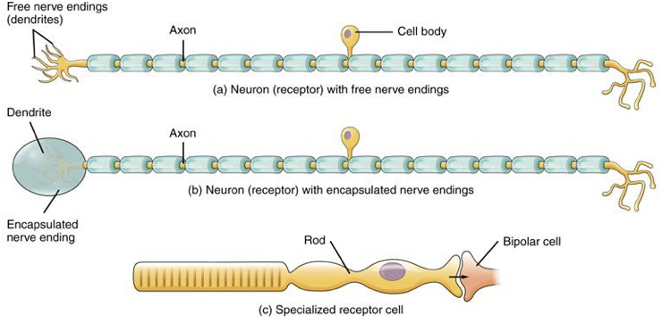
Cell Types:
Free nerve endings (for pain and temperature),
Encapsulated endings (for pressure and vibration),
Specialized receptor cells (for specific senses like taste and hearing).
Location:
Exteroceptors: Detect stimuli from the external environment (e.g., light, sound, touch).
Interoceptors: Respond to stimuli from internal organs (e.g., blood pressure, internal pain).
Proprioceptors: Sense limb position and movement within muscles and joints, crucial for coordination.
Functional Types: Sensory receptors respond to various forms of stimuli:
Chemoreceptors (taste and smell),
Osmoreceptors (detetion of solute concentrations of bodliy fluids)
Nociceptor (pain detection)
Mechanoreceptors (touch, hearing, and balance),
Thermoreceptors (temperature detection),
Photoreceptors (detection of light).
Sensory Modalities
Modality refers to the way that information is encoded
Submodalities: major five sense divided into specific categories
Major senses include taste, smell, touch, hearing, balance, and vision. This can be further categorized into:
General Senses: Distributed across the body, responsible for sensations like pressure, pain, and temperature—receptors are often found in the skin and deeper tissues.
Special Senses: Rely on specific organs (e.g., eyes for vision, ears for hearing) that gather information for specific modalities. Each sense can be subdivided into submodalities, such as types of touch (e.g., light touch, deep pressure) and sound frequencies in hearing.
Gustation (Taste)
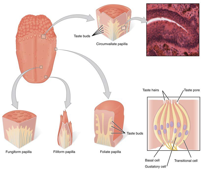
Taste Submodalities: The sense of taste has five major submodalities: sweet, salty, sour, bitter, and umami (savory). Each of these is detected by different types of taste receptors, which are specialized cells located in taste buds.
Taste Detection Process: Taste buds, found in four types of papillae (circumvallate, foliate, filiform, fungiform), react to dissolved food chemicals. When activated, these receptors generate neural impulses that travel to the brain, leading to the perception of taste and influencing food preferences and dietary choices.
Olfaction (Smell)
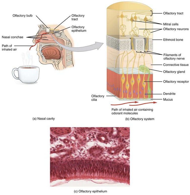
Mechanism: The sense of smell involves olfactory receptor neurons situated in the nasal cavity (olfactory epithelium). The dendrites of these olfactory sensory neurons extend into the mucosal layer to bind with odorant molecules, generating electrical signals that are transmitted to the olfactory bulb. From there, the signals bypass the thalamus and are relayed directly to the brain regions involved in processing smells, making olfaction unique among the senses.
Auditory System and Equilibrium
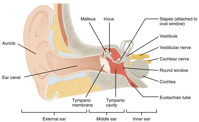
Audition (Hearing): Sound waves are converted into neural signals through the intricate structures of the ear:
External Ear: Comprises the auricle (pinna) which collects sound and the tympanic membrane (eardrum) that vibrates with sound waves.
Middle Ear: Contains three small bones known as ossicles (malleus, incus, stapes) that amplify sound waves before transmitting them to the inner ear.
Inner Ear: Houses the cochlea (responsible for hearing) and vestibular structures like the vestibule and semicircular canals, which are crucial for balance.

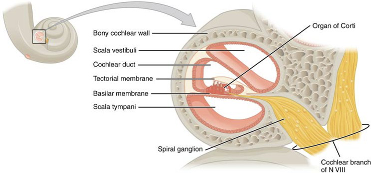
The cochlea is composed of:
spiral ganglia: a group of sensory neurons that transmit auditory information from the cochlea to the brain, facilitating the perception of sound.
stapes: a tiny bone in the middle ear that transmits sound vibrations from the incus to the oval window of the cochlea, playing a key role in the process of hearing.
scala vestibuli: the upper chamber of the cochlea filled with perilymph fluid, which plays a crucial role in the propagation of sound waves through the cochlear structure.
cochlear duct: the fluid-filled cavity within the cochlea, also known as the scala media, that contains endolymph and houses the organ of Corti, where sound transduction occurs.
scala tympani: the lower chamber of the cochlea that contains perilymph, extending from the round window at the base of the cochlea to the helicotrema at its apex, and plays a significant role in transmitting the pressure waves generated by sound vibrations.
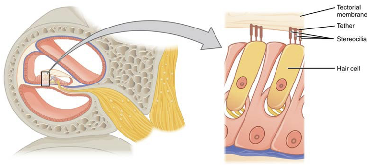
organs of corti: the sensory organ located within the cochlear duct, consisting of hair cells that convert sound vibrations into electrical signals, which are then transmitted to the auditory nerve for processing in the brain.
basilar membrane: the membrane within the cochlea that supports the organ of Corti and vibrates in response to sound waves
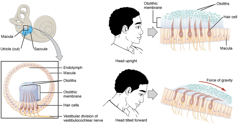
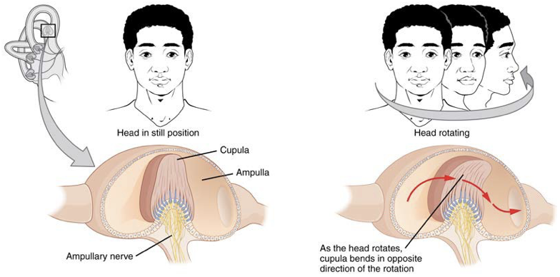
Equilibrium: The inner ear’s hair cells detected linear acceleration and head tilt in the utricle and saccule, while semicircular canals monitor rotational movements, contributing to the maintenance of balance and spatial orientation.
ampulla repsonds to rotational acceleration of the head, allowing the detection of changes in angular position, which is crucial for balance and coordination.
cupula: a gelatinous structure located within the ampulla of each semicircular canal that bends in response to movement, thereby stimulating the hair cells and facilitating the sensation of balance.
Somatosensation (Touch)
General Touch Sensation Types: These include pressure (how hard or soft something feels), vibration (detecting oscillations), temperature detection (warmth or cold), pain (noxious stimuli), proprioception (awareness of body position), and kinesthesia (sense of movement).
receptors are in skin but also founds in muscles, tendons, joint capsules, ligaments, and walls of visceral organs
Mechanotransduction: Specialized receptors are responsible for converting various mechanical stimuli into electrical signals. For instance, Merkel cells respond to light touch, while Pacinian corpuscles respond to deep pressure and vibration, allowing the brain to interpret different forms of tactile information.
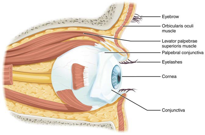
Vision
special sense of sight based on transduction of light stimuli received through eyes
palpebral conjunctiva: a thin, transparent membrane that covers the inner surface of the eyelids and extends to the surface of the eyeball, helping to protect and lubricate the eye.
lacrimal gland: where tears are produced
lacrimal duct: where tears flow through in medial corner of eye
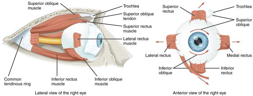
superior oblique rotates eye medially
inferior oblique rotates eye laterally
levator palpebrae superioris: elevates and retracts upper eyelid
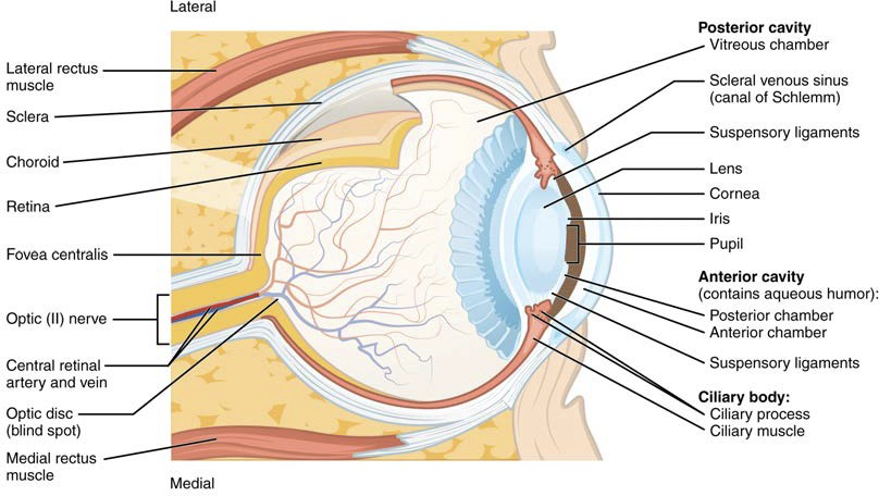
fibrous tunic
cornea, sclera
vascular tunic
choroid, ciliary body, iris
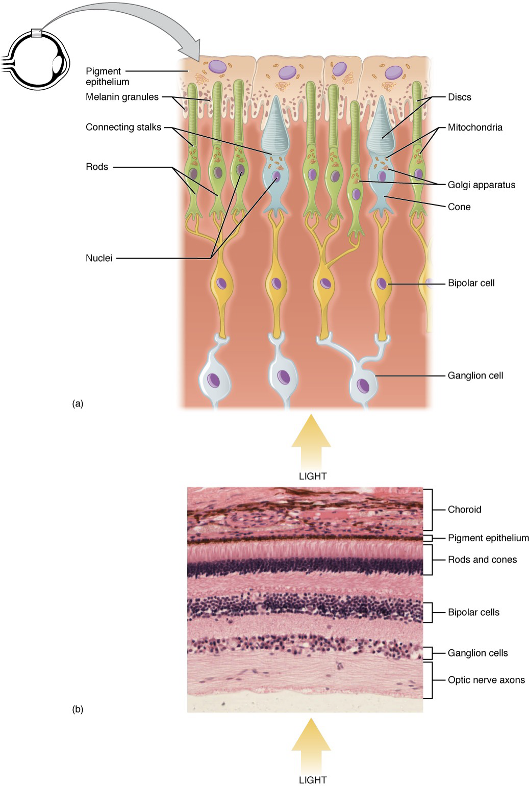
neural tunic (retina)
the innermost layer of the eye that contains photoreceptor cells responsible for converting light into neural signals.
cavities
aqueous humor
vitreous humor
Sensory Nerves
topographical arrangement: The topographical arrangement of the somatic nervous system refers to how sensory and motor pathways are organized within specific regions, facilitating efficient communication between the brain and various body parts.
Central Processing of Sensory Data
Sensory data is transmitted to the thalamus for processing, where it is integrated and routed to appropriate cortical regions based on the type of sensation being processed.
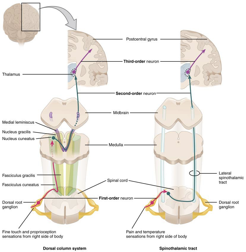
ascending pathways: The neural pathways that carry sensory information from the periphery to the brain,
Dorsal Column System: This pathway is responsible for transmitting touch, vibration, and proprioceptive information, decussating (crossing over) at the medulla.
begin at dorsal root ganglion cells
axons take on positional arrangement
separated into the fasciculus cuneatus and the fasciculus gracilis
fasciculis gracilis: contains axons from the lower limbs and lower trunk
targets nucleus gracilis
fasciculs cuneatus: contains axons from upper body
targets nucleus cuneatus
Spinothalamic Tract: This pathway processes temperature and pain sensations, decussating at the spinal cord level, reflecting how various types of sensory information are processed in the CNS.
begins with neurons in a dorsal root ganglion which then extend their axons to the dorsal horn of the spinal cord, where they synapse on second-order neurons and connects to the thalamus
neurons in thalamus then project their axons to the spinothalmic tract which synapse in the postcentral gyrus of the cerebral cortex
trigerminal pathway: carries somatosensory information from face, head, mouth, and nasal cavity
Integrative Processing
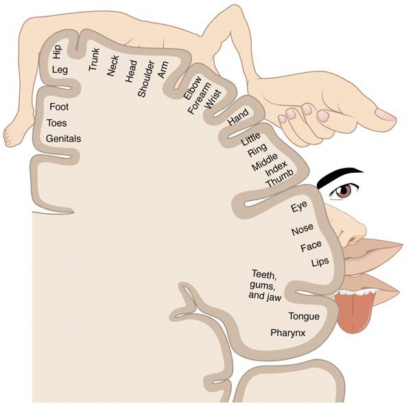
Cerebral Cortex: Different cortical regions are dedicated to processing various sensory inputs, structured in a topographic manner that mirrors body area representation (the somatosensory homunculus). This organization allows for efficient integration and interpretation of sensory information.
primary sensory cortex: Located in the postcentral gyrus, this region is crucial for processing tactile and proprioceptive information from the body, contributing to our perception of touch and spatial awareness.
association area: This region integrates information from multiple sensory modalities, facilitating higher-level processing such as perception, decision-making, and motor planning, thereby enhancing our ability to respond to complex stimuli.
multimodal integration area: This area is responsible for combining sensory inputs from different modalities, allowing for a more comprehensive understanding of our environment and facilitating coordinated responses.
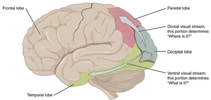
the primary visual cortex (V1) is surrounded by the visual association cortex (V2&V3)
Ventral and Dorsal Visual Streams: The dorsal visual stream primarily processes motion and spatial relationships ("where"), while the ventral stream focuses on object identification and significance ("what"), highlighting the complexity of visual information processing.
Motor Responses
Secondary motor cortices: These regions, including the premotor cortex and supplementary motor area, play a critical role in planning and coordinating voluntary movements, ensuring that sensory information is effectively translated into motor actions.
premotor cortex is is responsible in controlling movements of the core muscles to maintain posture during movement
supplemental motor area is responsible for planning and coordinating movement; manages sequential movements that are based on prior experience
frontal eye fields are responsible for moving the eyes in response to visual stimuli
Broca’s area is responsible for controlling movements of structures of speech production
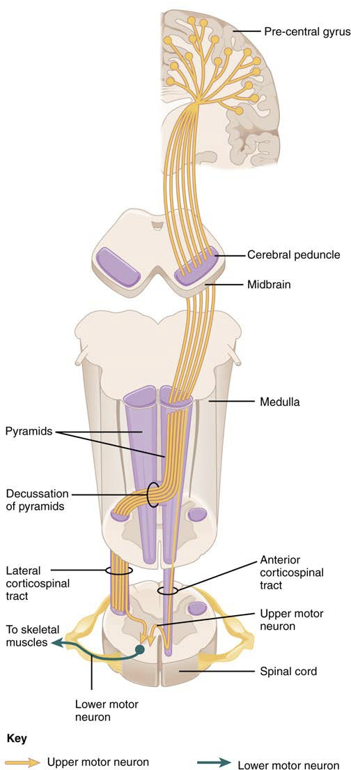
Primary motor cortex: located in the precentral gyrus, it is essential for the execution of voluntary movements by sending signals to the muscles.
Motor Pathways: The integration of sensory input and motor output occurs through specific pathways descending from the brain:
Corticobulbar Tract: Responsible for controlling the muscles of the head and neck, facilitating actions such as facial expressions and swallowing.
Corticospinal Tract: Primarily governs the control of limbs and trunk movements, utilizing large pyramidal cells (Betz cells) located in the primary motor cortex to execute voluntary movements.
Extrapyramidal system
tectospinal tract: important for postural movements
reticulospinal tract: influences trunk and proximal limb muscles related to posture and locomotion
vestibulospinal tract: allows posture, movement, balance to be modulated by input from the vestibular system, providing the body with the ability to maintain stability during head movements.
rubrospinal tract: facilitates voluntary movement, particularly in the upper limbs, by providing excitatory input to the flexor muscles and inhibitory input to the extensor muscles.
Types of Reflexes
Withdrawal Reflex: This reflex represents a protective mechanism where a sensory neuron activates a motor neuron leading to muscle contraction while an interneuron simultaneously inhibits the contraction of antagonistic muscles, exemplifying coordinated muscular reflex actions.
Stretch Reflex: This reflex maintains muscle length by eliciting contraction in response to stretch; it is commonly tested during physical examinations using simple techniques like the patellar reflex (knee-jerk). This reflex is indicative of the integrity of the nervous system.
Corneal Reflex: This reflex occurs when the cornea is stimulated, resulting in closure of the eyelids as a protective response; it involves sensory neurons from the trigeminal nerve and motor output from the facial nerve, ensuring the eye is safeguarded against potential harm.
Conclusion
A comprehensive understanding of the somatic nervous system revolves around the integration of both sensory and motor functions, with feedback loops playing a vital role in refining control over voluntary and reflexive movements. This underscores the intricate and dynamic interactions within the nervous system that facilitate human movement and sensory perception.
Additional Categories
Clinical Relevance
Understanding the somatic nervous system is vital in clinical settings as it helps healthcare providers diagnose and treat neurological disorders affecting motor control and sensory perception.
Conditions such as multiple sclerosis, peripheral neuropathy, and spinal cord injuries can severely impact somatic functions, highlighting the importance of early diagnosis and intervention.
Developmental Aspects
The somatic nervous system develops throughout early life, influenced by genetic and environmental factors. Proper sensory and motor integration during this period is essential for normal physical and cognitive development.
Neuroplasticity within the somatic nervous system allows for adaptations following injury and during learning processes, showcasing its resilience and capacity for recovery.
Comparative Anatomy
Studying the somatic nervous system across different species provides insights into evolutionary adaptations of motor and sensory capabilities.
Examining variations in somatic structures can reveal how different organisms have evolved unique solutions for interacting with their environment, such as specialized limbs or advanced sensory processing pathways.