cognitive; week 6; fMRI and cognition
what is cognitive neuroscience?
Cognitive psychology – the study of human mental processes
Tools: tasks that employ specific cognitive processes
Measures: accuracy, reaction times, eye-movements
Neuroscience – the study of the brain and nervous system
Tools: brain imaging tools (EEG, fMRI, etc)
Measures: EEG signal (ERP), BOLD signal, etc
Cognitive neuroscience:
The study of the relation between brain structures/activity, and cognitive functions
The study of cognitive processes using neuroscientific measures and knowledge
Example: Face N170
Studies show a larger N170 response to faces than objects
Studies examine the properties that affect face processing (e.g., inversion, unaffected by familiarity) and how it affects N170.
Researchers conclude that N170 reflect the structural encoding of faces, prior to their identification (Eimer, 2011)
Then, other researchers use group difference in N170 to conclude about face processing in different groups (Feuerriegel et al., 2015)
Some researchers suggested an alternative explanation of N170: that N170 is merely associated with low-level features in face pictures.
fMRI refresher
MRI
Atoms (in this case, hydrogen atoms) are like constantly spinning magnets
These atoms align when in a scanner’s magnetic field.
Then, you send in radio waves to make them face in a new direction
When they relax and return to their previous alignment, they emit energy (i.e. ‘resonance’)
This energy is what the scanner uses to create an image.
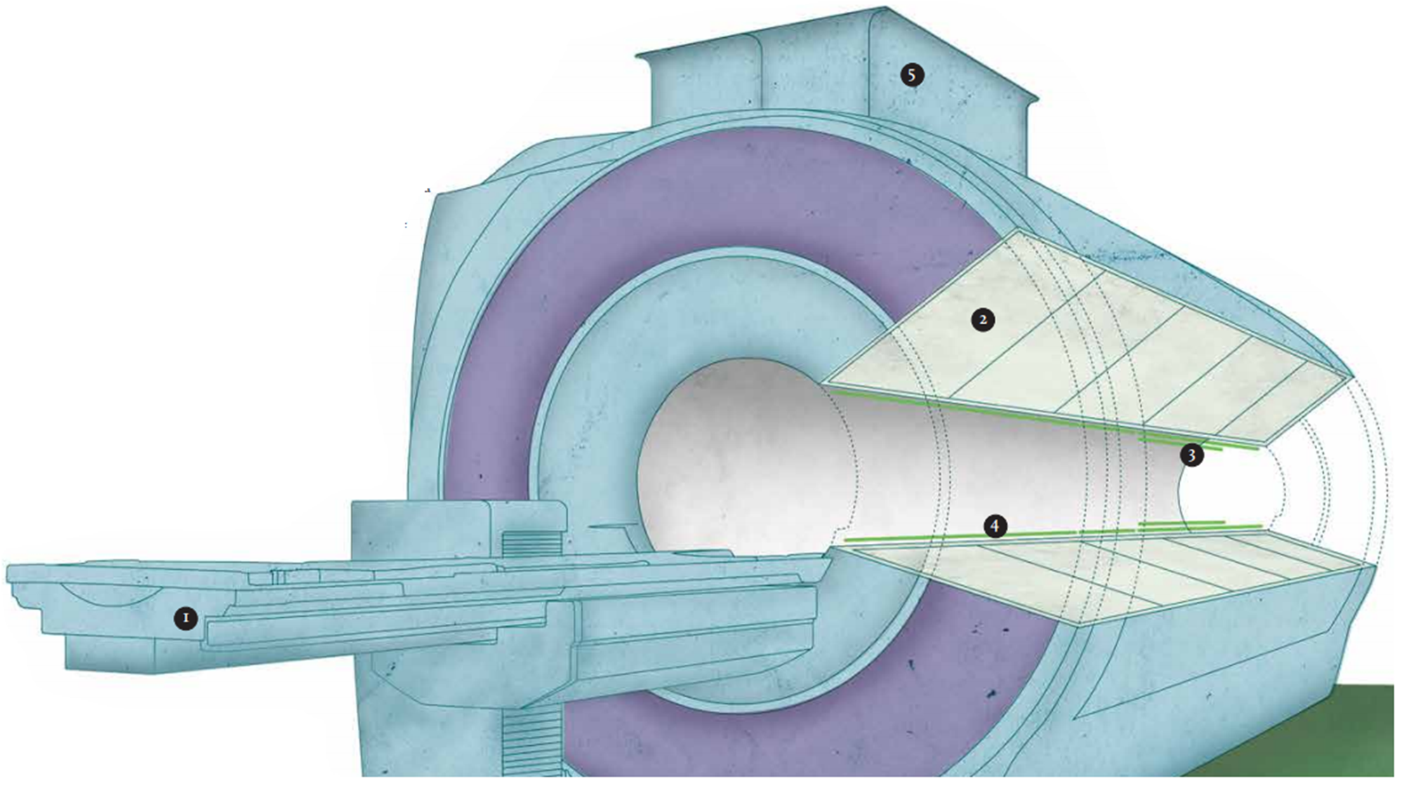
MRI components
1. Patient Platform - This is where the patient lies in the exact centre of the magnetic field. The water and fat in our bodies means that we’re full of hydrogen atoms, and it’s these that MRI targets.
2. Magnet - Magnets commonly used in MRI scanners today are 0.5–3.0 tesla strong, but some research uses magnets as powerful as 60 T (a fridge magnet is about 0.005 T).
3. Radio frequency coil - This directs a radio wave at the area you want to examine and detects the waves bounced back.
MRI Scans
Static structure of the brain
Hydrogen atoms in different tissues (such as fat and water) have different relaxation times and can be identified separately.
The lower the water content of an area, the fewer hydrogen atoms there will be emitting signals.
The weaker the signal, the darker the area appears on the scan.
The result is shades of grey: fat is quite light, but bone is dark.
functional magnetic resonance imaging (FMRI)
Not just structure, but function
Based on the same technique as MRI (using magnets and radio waves)
But interested in blood flow in the brain
Blood contains hemoglobin which contains iron (which is magnetic)
fMRI
Deoxygenated blood is affected by a magnetic field differently than oxygenated blood.
When neurons are active, they burn energy. This is automatically replenished via oxygen carried by hemoglobin in the blood stream
Active parts of the brain contain more oxygen-rich blood
By measuring the BOLD (Blood Oxygen Level Dependent) response in the MRI scanner, we can work out which parts of the brain were active recently.
How fMRI works
oxygen is delivered to neurons by haemoglobin in capillary red blood cells
more haemoglobin in areas of the brain when it needs to replenish the oxygen used by active neurons
BOLD response
Blood Oxygen Level Dependent
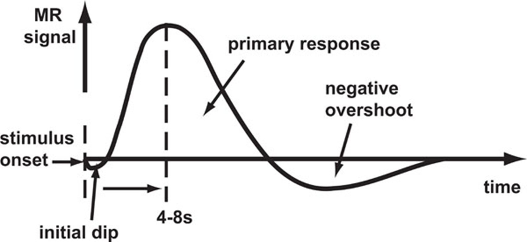
we’re most interested in the biggest part of the graph
advantages of fMRI:
excellent spatial resolution (get structural data within same session)
non-invasive
tells us which parts of the brain are used in tasks
Disadvantages of fMRI
poor temporal resolution
the experience is noisy, have to stay very still, claustrophobic
BOLD isn’t a direct measure of activity, and care should be taking interpreting it
expensive
can’t have any metal based equipment for stimulus presentation
What can fMRI tell us about cognition?
How does culture affect face processing?
Adams et al. 2010
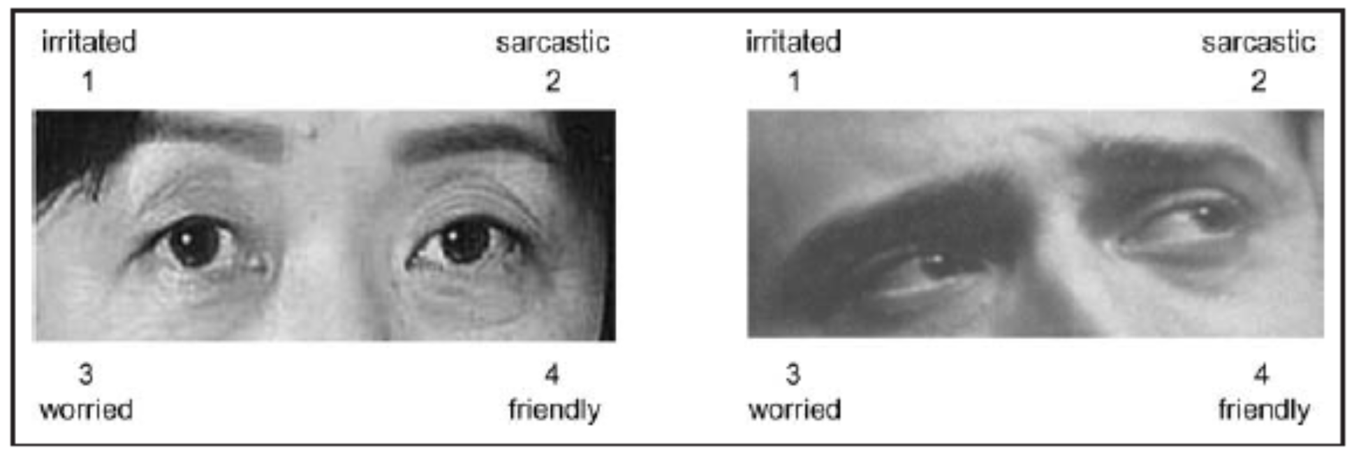
reading the mind in the eyes, what is the expression or feeling of the eyes? they’re only shown the eyes (and eyebrows) and no other feature
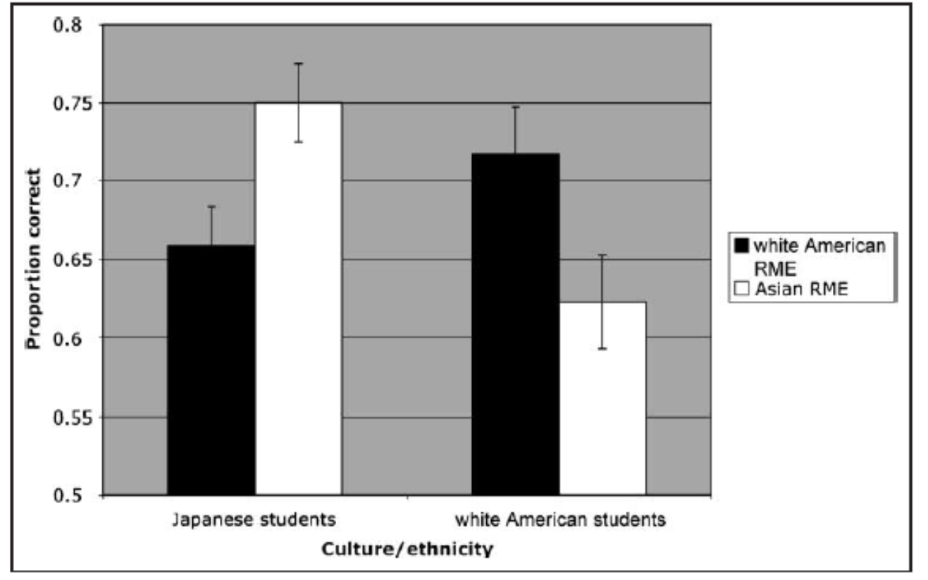
behavioural results: among Japanese students, the proportion of correct responses was lower with white Americans than Asians, this is the own-race effect, Americans also showed this effect
fMRI results: posterior Superior Temporal Sulcus (pSTS)
Why focus on this one area? It has been shown to be sensitive to things understood to be important for comprehension of human intention such as to lip reading, mouth movement, body movement, eye gaze, hand action.
Single-cell recordings in monkeys
Neurophysiological and neuroimaging studies in humans
Sensitive to implied motion and more generally to stimuli that signal the actions of another individual (Allison et al., 2000).
Conclusion: pSTS activity reflects high-level reasoning and action interpretation
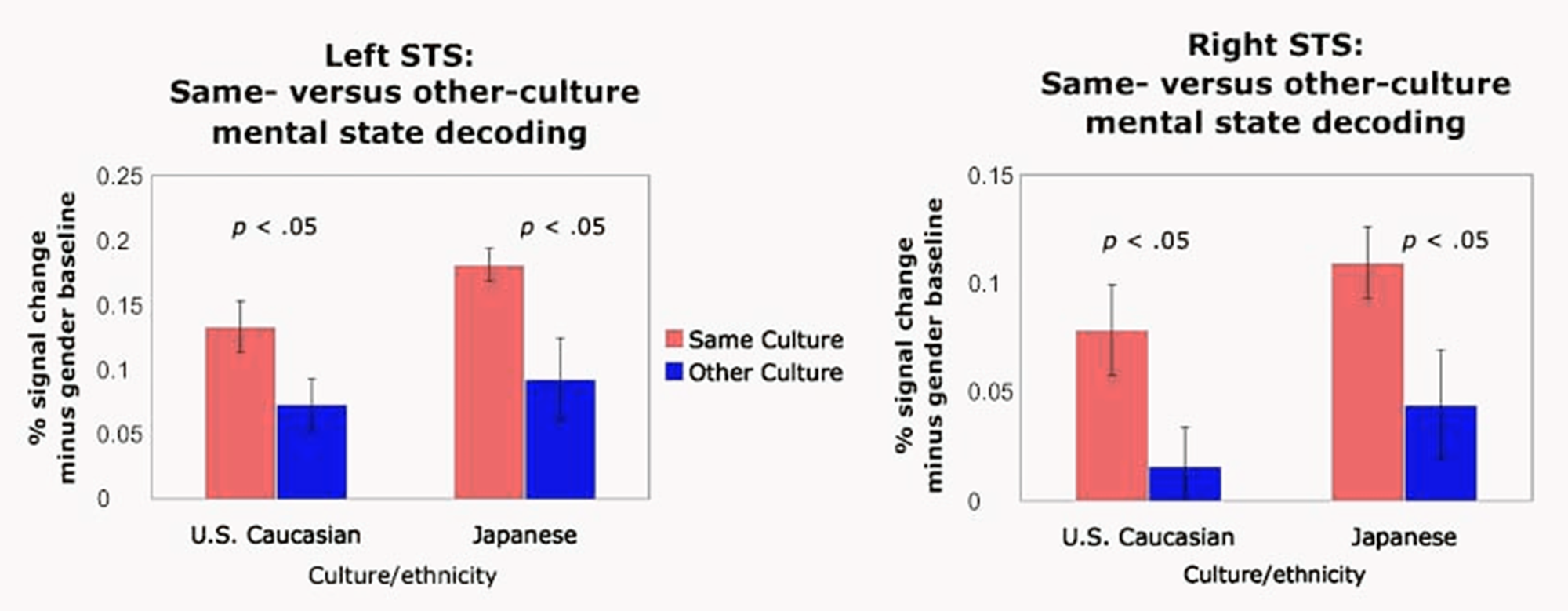
in the behavioural results there was an own-race effect, and within the fMRI results (all cases with both sides of the brain and both races) there was a more activity of the pSTS when comprehending their own race (own-race effect)
Adams et al. 2010 conclusions
Behavioural findings:
Both Japanese and white American participants showed own-race effect when decoding mental state from the eyes
fMRI findings:
Performance pattern was mirrored by culturally tuned neural activity in the bilateral pSTS
Overall, there was a high-level of consistency in neural responses between Japanese and white American participants when decoding mental states from the eyes
So what did fMRI tell us?
Adams et al. 2010
Own race bias in the ability to decode mental states can be seen via consistent brain pSTS activity
pSTS activity = high-level reasoning and action interpretation
Own race bias in mental state decoding is associated with different abilities in reasoning and interpreting stimuli for same versus other race (not, for example, low-level perception)
Discussion
The fMRI records activity from all of the brain. Why focus only on a single (or a small number of) specific area(s)?
The researchers compared activity to a “gender discrimination” task
they gave pps two kinds of tasks, including what we have already talked about and to discriminate what is the gender of the person you are seeing, when calculating the activity they subtracted the gender related activity and looked at the difference between these two tasks- they do this because neuroscience is sensitive to alpha inflation, where a result comes out significant just because (95% sig level, so 100 regression ran = approx 5 random significant results), no fMRI study compares the activity to nothing, all stimuli results in activation of parts of the brain, so what they wanted to do was create the same stimuli that activates the same areas regardless if they are Japanese or American, so by comparing also to gender we remove activity that we don’t care about (somehow)
Discussion
the fMRI records activity from all of the brain. Why focus only on a single (or a small number of) specific areas?
Neuroscience is sensitive to “alpha inflation” due to multiple comparisons. If we compare numerous areas, we’ll eventually find a statistical difference due to type-1 error.
The researchers compared activity to a “gender discrimination” task. Why?
Any stimulus results in a lot of activation that is unrelated to the target process.
Gender discrimination is unrelated to people’s intent. By comparing both tasks, we ensure that the difference doesn’t stem from low-level differences.
Every task needs its own baseline (which activity do you want to remove?).
Using cognitive neuroscience to study individual differences
Do autistic individuals and neurotypical individuals process emotional images in the same way?
Kane et al. (2016)- in the first task, you need to process the person’s emotions in order to get the answer right, but in the second condition you can see an emotion but you are not directly asked anything regarding it. After this, they compared the performance between autistic and nuerotypical participants. They preform exactly the same. But they were interested in looking at the activity in the brain whilst doing the task, rather than if they go the answers correct.

Results
1. No behavioural differences in accuracy or RT
pSTS and mPFC results, the areas strongly activated, the areas for the explicit tasks in autistic individuals showed almost identical activity to neurotypical individuals, however, when liking at the implicit area the overall activity is lower for autistic individuals
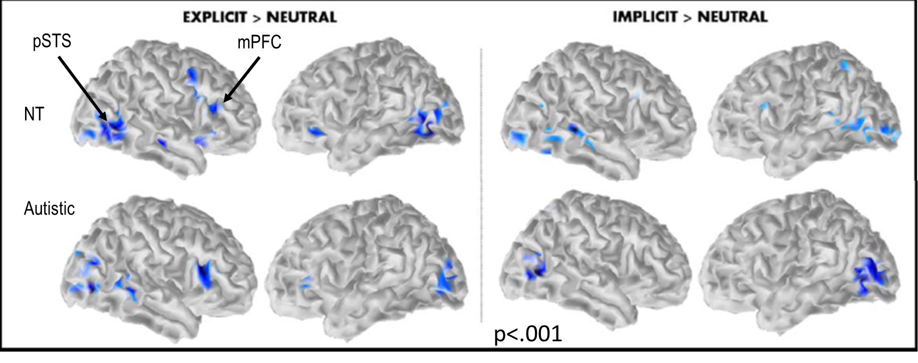
Kana et al. 2016- main results
Both the autistic and neurotypical participants activated a similar network of brain regions when explicitly asked to identify emotional expressions
Autistic participants had reduced activation in the MPFC and pSTS relative to neurotypical participants for implicit emotion processing but not during explicit emotion processing
So what did fMRI tell us?
Autistic people recruit task-specific brain regions for processing emotions when explicitly asked to do so
Implication: Perhaps more explicit instructions for autistic individuals when emotion processing is required would be helpful.
Increased activity in the identified brain regions in neurotypical people may indicate higher likelihood of automatic emotion processing.
Implication: Maybe autistic people don’t have an inherent difficulty recognising emotions but are less likely to do so unprompted or whilst engaging in other cognitively demanding tasks.
A second-person cognitive neuroscience & hyperscanning
The social brain network- undergoes significant development during adolescence, no specific area is pin pointed, it rather works as a social network and multiple areas are implicated in social activity and processing
Third person Vs second person
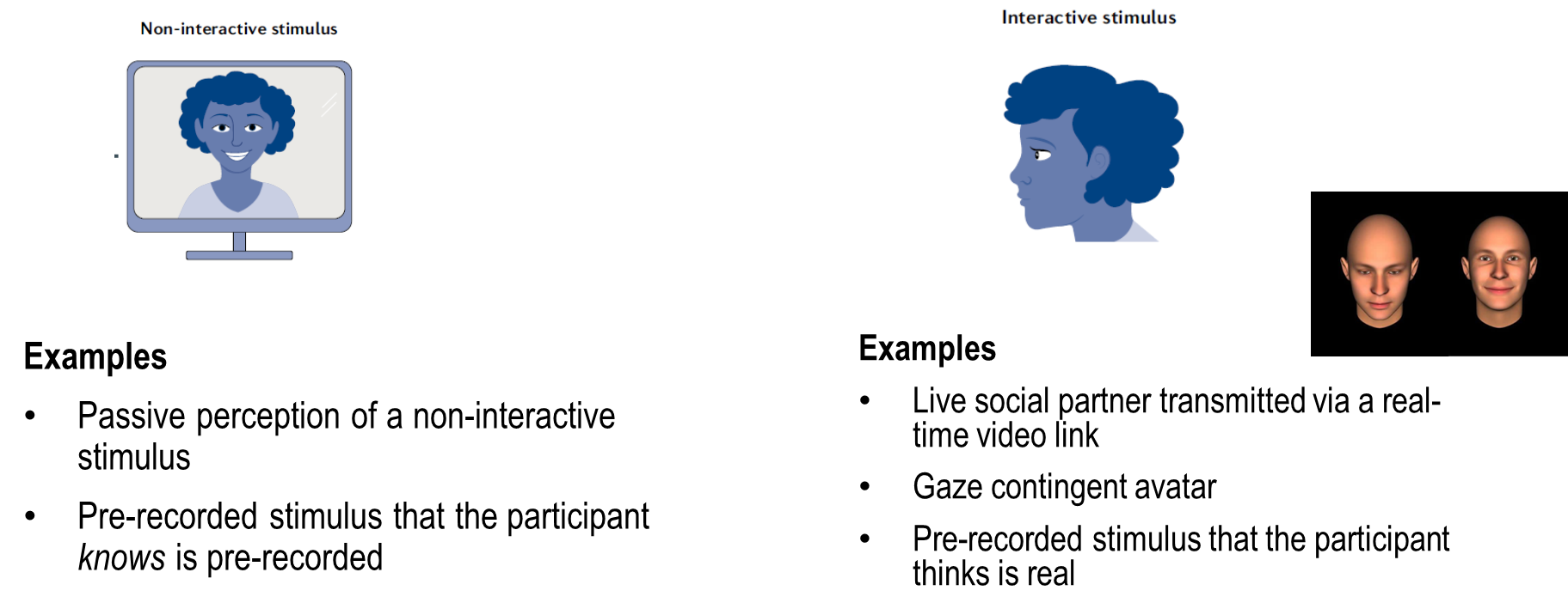
third person is standard when looking at activity in the brain
Hyperscanning- dual brain studies, they respond to the same stimuli so you can compare the activity within both people’s brains more accurately, both people get scanned at the same time
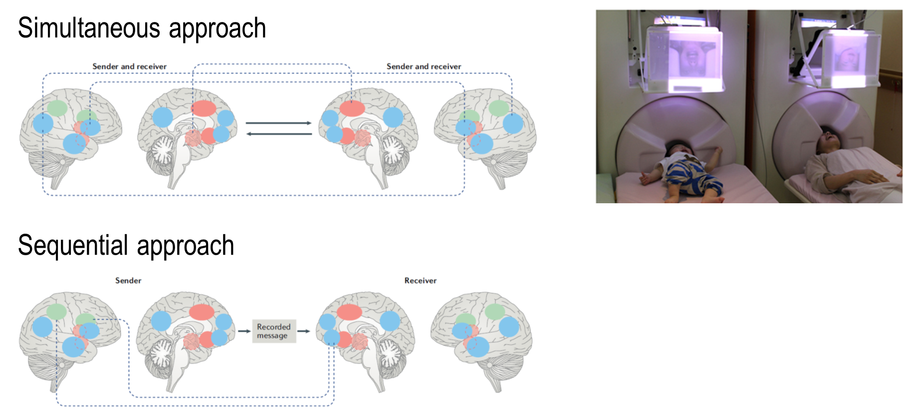
-sequential approach
one person goes into scanning, gets their activity recorded, then shows it to the second person so they respond to the activity as well as the reaction of the first person
Flow of affective information between communicating brains
Anders et al (2011)
participants: 6 heterosexual romantic couples
women’s task:
express the following emotions one by one:
joy
anger
fear
disgust
sadness
men’s task:
watch your partner’s face and “try to feel with her”
Results
The researcher examined whether they could predict (classification accuracy) the brain activity of the men based on the brain activity of their partner.
Some area were co-activated, resulting in high classification accuracy.
Classification accuracy was highest when the men viewed their partners’ video rather than other videos.

Anders et al. 2011 conclusions
The activity in one person’s brain can influence the activity in another person’s brain
Information can be successfully transferred
Information is better transferred between partners
This paradigm provides a tool that will open a new perspective in social neuroscience
which brain areas are involved in special cognition?
What can fMRI tell us?
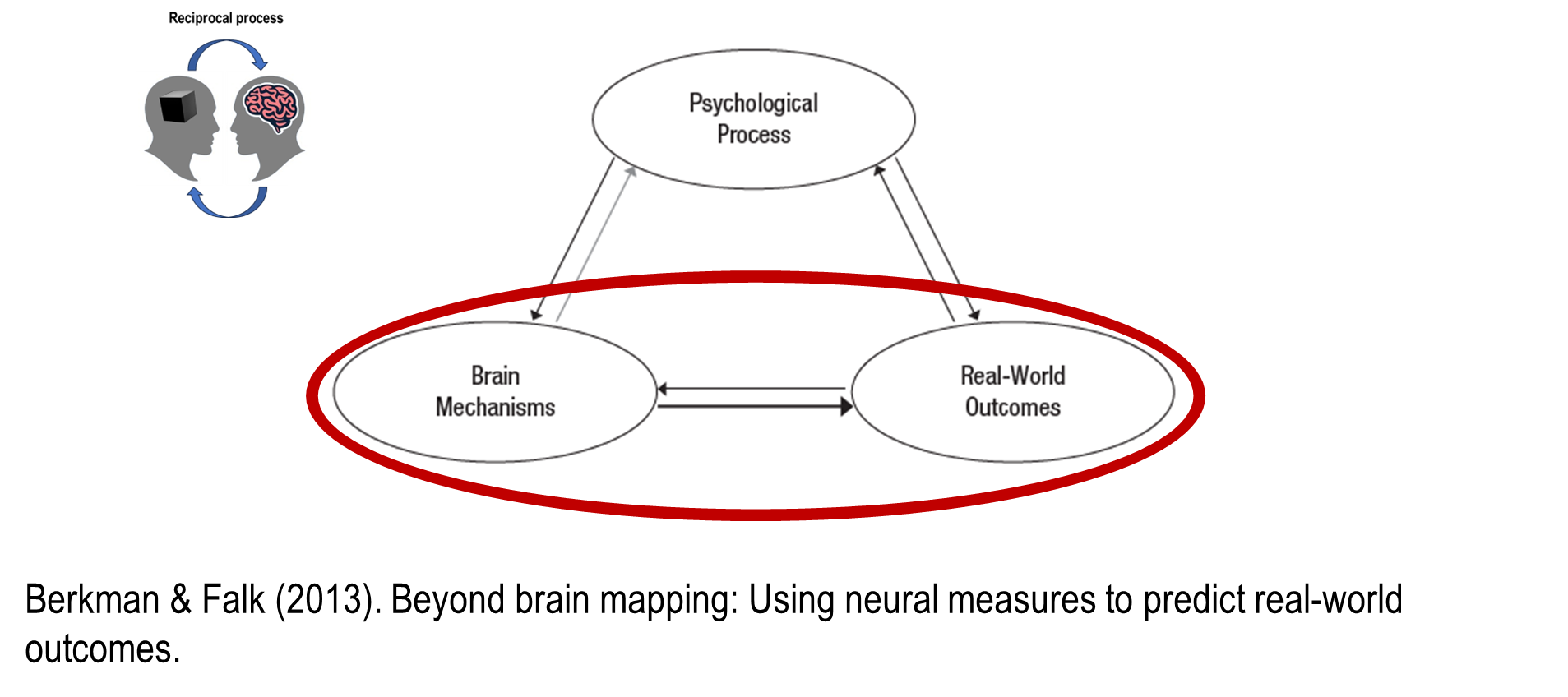
the traditional neuroscience approach is using knowledge about psychological processing says things about brain mechanisms and vice versa
how do brain mechanisms help us predict real-world outcomes?
brain-as-predictor approach
Brain-as-predictor framework- how does it work?
Step 1: Generate a hypothesis of which brain regions are involved in a particular cognitive process of interest
Step 2: Collect data in which neural activation in hypothesised regions is measured and data on behavioural outcomes is recorded
Step 3: Test whether the activity in brain regions specified in Step 1 predicts behaviour outcomes measured in Step 2
Take-home messages:
fMRI can tell us about…
The brain activity that underlies mental processes
fMRI can be used to…
Study mental processes and what affects them
Make predictions about behaviour outside of the laboratory
Reading
Abstract
Psychologists have traditionally used self-report measures and performance on laboratory tasks to achieve this end. However, these measures are limited in their ability to predict behaviour in certain contexts.
brief overview of brain-as-predictor approach
This integrates traditional neuroimaging methods with measures of behavioural outcomes that extend beyond the immediate experimental session.
Previously, neuroimaging experiments focused on understanding basic psychological processes directly observed in the laboratory. However, brain measures can predict outcomes (e.g., purchasing decisions, clinical outcomes) over longer timescales in ways that go beyond what was possible with self-report data alone.
This approach can be used to reveal the connections between neural activity in laboratory contexts and longer-term, ecologically valid outcomes.
Introduction
Less than a quarter of pilot episodes become full shows, and the majority that do are cancelled within the first few years. Why are viewers and network executives so poor at judging which shows people will watch?
One answer is that the mental processes that give rise to behaviours, such as tuning in to a TV show, are not always accessible to conscious awareness.
A similar argument can be made about why it is so hard to predict the success of health interventions or efforts to get people to save for retirement: People are notoriously limited in their ability to consciously identify why they do what they do. However, the mental processes underlying behaviour are nonetheless represented in the brain.
What does the reading article argue?
It argues that knowledge gained from decades of work in cognitive neuroscience about the mapping between mental process and brain function can be leveraged to predict meaningful outcomes beyond the laboratory.
We found that viewers’ brain activation while watching a set of commercials in a “neural focus group” predicted the success of the commercials in the media markets where they were aired, and did so better than viewers’ reports of the ads’ effectiveness.
This is one example of a more general brain-as-predictor approach, which treats neural measures (e.g., activation, structure, connectivity) as independent variables in models that predict longitudinal outcomes as dependent variables.
The adoption of the brain-as-predictor approach represents a paradigm shift for research in neuroscience, complementing traditional brain-mapping studies in which psychological processes are manipulated and the resulting neural activity is observed as the dependent measure.
This approach also builds on research on the neural bases of individual differences in personality and responsiveness to clinical treatments. The brain-as-predictor approach differs from approaches taken in this earlier work, however, in the level of specificity of the hypothesized neural systems and targeted outcomes.
Describe this:
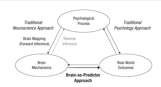
The brain-as-predictor approach.
Traditionally, psychologists have been interested in mapping the relationship between psychological processes (e.g., cognitions, emotions) and real-world outcomes (e.g., health behaviours, discrimination). Whereas, neuroscientists have used neuroimaging tools to map the relationship between psychological process and brain mechanisms. The brain-as-predictor approach integrates these methods by using brain systems that have been linked to specific psychological processes to predict meaningful outcomes beyond the laboratory.
It offers new ways to explain unaccounted variance in behavioural outcomes and to test whether hypothesized psychological processes (via their neural associates) are predictive of those outcomes.
Bidirectional arrows emphasize that each construct is likely to affect the others and that the brain-as-predictor approach complements existing methods for studying the other relationships shown.
The arrows indicate conceptual relationships between independent and dependent variables rather than causality; manipulation of brain function (e.g., using transcranial magnetic stimulation or in clinical lesion studies) is necessary in order to establish causal relationships between brain measures and behaviour.
Potential for theoretical and applied advances
In the brain-as-predictor framework, the brain is viewed as an additional window into psychological processes that compliments other measures, such as self-reports and other biological measures; its specific role in determining behaviour can be examined in the context of those other measures to advance theory and application.
For example, in our work predicting the success of ad campaigns, we have used neural measures as an alternative way to study psychological processes that unfold during exposure to ads and that are difficult to capture using other methods.
Our results illustrate how neuroimaging can add to the understanding of social influence and can also be applied practically to the problem of designing more efficient health campaigns.
The brain-as-predictor approach also improves the ecological validity of neuroimaging experiments by connecting neural measures directly to outcomes beyond the laboratory.
For example, a study that predicts outcomes for the treatment of problem gambling on the basis of activation in regions involved in self-control would provide confirmatory support for the involvement of those regions in self-control and would also suggest ways to improve interventions. In this way, the brain-as-predictor approach broadens our ability to test theory and facilitates the translation of basic neuroscience results.
examples of the brain-as-predictor approach
Beyond informing psychological theory, the wide range of outcomes that can be brought into the brain-as-predictor framework represents a compelling new way for neuroscience to interface with other fields, such as public health, political science, marketing, sociology, and communication studies. The following sections highlight the advantages of the brain as-predictor framework in several areas in psychology.
cognition
An early study employing the brain-as-predictor approach pre dicted intelligence from brain measures. A model of intelligence based on brain structure and function measured separately in one group explained 45% of the vari ance in intelligence in a second, independent sample. Identify ing regions of interest (ROIs) in advance and examining their involvement in a separate sample moves the focus of research from brain mapping to testing the predictive validity of mea sures from specified constellations of brain regions. Cognitive neuroscientists have also used neural signals to predict the trajectories of skill acquisition and age-related cognitive decline and to understand the overvaluation of short-term benefits relative to long-term costs
health
The brain-as-predictor approach has also been applied iteratively to improve the design and selection of health communications and uncover the neural foundations of their effectiveness.
For instance, our work predicting the population-level success of ad campaigns built on previous studies from our lab. The earlier work identified brain regions associated with persuasion and used activity in those regions to predict increases in sun screen use and reductions in smoking in groups exposed to relevant public service announcements.
Throughout this research, we used a test-validate procedure, in which brain-mapping results in one domain (e.g., regions associated with individual behavior change) were tested as predictors in subsequent, conceptually related studies (e.g., studies investigating individual behavior change in a different domain or the population-level success of different ad campaigns).
Samples used in brain-as-predictor approach:
The use of different participant samples to identify and subsequently interrogate ROIs offers the advantage of conceptual replication across samples. It can also be used in a “neural focus group” framework, in which neural activation in a small group predicts outcomes in a larger population.
However, we note that independent samples of participants are not always necessary; ROIs may also be defined using an independent psychological localizer task, which isolates the brain regions associated with a process of interest for each subject within a single group of subjects.
For example, Chua et al used one task to identify neural activity associated with self-related processing in a sample of smokers, and then extracted activity within those regions during a tailored health-message intervention to predict subsequent quitting within the same group.
How can the brain-as-predictor approach can also be used to examine relationships between basic social, cognitive, and affective processes and health-relevant outcomes to reveal how these psychological processes get “under the skin.”
For example, neural activity during an emotion-regulation task predicted daily patterns of release of the stress hormone corti sol in older adults. Another study showed that activation within the brain’s reward system in response to appetitive foods and erotica predicted changes in body mass index and risky sexual behaviour (respectively and separately) across 6 months. These studies highlight the potential of brain-as-predictor approaches to link multiple levels of analysis, to examine outcomes beyond observed behaviour, such as neuroendocrine or immune markers, and to improve prediction.
Economic decisions
Neuroimaging data have also been used to predict consumer choices and donation behaviour outside of the scanner on the basis of neural responses during protocols in which participants were exposed to stimuli without being asked to judge them. These data suggest that neural signals encode information that predicts sub sequent behaviour even in the absence of a specific choice or evaluation. Consumer-choice studies have also suggested that neural data can predict broader population responses.
Clinical, neurological and addiction outcomes
Neural activity during basic laboratory tasks also predicts clinically relevant outcomes. Examples include using brain activation prospectively to predict responsiveness to therapy, risk for depression, medical outcomes, and relapse in illicit drug users.
Along these lines, we used the brain-as-predictor approach to under stand the mechanisms that lead to successful regulation of cravings in the context of smoking cessation. Neural activation in a self-control net work during a self-control task administered at baseline moderated the subsequent hour-to-hour relationship between craving and smoking in the early weeks of quitting.
These data provide support for the hypothesis that breaking the link between cravings and smoking involves self-control, which is instantiated in specific, common networks in the brain. This study illustrates the integration of neural measures with experience-sampling data and the deployment of multilevel models containing both kinds of data.
This logic can also be extended to incorporate neural data from hypothesized ROIs into structural-equation models, nonparametric models, and other statistical models as they are developed
A guide to the brain-as-predictor approach
procedure:
The studies reviewed here, and others like them, provide clues about how to apply a brain-as-predictor approach to study a range of outcomes. However, this approach has not yet been formally defined and differentiated from others. We suggest the following three-step approach.
First, hypothesis generation, in which candidate brain regions or networks are identified and a priori ROIs defined using any means for identifying neural regions associated with the hypothesized psychological constructs. In the study described in our opening example, we hypothesized on the basis of prior results that neural activity in the brain’s medial prefrontal cortex would predict the success of ad campaigns, meta-analyses on the process of interest, or independent tasks within the same sample analyzed with traditional univariate methods or newer multivariate or machine-learning techniques can also be used to identify ROIs.
Second, data collection, in which neural activation in hypothesized regions is measured and data on longitudinal outcomes are subsequently collected using methods including but not limited to experience sampling, single-session follow ups, or behavioural observation.
Third, hypothesis testing, in which the validity of the hypothesized regions to predict longitudinal outcomes is tested using a predictive statistical model that specifies brain measures as predictors
convergent and discriminant validity
A critical consideration is whether neural data contain reliable, predictive information beyond what could be obtained otherwise. Demonstration of discriminant validity requires gathering not only brain data in the second step but also other data that might be predictive (e.g., self-report, behavioural, or endocrine data) and assessing whether the neural data provide additional predictive power. For instance, in the earlier example in which neural responses to health communications predicted subsequent reductions in smoking, neural activity doubled the amount of variance in behaviour change explained, relative to a model containing self-report measures alone.
In addition, brain and self-report measures overlap in the variance that they explain, which may provide insight into the processes contributing to the predictive relationship (i.e., convergent validity). For example, if the relationship between brain activity in the ROIs and the behavioural outcome were mediated by self-reports of motivation to quit, it would suggest a potential role for this network in motivation and point to a new intervention target.
In both cases, psychometric reliability of neuroimaging data is a critical consideration when comparing brain measures with other types of variables. Neuroimaging data can have high test retest reliability depending on factors including the hardware used to obtain the data, the length of the test-retest interval, and the complexity of the cognitive processes tested. However, the reliability of neuroimaging data, like that of any other kind of data, must be evaluated in light of the study design and other available measures.
predictor selection
In the brain-as-predictor approach, unlike traditional neuroimaging approaches that generate whole-brain maps as output, the neural predictor must be specified in advance. Neural predictors presumed to be involved in key mental processes are chosen on the basis of psychological theory and prior brain-mapping results. Because predictor regions represent the operationalization of a mental process that will be used for theory testing, their careful selection is critical, akin to selecting a behavioural task or self-report measure to tap a con struct. In this sense, the brain-as-predictor approach relies on the same scientific logic as any other predictive approach in psychology (e.g., predicting behaviour change from intention) but with a different independent variable.
iterative process
The brain-as-predictor approach is only one part of an iterative cycle of exploratory and confirmatory hypothesis testing designed to advance theory: Brain-mapping and brain as-predictor approaches can be used together to triangulate the relationships among neural, mental process, and behavioural variables. Traditionally, studies have identified candidate regions for a given psychological process (e.g., self-control); brain-as-predictor studies employ confirmatory predictive analyses that test the involvement of ROIs in that process and identify conditions under which brain activity does and does not predict the outcome (e.g., breaking the link between craving and smoking). Brain-as-predictor logic can also be used in neuroimaging studies that employ additional tools designed to facilitate causal inference and in experiments that manipulate treatment condition and observe subsequent outcomes, with neural function as a hypothesized mediator.
conclusion
Traditional neuroscientific results can be leveraged to uncover unique predictive brain-behaviour connections. Though the brain-as-predictor approach is being used more often, researchers rarely call attention to whether neural measures are treated as predictor or outcome variables. Such acknowledgment is essential from a theory-building and -testing perspective. For example, meta-analyses of both brain-as-predictor and brain mapping (brain-as-outcome) results would be best served if studies were easily classified as one or the other. In addition, brain-as-predictor “best practices” will emerge more efficiently if researchers can easily track their use in the published literature. Further, the applicability of the approach to fields outside of neuroscience (e.g., medicine, political science) will be most apparent when the predictive capacity of brain activity above and beyond other measures is explicitly quantified. We have articulated this brain-as-predictor approach using examples from functional neuroimaging, but the same principles can apply to brain structure, peripheral-nervous-system function, genes, and other biological measures. Future extensions will allow scientists to use neural markers as longitudinal predictors of diverse outcomes across a range of fields. Extending the reach of neuroscientific meth ods beyond exploratory brain mapping allows for stronger theory testing, sheds light on fundamental neuroscientific questions, and enables prospective prediction of outcomes inaccessible by other means