Unit 5: Cell Division Study Guide
Chromosomes and Karyotypes
Introduction
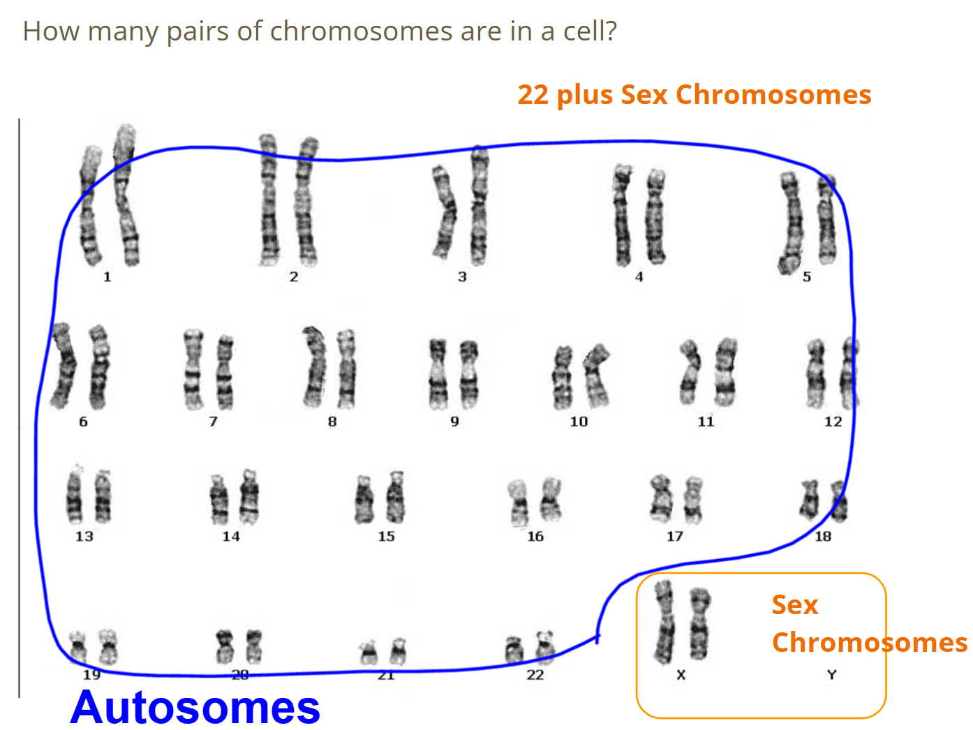
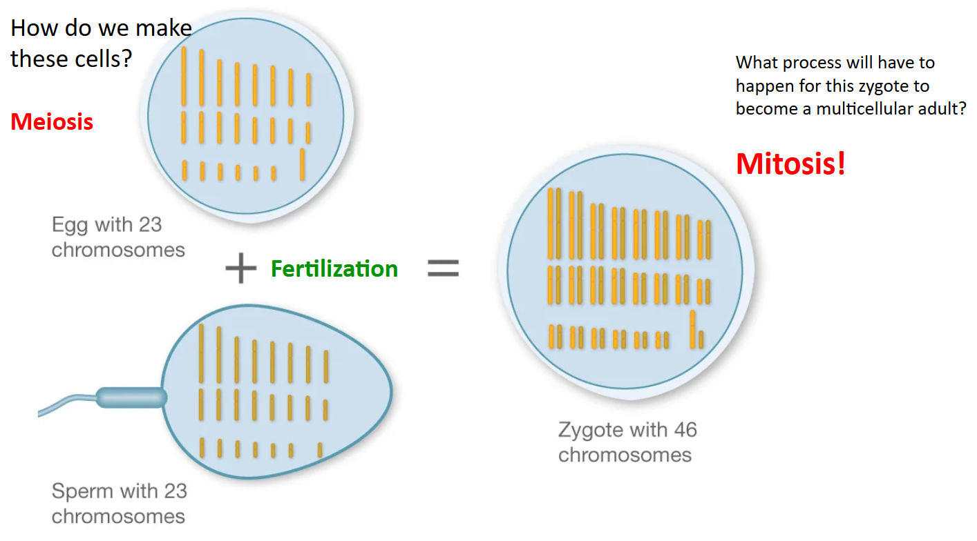
Humans have 46 chromosomes
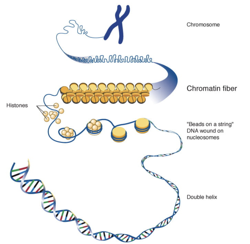
Chromosomes are made out of DNA and Histone Proteins
Problems with DNA
DNA is difficult to move around without getting tangled
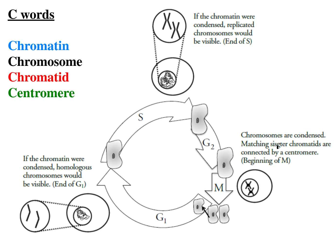
Need it long and stretched out to copy it or to make proteins: Chromatin
Chromatin = Everything except cell division
Need it bundled (condensed) to move it around: Chromosome
Chromosome = Cell Division
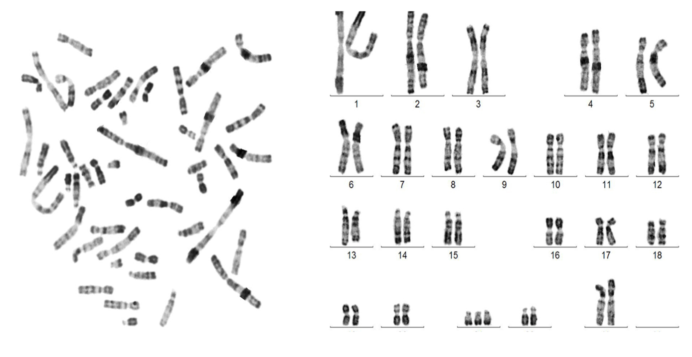
Karyotypes
Karyotype: A visual display of an organism’s chromosomes
Allows us to learn about chromosome abnormalities
Too few, too many, too short, too long
Allows us to determine biological sex
Making a karyotype
Take pictures of the chromosomes in a cell (when they’re in chromosome form) and arrange them by size
Largest pair is labeled chromosome #1
Variations in Sex Chromosomes
Some people have a chromosome pattern other than the usual XY or XX
Could have XO or XXY
Internal and external sex organs can be either male or female but they may not go through full physical development at puberty
Types of Intersex Variations
Klinefelter Syndrome
Individual assigned male at birth is born with an extra X chromosome (XXY)
Do not produce the typical level of testosterone (a hormone responsible for testes and body hair)
Turner Syndrome
Individual identified as female at birth is missing an X chromosome (XO)
Infertile and height is shorter than average
The Cell Cycle
Introduction
DNA replication has to happen because each new cell that is produced needs a complete and accurate copy of the DNA or it will likely die
DNA replication occurs any time when a cell or nucleus divides
Causes of Cell Division
Growth
Repair
Immune System
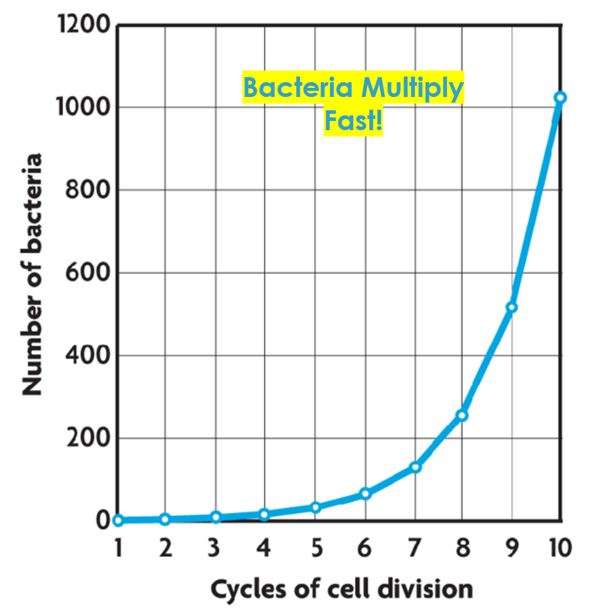
Binary Fission
Cell Division in Prokaryotes (Bacteria and Archaea)
Asexual Reproduction
Results in 2 genetically identical daughter cells
Eukaryotes
Mitosis: division of the nucleus
Growth and Repair
Meiosis: division of the cytoplasm
Make gametes (egg or sperm)
Mitosis
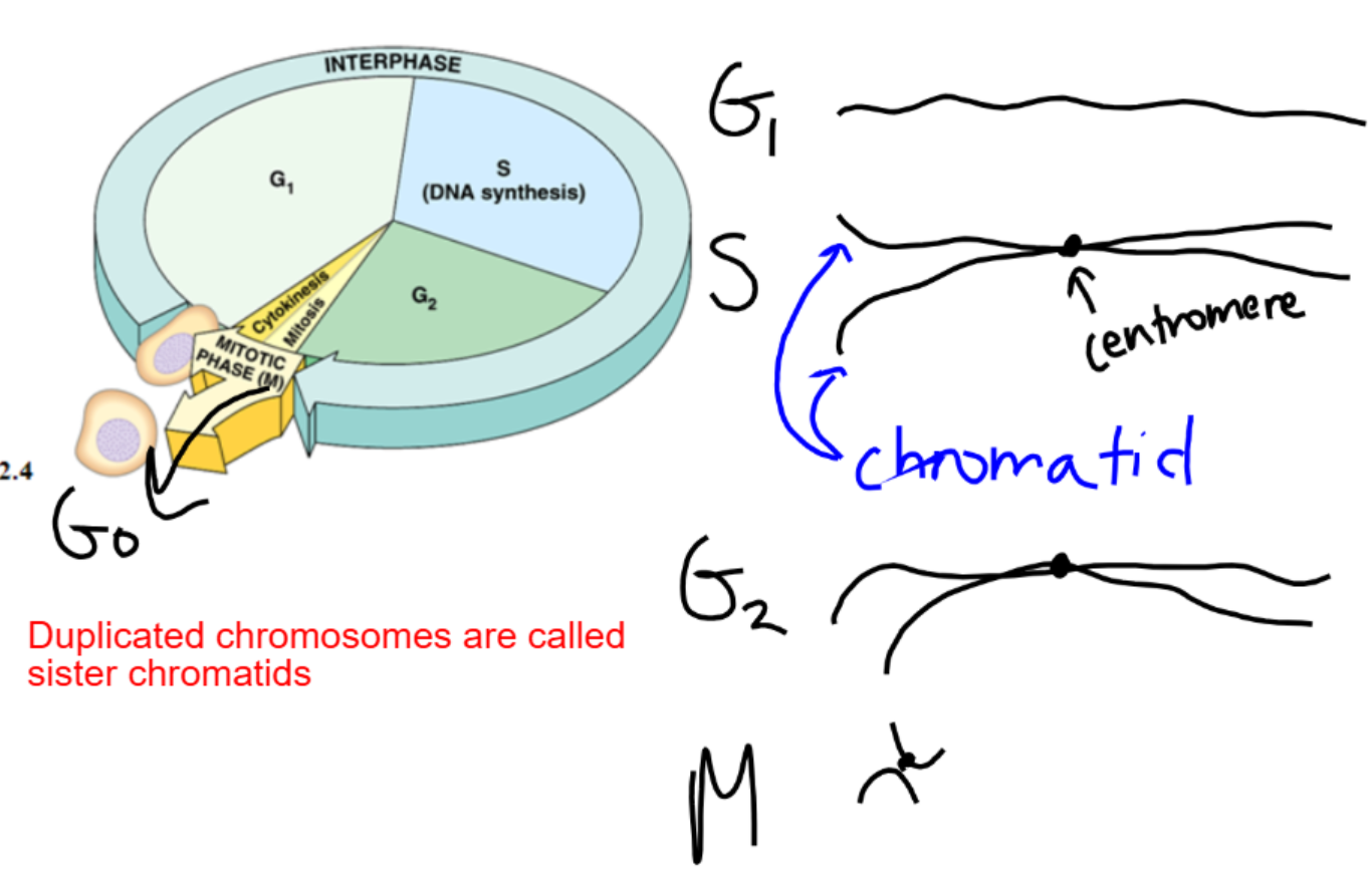
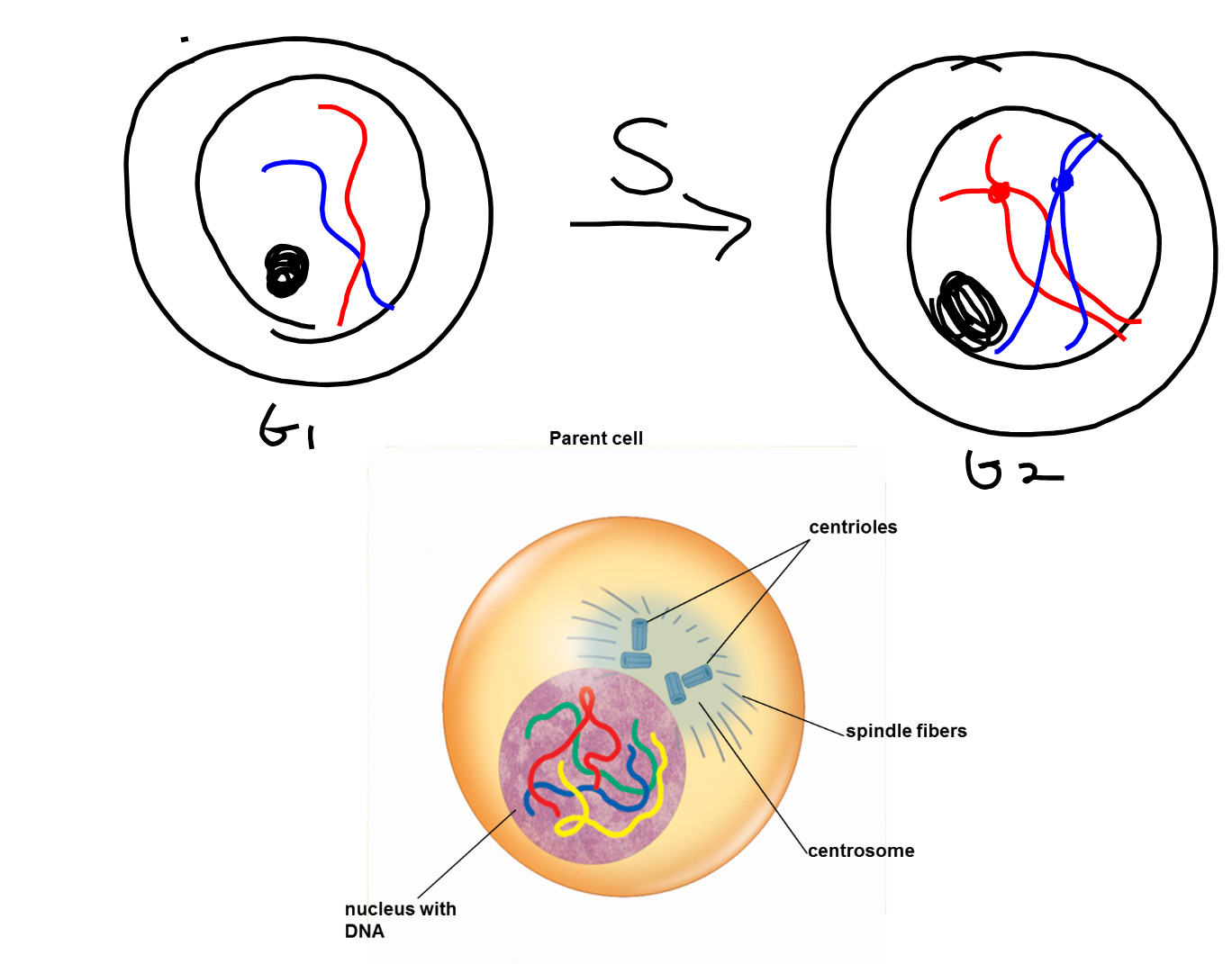
Interphase
Cell grows
Cell replicates its chromosomes
Prepares for cell division
Prophase
Chromosomes condense
Spindle fibers form
Nucleus and nucleolus disappear
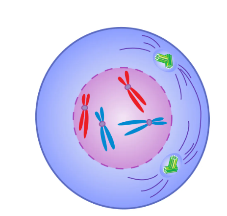
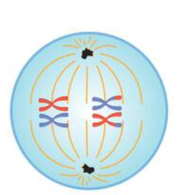
Metaphase
Chromosomes line up in the middle
Spindle fibers attach to centromeres
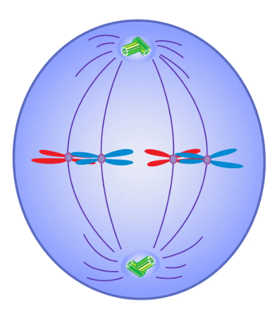
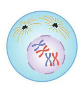
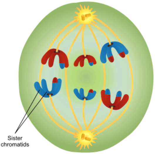
Anaphase
Centromeres split
Sister chromatids separate to opposite sides of the cell
Telophase
New nuclei form
Chromosomes begin to uncoil
Cytokinesis
Cytoplasm is split
2 genetically identical daughter cells (diploid)
Meiosis
Introduction
Meiosis is the process that produces egg and sperm (genetically unique)
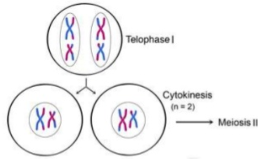
Meiosis 2 is just like mitosis
Mitosis vs. Meiosis
Mitosis
Diploid
Each cell has 2 complete sets of chromosomes (46)
2 identical diploid cells
Meiosis
Haploid
Each cell has 1 set of chromosomes (23)
4 unique haploid cells
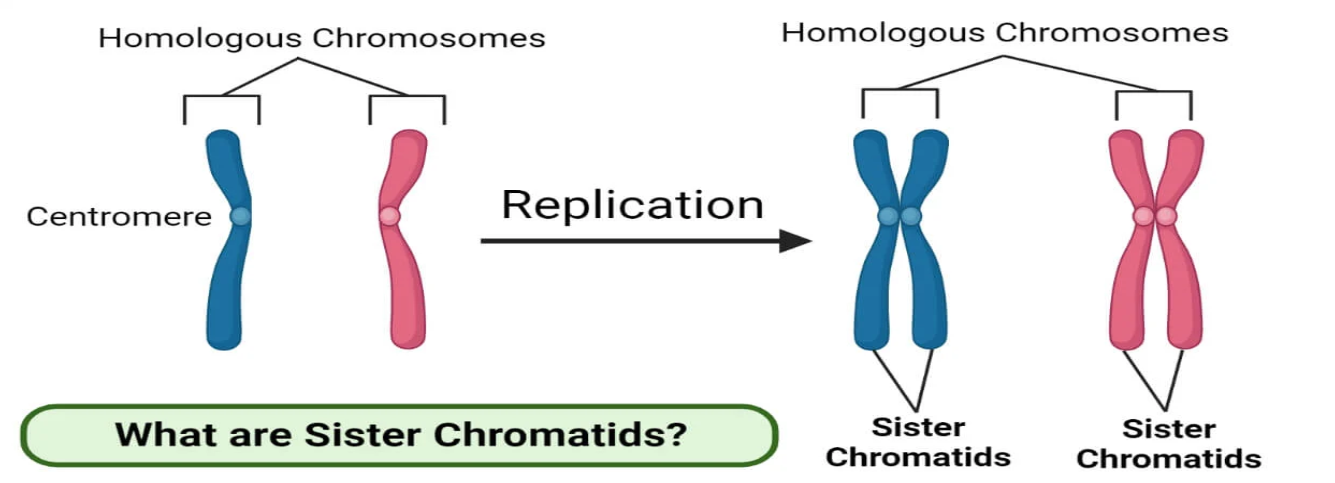
Homologous Chromosomes vs. Sister Chromatids
Homologous: Chromosomes are approximately the same size, and have the same types of genes in the same location
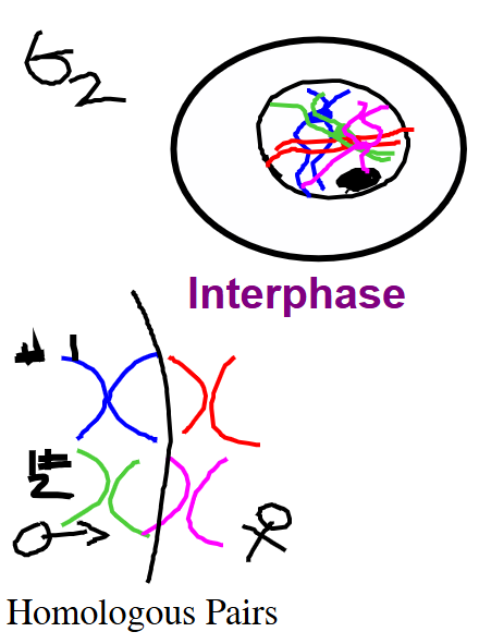
Interphase
Cell grows
Cell replicates its chromosomes
Prepares for cell division
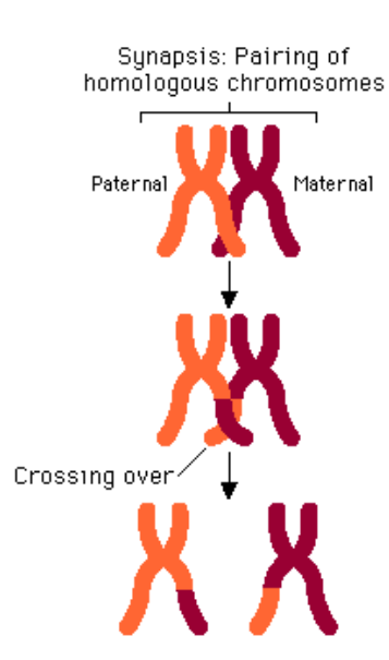
Prophase 1
Synapsis (homologues pair up)
Tetrads form
Crossing Over
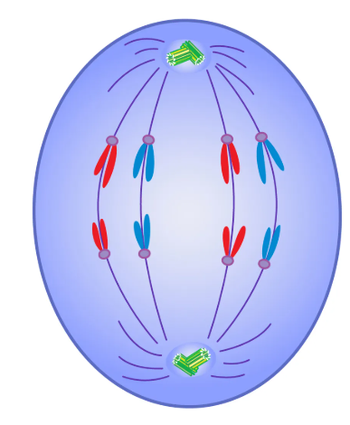
Metaphase 1
Homologous pairs line up
Each side of the equator has chromosomes from both parents
Anaphase 1
Homologous pairs split and travel to poles
Centromeres do NOT break
Independent Assortment: Random alignment of homologous chromosome pairs at metaphase plate, which leads to genetive diversity
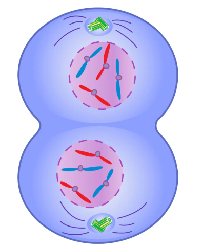
Telophase 1 and Cytokinesis
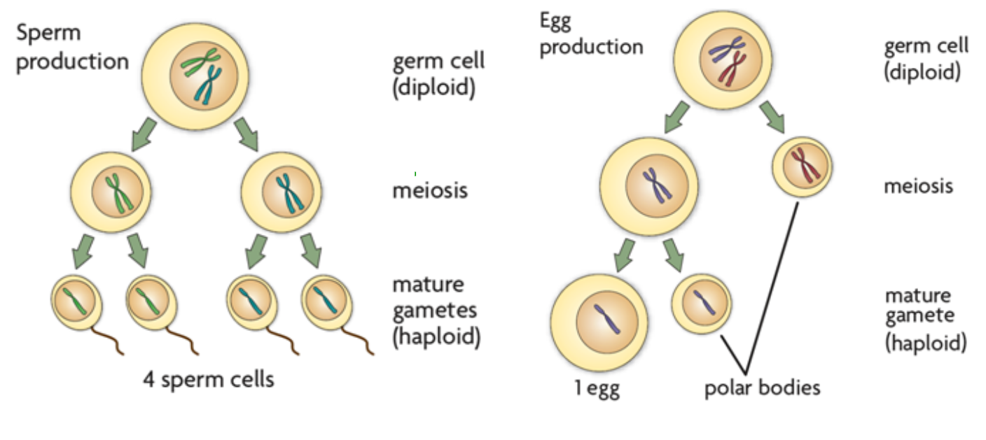
Meiosis in Male and Female
Cancer
Introduction
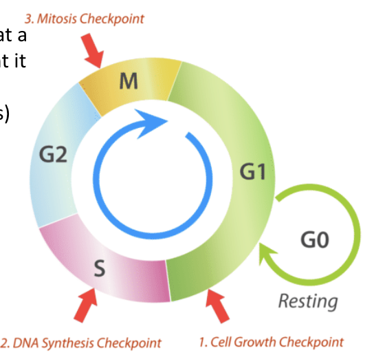
Checkpoints exist throughout the cell cycle
If a cell does not meet a certain standard at a checkpoint, it will die (apoptosis)
What is Cancer?
Cancer is uncontrolled cell division
Cancerous cells should stop dividing and/or undergo apoptosis
Mutations in genes that code for proteins that regulate the cell cycle prevent the proteins from functioning properly
Cells divide rapidly and uncontrollably, forming a mass called a tumor
Benign vs. Malignant Tumors
Benign Tumor
Tumor cells grow only locally and cannot spread by invasion or metastasis
Malignant Tumor
Cells invade neighboring tissues, enter blood vessels, and metastasize to different sites
Causes of Cancer
Genetics
Malnutrition
Environment
Lifestyle