108.5 Microscopic Examination Chapter07 2022.23
Topic: Microscopic Examination of
Urine
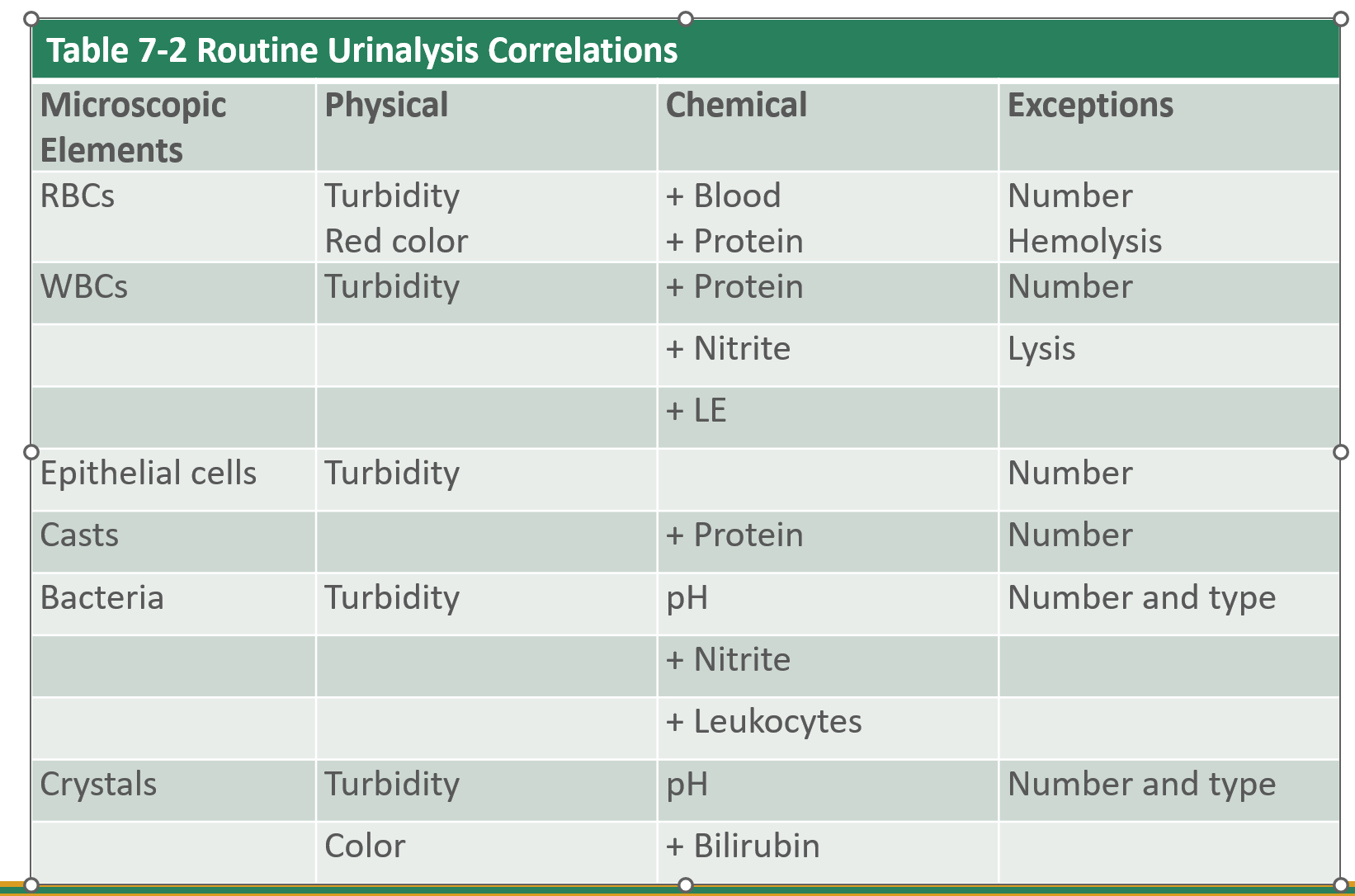
Elements in urine during a microscopic examination
Red blood cells (RBCs)
WBC
Epithelial cells
Casts
Bacteria
Yeast
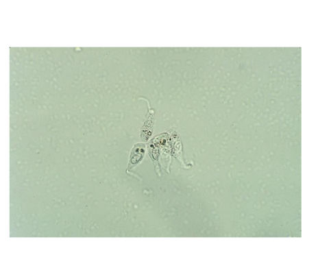
Parasites
Mucus
Spermatozoa
Crystals
Artifacts
Specimen volume/Centrifugation
12mL quantities
centrifuge 10 to 15mL of urine
Centrifuge for 5 min at 400 RCF
cap all specimens
RCF =
Sediment Preparation/ Volume of sediment examined
Volume of sediment = 0.5 to 1.0mL/ 12:1 ratio ( 12 part centrifuged / 1 part sediment)
Urine should be aspirated off rather than poured off to control amount poured off
conventional glass slide method
20uL sediment
22 × 22 glass cover slip
do not overflow cover slip ( heavier elements such as casts flow outside)
Reporting the examination
Casts = average per lpf (light power field)
RBC,WBC = average per hpf( high power field)
Epithelial cells, crystals in semiquantitative terms such as few, moderate, many, (1+, 2+. 3+, 4+) lpf or hpf
Correlating results
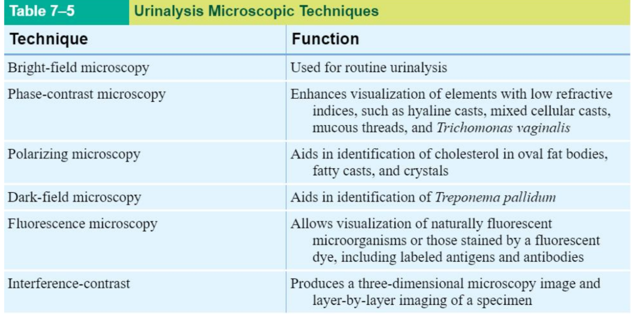
Sediment Examination Techniques
Factors that affect Sediment appearance
Cells and casts in various stages of development and degeneration
Distortion of cells and crystals by chemical content of the specimen
the presence of inclusions in cells and casts
Contamination by artifacts
Sediment Stains
Use to identify cellular structures such as ( Nuclei, Cytoplasm, Inclusions)
Stains = Supravital, Acetic Acid, Lipid, Gram, Hansel, Prussian Blue
Supravital
Most frequently use
Sternheimer-Malbin stain = crystal violet/ Safranin O
Dye absorbed well by WBC, epithelial cells, and casts
help differentiate WBC and renal tubular epithelial cells
Acetic Acid
Enhances nuclear detail of WBC and epithelial cells
RBC are lysed
Lipid
Appearance of free fat droplets and lipid containing cells and casts in the sediment
Use Oil Red O and Sudan III
Triglycerides/ neutral fats = stain orange- red
Cholesterol doesn’t stain
Gram
Use to differentiate gram + (blue) and gram - (red)
Help to identify bacterial Casts
Hansel Stain
Use to stain urinary eosinophils
consists of methylene blue and eosin y in methanol
Prussian Blue Stain
Hemosiderin granules
Stain for iron
Microscopy
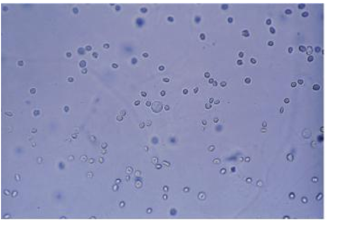
RBC
Smooth, nonnucleated, biconcave disks ~7 µm
Crenated in hypersthenuric urine
Ghost cells in hyposthenuric urine
Dysmorphic RBCGlomerular bleeding
Strenuous exercise
Acanthocytic with multiple
protrusions
Hypochromic, blebs
Aid in diagnosis
RBC Clinical Significancedamage to glomerular membrane or vascular
injury to the genitourinary tract
Cloudy, red to brown urine, advanced disease,
trauma, acute infection, coagulation disorders
Microscopic hematuria=early glomerular disease,
malignancy, strenuous exercise, renal calculi
confirmation
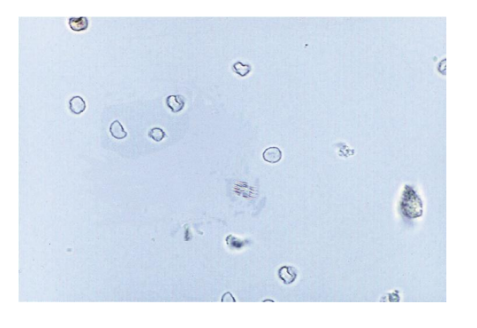
WBC
12 µm
Neutrophil is predominant
Identify under high power
Glitter cells
Hypotonic urine
Brownian movement
Swell; granules sparkle
Light blue if stained
Nonpathologic

Glitter cell
Eosinophils (WBC)
Drug-induced interstitial
nephritis
Renal transplant rejection
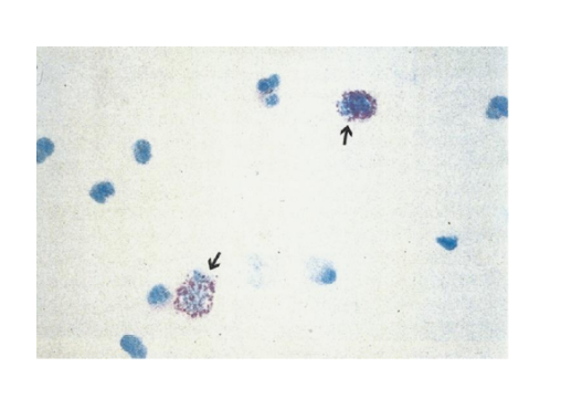
Mononuclear (WBC)
Lymphocytes, monocytes,
macrophages, histiocytes are
rare
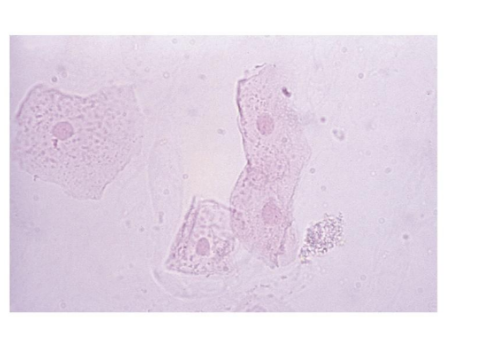
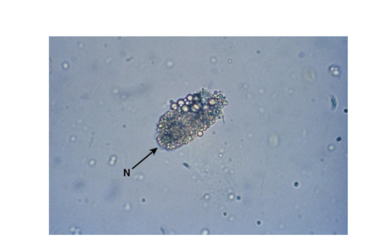
Epithelial Cells
Squamous = vagina, male and female urethra
‒First structures observed
Transitional (urothelial)- bladder, renal pelvis, calyces, ureters, upper male urethra
Renal tubular epithelial (RTE)- renal tubules
Squamous
Largest cell in urine
Good for focusing microscope
Rare, few, moderate, many
lpf or hpf per laboratory
Normal sloughing
Contamination if not midstream clean-catch
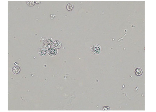
Transitional Epithelial Cells
Spherical: absorb water in bladder and become large and round
Polyhedral: multiple sides
Caudate: has a tail
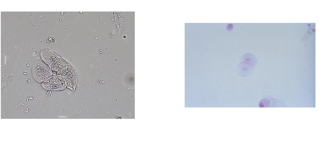
Renal Tubular Epithelial Cells in Proximal Convoluted Tubule Cells
Larger than other RTEs
Columnar, convoluted, rectangular
May resemble casts
Coarsely granular cytoplasm
Examine for presence of nucleus
RTE ( Distal Convoluted Tubule Cells
Round or oval shaped,,smaller
May be mistaken for WBCs or spherical transitional cells
Observe the eccentrically placed nucleus to differentiate from spherical transitional