Unit 1: Foundations
Prehistoric ancestors
~7000 years ago
knew that brain is vital to life
skull surgeries
evidence of trepanation: a surgical procedure involving drilling holes into the skull, which heals afterwards
Ancient Egypt
~5000 years ago
heart is the seat of soul and memory (not the head)
brain was disregarded during mummification while other organs were preserved
Ancient Greece
correlation between the brain structures and their respective functions
Hippocrates (460-379 BCE): brain is involved in sensation and intelligence
Aristotle (384-322 BCE): brain serves as a “radiator” that controls blood
The Roman Empire
Galen (130-300 CE): Greek physician
animal dissection
differentiated between the cerebellum, cerebrum, and ventricles
attempted to determine function through studying structure
The Renaissance Period
fluid-mechanical theory of brain function ventricles pump fluid to move body
reason muscles bulge when flexed
supported by Rene Descartes
believed this only worked for animals, not humans
philosophical mind-brain problem: human mind distinct from the brain
Seventeenth and Eighteenth Centuries
gray and white matter in brain
really two shades of pink, but organ turned gray when left out
determined the connection of the white matter ot the nerves of the body
different color and different functions serve as indications of different functions
End of Eighteenth Century
brain fully dissected
recognized CNS and PNS
gyri: raised portions of brain tissue
sulci: shallowly indented portion of brain tissue
fissures: indents deeper than sulci
eventually, lobes
The Nineteenth Century
nerves as wires:
Benjamin Franklin had been studying electricity
Galvani and Bois Raymond discover that muscles twitch when stimulated with electricity → nerves, like wires, conduct signals to and from the brain
There must be electricity in the body to control muscles
Important Scientists
Bell and Magendie:
discover dorsal and ventral roots carry information in opposite directions
ventral = front
dorsal = back
Charles Bell
cerebellum: origin of motor fibers
cerebrum: destination of sensory fibers
Franz Joseph Gall
phrenology: bumps on the surface of the skull reflect brain surface and related personality traits
has since been disproven
Paul Broca
localization of function in the brain
presented with a patient who could understand speech but could not speak
discrete region of the human cerebellum for speech
Broca’s area: located in the left frontal lobe, responsible for speech production
Regional specialization in different species —> heightened senses
nervous system of different species may share common mechanisms
Historically, why were animals considered good models for learning about the brain?
Different animals allow scientist to study different specializations.
What are the five broad divisions of the neuroscience today?
Molecular: study chemical messengers
Cellular: How do molecules work together?
Systems: visual, motor, sensory
Behavioral: studies memories and dreams
Cognitive: self-awareness, language, and imagination
What is the difference between clinical and experimental neuroscience?
Clinical: has MD component, deals with patients and treatment
Experimental: laboratory, computational, study structures, etc.
Animals in research: What is the difference between animal welfare and animal and animal rights?
animals are renewable natural resources
the more basic the process under investigation, the more distant the evolutionary relationship with humans
(simple —> complex: nematodes, insects, snails, squid, rodents, monkeys)
animal welfare: moral responsibilities
must be worthwhile experience and well planned with access to needs
consider alternatives first
animal rights: ask questions
ex.) death of mouse = death of humans?
Glial cells: insulate, support, and nourish neurons
Glial cells “Give”
Neurons: process information, sense environmental changes, communicate changes to other neurons, command body response
Histology: study of tissue structure
What is the difference between the Nissl stain and the Golgi stain?
Nissl stains: stain the dense bodies (Nissl bodies) surrounding Neuron nuclei
distinguished between neurons and glia cells, studying arrangements of neurons in brain
Golgi stains: distinguish main parts of neuron
soma and neurites (axons and dendrites)
Neuron Doctrine: cell theory applies to neurons
cell is the smallest living organisms (dispose waste, consume nutrients, use O2, etc.)
not proven until 1950s with electron microscope
Cajal’s Contribution: neurons communicate by contact, not continuity
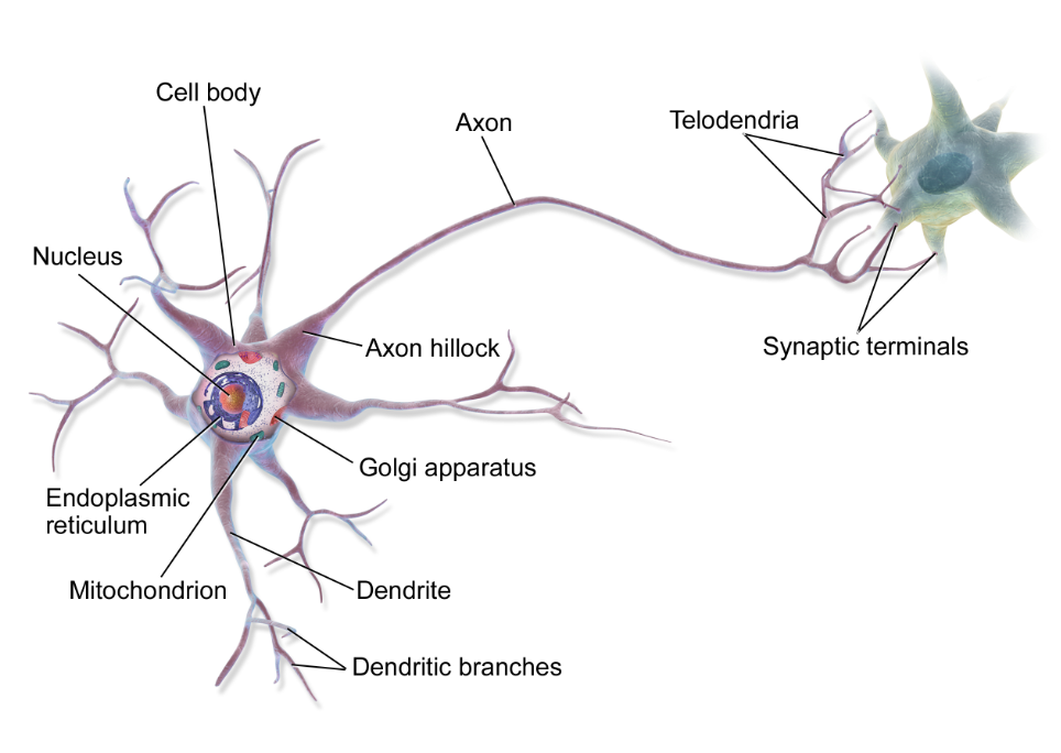
The Soma:
cytosol
organelles
cytoplasm
The Nucleus:
Gene expression
Transcription
RNA processing
Axon:
Axon hillock (beginning)
Axon proper (middle)
Axon terminals (end)
Endoplasmic reticulum does not extend to the end of the axon
unique protein composition
The axon terminal:
no microtubules
lots of synaptic vessels
membrane proteins
lots of mitochondria
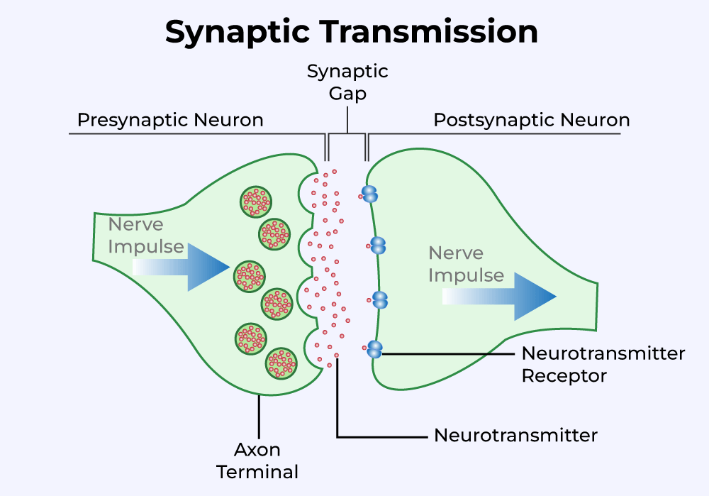
The Synapse:
electrical to chemical to electrical transmission
synaptic transmission dysfunction —> mental disorders
neurotransmitters are like the key to a locked door, makes the postsynaptic neuron electrical
Four ways to classify neurons
Number of neurites: unipolar, bipolar, multipolar
Shape: stellate (star burst) and pyramidal
Connections in the CNS: sensory, motor, and interneurons
Type of neurotransmitter
Glial Cells
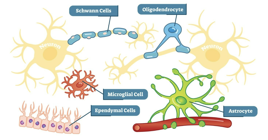
Astrocytes cells:
most numerous
influence neuron growth
chemical regulator of extracellular space —> blood-brain barrier
Ependymal cells:
line ventricles
make cerebrospinal fluid
cilia moves cerebrospinal fluid
Myelinating cells:
insulate cells, like rubber on wire
Shwann cell (PNS)
Oligodendrocytes (CNS)
Nod of Ranvier: region where axon is exposed between cells
Microglia:
smallest
mobile
work as macrophages to clear dead neurons, infections, etc.
Neuron Membrane and Action Potential
Parts of Neuron and Primary Chemicals
Cytosol (extracellular fluid)
water
Potassium (K+)
Anions (A-)
Extracellular Fluid
water
Sodium (Na+)
Chloride (Cl-)
Phospholipid Membrane
Lipid bilayer
Polar head - hydrophilic
Nonpolar tails: hydrophobic
Proteins
enzymes: speeds up reaction
Cytoskeleton: holds shape
Receptors: recieves messages from outside cell
Channel Proteins: serve as hallways and don’t require energy
Ion Pumps: requires energy to move ions in an out of cell
Ion Diffusion
Concentration gradient
C
The Structure of the Nervous System

Brain consists of the cerebrum, cerebellum, and brain stem
Cerebrum: touch, vision, hearing, speech, reasoning, emotions, learning, and fine control movements
covered in:
gyrus: raised sections
sulcus: lowered sections
longitudinal fissure: deep crevice
Cerebellum: coordinate muscle movements, maintain posture, and balance
Brain stem: connects to cerebrum and cerebellum to the spinal cord to control breathing, body temperature, sleep cycles, digestion, sneezing, coughing, vomiting, and swallowing
covered in dura: tough tissue covering
lesser coverings:
arachnoid: in between the two, web-like connections, space containing cerebral spinal fluid
pia: most delicate, adds shine
bilaterilization: each side of brain controls opposite side of body
astereognosis: inability to determine 3d shape by touch
graphesthesia: inability to determine writing on skin
apraxia: inability to perform certain motor tasks when asked
anterograde amnesia: inability to form new memory
retrograde amnesia: inability to recall old memories
differences between left and right hemispheres
left tends to be more developed, associated with math, logic, and language
right is smaller than the left hemisphere, associated with spatial awareness, facial recognition, sensory, and emotions
describe the primary blood supply for the brain
arising from the aorta
vertebral arteries
internal carotid arteries
circle of willis: main blood vessel that supplies brain with blood
jiopji[
/
/