Unit 3: Skeletal and Muscular System Exam
Objective 1: Identify the major components of the skeletal system and describe their functions
Major functions of the skeletal system: support and protection (organs); body movement - muscles “pulling” on bones
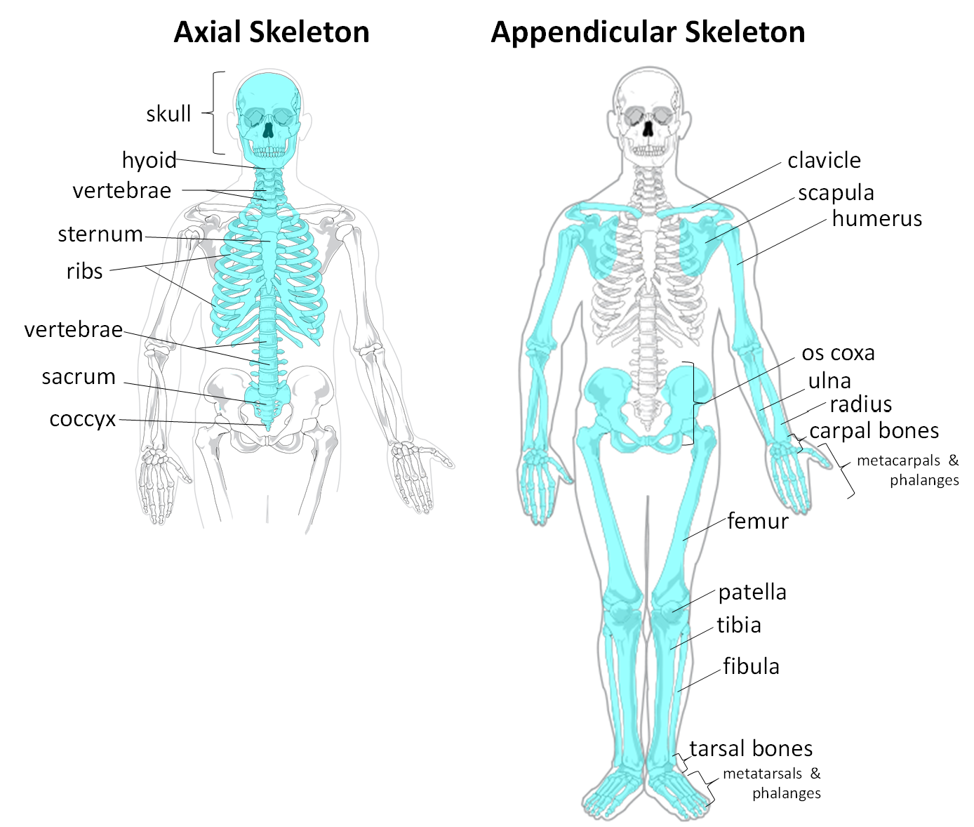
Skeletal system consists of 2 majors divisions: axial and appendicular
Objective 2: Identify the organic and inorganic components of the bone matrix
Bone tissue is also called osseous tissue
Organic components:
collagen
osteoblasts (bone forming cells)
osteoclasts (bone reabsorbing cells)
Inorganic components:
calcium phosphate, in storage: calcium, magnesium, sodium and potassium (salts)
Objective 3: Differentiate between red and yellow bone marrow
Red bone marrow: produces blood cells; found in flat bones
Yellow marrow: fat storage; found in medullary cavity of long bones
Objective 4: Define hematopoiesis
Hematopoiesis: the formation of blood cells; occurs in the red bone marrow
Objective 5: Identify the parts of the long bone

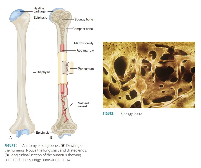
Epiphysis: the ends of the bone; has a thing layer of compact bone, while internally the bone is cancellous (spongy bone); capped with articular cartilage
Epiphyseal Line: also called the growth plate; found at epiphysis’ of long bones
Diaphysis: shaft of the long bone; made of compact bone with a central cavity
Articular/Hyaline Cartilage: found at the ends of long bones; smooth, slippery, and bloodless; allows for smooth movement of bones
Periosteum: vascular, sensitive life support covering; provides nutrient-rich blood for bone cells; source of bone-developing cells during growth or after a fracture; outermost layer of bone
Cancellous (spongy) Bone: bone with a spongy appearance containing red marrow
Compact Bone: dense bone found in the diaphysis; repeated pattern arranged in concentric layers of solid bone; layer just beneath the periosteum
Medullary Cavity: lightens bone weight and provides space for yellow marrow
Endosteum: tissue that lines the medullary cavity
Objective 6 & 7: Locate and identify the structures that make up the axial and appendicular skeleton
Axial: skull (occipital bones and mandible), spine, sternum, ribcage, sacrum, coccyx, hyoid, and ossicles
Appendicular:
- Pectoral girdle: scapula, clavicle, humerus, radius, ulna, carpals, metacarpals, and phalanges
- Pelvic girdle: hips, pelvis, femur, patella, tibia, fibula, tarsals, metatarsals, and phalanges

Objective 8: Identify the parts of the synovial joints
Bone
Synovial Membrane: produces synovial fluid
Synovial Fluid: stored in articular cavity; contains nutrients for cells (cartilage); movement of joints pushes fluid into cartilage to deliver nutrients
Articular/Hyaline Cartilage: lines the epiphyses of the bones of the joints; allows for smooth movement
Joint/Articular Capsule (CT): membrane that surrounds the joint

Objective 9: Describe the parts of the knee joint (bones and ligaments)
Bone of the knee joint:
Femur: thigh bone
Tibia: medial leg bone
Fibula: lateral leg bone
Patella: knee bone
Lateral/Medial Condyle: bony protrusions of the femur
Ligaments of the knee joint:
Posterior Cruciate Ligament: Posterior ligament connecting the femur to the tibia
Anterior Cruciate Ligament: Anterior ligament connecting the femur to the tibia
Fibular (Lateral) Collateral Ligament: strap-like ligament that stabilizes the hinge motion of the knee preventing excessive lateral movement; thinner and rounder, attaches femur to fibula
Tibial (Medial) Collateral Ligament: strap-like ligament that stabilizes the hinge motion of the knee preventing excessive medial movement; attaches femur to tibia
Patellar Ligament: continuation of the quadriceps tendon; attaches the patella to the tibia
Other parts of the knee joint:
Medial/Lateral Meniscus: cartilage between the femur and the tibia; stabilize the joint and act as shock absorption
Quadriceps Tendon: connects the quadriceps muscle to the patella
Objective 10: Describe the specific movements that occur at the major joints of the body
Diarthroses (Synovial) Joint: Freely-moving joint allowing for extensive flexibility and movement
Ball and Socket: ball shaped surface that fits into concave surface; allows for complete ROM (widest range of all joints)
- shoulder and hip jointsCondyloid (Ellipsoidal): oval shaped surface that fits into elliptical surface; moves in 2 planes at right angles to one another
- wrist joint (b/t carpals and radius)Gliding (Plane): articulating surfaces, usually flat; gliding movement (non-axial)
- b/t wrist bonesHinge: spool-shaped surface that fits into a concave surface; moves in 1 plane about a single axis (like a door hinge)
- elbow, knee, ankle, and finger jointsPivot: arch-shaped surface that that rotates around a rounded pivot; rotational movement
- b/t axis and atlas vertebraeSaddle: saddle-shaped surface that fits into a socket curved in the opposite direction; movement similar to condyloid joint
- thumb joint
Objective 11: Differentiate among the types of skeletal joints based on structure and function
Synarthroses (Fibrous): immovable joint; consists of edges of bones connected by dense fibrous connective tissue
- skull bones, radius to ulna, tooth to jawboneAmphiarthroses (Cartilaginous): semi-moveable joint, limited movement and light flexibility; consists of two bones connected by cartilage
- intervertebral discs between vertebrae, pubic symphysisDiarthroses (Synovial): freely-moveable joint, extensive movement and flexibility; consists of bones connected by joint capsule, bones are technically not in contact w/ each other
- knee, shoulder, and elbow joints
Objective 12: Define osteoporosis
Osteoporosis: A condition where bones become weak and brittle (bone building can’t keep up w/ bone depletion)
Objective 13: Identify the three types of muscle tissue found in the human body and discuss their structure, function, and location
Skeletal Muscle: striated, multi-nucleated, non-branched, contains many mitochondria, composed of contractile fibers; allows for voluntary movement; found all over the body
Cardiac Muscle: branches, striated, single nucleus, contains many mitochondria; allows for involuntary contraction of the heart; found in the heart
Smooth Muscle: non-striated, single nucleus, tapered, appears “smooth”; responsible for involuntary muscle movement; found in walls of hollow organs (stomach, intestines, liver, etc.)
Objective 14: Explain the role of agonist and antagonist muscles
Agonist Muscle: the muscle that’s contracting during movement
Antagonist Muscle: the muscle that’s relaxing/lengthening during movement
Objective 15: Describe the microscopic structure of the skeletal muscle tissue
From superficial to deep:
Tendon connects muscle to bone
Epimysium: membrane covering the entire muscle
Perimysium: membrane covering fascicle
Fascicle: bundle of muscle fiber; makes up the entire muscle
Endomysium: connective tissue outer-membrane covering muscle fiber/cell
Muscle fiber/cell (myoCYTES): composed of myoFIBRILS
Sarcolemma: name of a muscle cell’s membrane, NOT the outer CT membrane that covers the cell; helps transduce electrical neural signals
MyoFIBRIL: make up muscle cells; contains myoFILAMENTS that make up contractile unit of the cell
MyoFILAMENT: protein fiber that makes up the myoFIBRIL; two major types are actin (thin filament) and myosin (thick filament)
Sarcomere: contractile unit of the muscle cell; made up of thick and thin myofilaments (myosin and actin respectively)
I-Band: portion of the muscle made of thin filaments as seen vertically
A-Band: portion of the muscle made of thick filaments as seen vertically
Z-Line: boundary line between sarcomeres
Sarcoplasmic Reticulum: network of tubes that run parallel to the myofilaments (actin & myosin); storage site of Ca+
T-Tubules: tubules that run perpendicular to myofilaments; allows electrical impulse to travel across cell
Troponin: protein that covers the binding site on actin (troponIN = on actIN)
Tropomyosin: protein that is attached to troponin; responsible for moving troponin off actin (tropoMyosin = Move)
Muscle (covered by epimysium) → fascicles (covered by perimysium) → muscle fiber/cell (covered by endomysium) → myofibrils → myofilaments (actin & myosin)

Objective 16 & 17: Explain how muscle contraction occurs at the level of the sarcomere & explain how an impulse generated by the CNS results in the contraction of a skeletal muscle
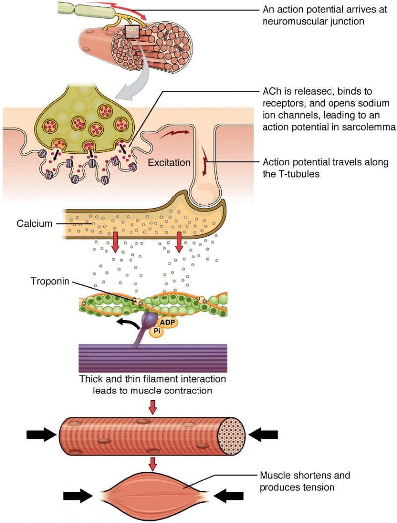
Sliding Filament Theory:
Signal travels down motor neuron
Motor neuron released ACh into synaptic cleft b/t neuron and muscle cell
ACh binds to receptors in sarcolemma (muscle cell membrane)
Signal is transduced; electrical impulse travels in sarcolemma
Signal travels into the cell via t-tubules
Impulse stimulates sarcoplasmic reticulum (network) to release Ca+
Ca+ binds to troponin on actin, which causes:
Tropomyosin moves off of myosin’s binding site on actin
Myosin head binds to binding site on actin (requires ATP)
Myosin pulls actin toward the center of the sarcomere
Myosin head detaches from actin to release the actin (requires ATP)

Objective 18: Discuss the impact of botulinum toxin at the neuromuscular junction
Botulism toxin is produced by bacteria. Can be consumed or injected via Botox. Botulism toxin prevents the release of ACh, inhibiting signals from motor neurons to muscle cells, causing paralysis
Objective 19: Name the major muscles of the human body, general location, and describe their movements
Movements
Flexion: bending a limb; decreasing angle of a joint
Extension: straightening a limb; increasing angle of a joint
Rotation (Medial/Lateral)
Pronation: rotation of an arm/leg so palm is down or sole out
Supination: rotation of an arm/leg so palm is up or sole in
Abduction: movement away from the midline of the body (raising arms = alien ABDUCTION)
Adduction: movement toward the midline of the body
Inversion: movement of sole towards the midline of the body
Eversion: movement of sole away from the midline of the body
Protraction: movement forward
Retraction: movement backward
Elevation: raise up
Depression: push down
Major Muscles
Pectoralis Major: anterior chest; flexes shoulder, adducts shoulder, medially rotates shoulder
Rectus Abdominus: anterior trunk, from mid-rib to pelvis; flexes spine
External/Internal Oblique: anterior trunk, lateral to rectus abdominus, internal is deep to external; sagittal flexion of spine, laterally flexes and rotates spine to one side (transversal)
Trapezius: posterior neck/trunk, runs almost the length of the spine; elevation of scapula (upper fibers), adduct scapula, depression of scapula (lower fibers)
Deltoid: superior shoulder; abducts shoulder, anterior fibers flex and medially rotate shoulder, posterior fibers extend and laterally rotate shoulder
Biceps Brachii: anterior proximal arm; flexes elbow, supinates forearm, flexes arm (long head of biceps)
Triceps Brachii: posterior proximal arm; extends elbow, extends shoulder
Gluteus Maximus: posterior proximal leg; extends hip, laterally rotates hip, abducts hip
Psoas: anterior deep pelvis; flexes hip, flexes trunk
Rectus Femoris (Quadriceps): anterior proximal leg; extends knee, flexes hip
Biceps Femoris (Hamstrings): posterior proximal leg, deep to gluteus maximus; flexes knee, extends hip
Gastrocnemius: posterior distal leg; plantar flexes ankle
Objective 20: Explain the meaning of the terms insertion and origin and describe how skeletal muscles attach to the bony skeleton