knowt logo

Lecture 5: Alteration in Neurological Function

  • Brain Trauma:

    • traumatic brain injury- a traumatic insult to the brain

      • resulting physical, intellectual, emotional, social, and vocational changes (idk if that means it does or it can result in)

      • caused 50% by motor vehicle accidents, 21% falls, 29% by violence and sports

    • head injuries are broadly categorized into blunt and open trauma

      • blunt trauma (closed, nonmissile)

        • head strikes hard surface or a rapidly moving object strikes the head

        • the dura remains intact and brain tissues are not exposed to the environment

        • the trauma causes focal (local) or diffuse (general) brain injuries

      • open trauma (penetrating, missile)

        • injury breaks the dura and exposes the cranial contents to the environment

        • the trauma causes primarily focal injuries ex. weapon (idk what weapon is doing here)

    • coup injury

      • injury directly below (i think below as in below the cranium) the point of impact

      • objects such as a baseball bat striking the front of the head usually produce only coup injuries

    • contrecoup injuries

      • injury on the pole opposite the site of impact

      • objects striking the side of the head produces contrecoup injury

      • the injury is caused by the skull on the other side. the brain moves and collides with the hard surface of the cranium

        • i think for a fall or impact, the initial impact puts energy into the skull in the opposite direction (for every action there is an equal and opposite reaction and also maybe rebounding?) and then it moves and collides w the other side of the cranium). thats how i understand it at least

    • compound fractures

      • compound fractures are caused by objects striking the head with great force or by the head striking an object forcefully

  • focal brain injury:

    • focal brain injury is specific observable brain lesion

      • epidural hemorrhage, subdural hematoma, and intracerebral hematoma

        • hemorrhage- active bleeding, hematoma- accumulated blood post (or during) active bleeding (usually once it stops or slows down)

        • epidural being the only hemorrhage is probably random

    • force of impact typically produces contusions (bruise produced by bleeding into the skin)

    • contusions can cause

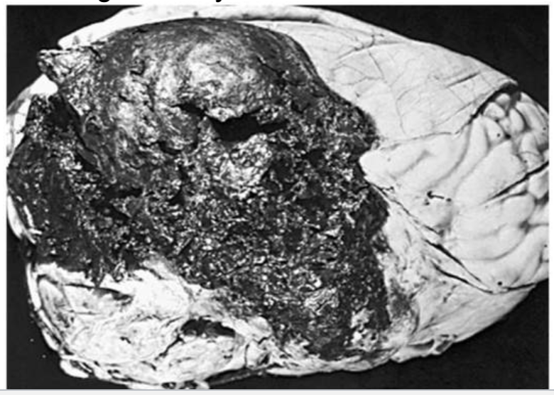
      • extradural (epidural) hemorrhages or hematomas (collection of blood in soft tissue)

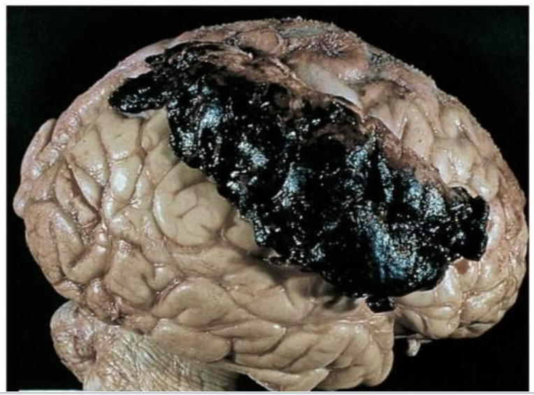
      • subdural hematomas

      • intracerebral hematomas

extradural/epidural hematoma: skull fracture w tear of the middle meningeal artery and veinsubdural hematoma
  • diffuse brain injury:

    • diffuse brain injury or diffuse axonal injury (dai) results from

      • shaking, inertial effort

      • acceleration/deceleration (ex. rotational acceleration can cause shearing, tearing, or stretching of nerve fibers that can lead to axonal damage)

        • axonal damage- shearing, tearing, or stretching of nerve fibers

      • severity of damage corresponds to the amount of shearing force applied to the brain and brain stem

    • dai categories:

      • mild concussion

      • classical concussion

      • diffuse axonal injuries (dai): mild, moderate, and severe

        • first 2 are categories of diffuse brain injuries (dai seems to be used a little more broadly in these slides) while the last is specifically categories of dai

  • mild concussion:

    • temporary axonal disturbances causing attention and memory deficits but no loss of consciousness

    • 3 forms of mild concussion:

      • 1- confusion, disorientation, and momentary amnesia (partial or total loss of memory)

      • 2- momentary confusion and retrograde amnesia develops after 5-10 min

      • 3- confusion w retrograde and anterograde amnesia develops after impact

  • classic cerebral concussion:

    • grade 4 (might be connected to 1,2 & 3 from the last slide bc i think theyre all grades of concussions just 4 is classic while 1-3 is mild)

      • disconnection of cerebral systems from the brain stem and reticular activating system

      • physiologic and neurologic dysfunction without substantial anatomic disruption

      • loss of consciousness (<6 hrs)

      • anterograde and retrograde amnesia

  • diffuse axonal injury:

    • produces a traumatic coma lasting more than 6 hrs bc of axonal disruption

    • 3 forms of dai

      • mild- post traumatic coma hasts 6-24 hrs

      • moderate- actual tearing of some axons in both hemispheres

      • severe- involves severe mechanical disruption of many axons (extending to diencephalon)

        • diencephalon- part of the brain right above the brain stem, contains thalamus, hypothalamus etc.

  • clinical manifestation of focal brain injury:

    • loss of consciousness (no longer than 5 min)

    • loss of reflexes

      • results the individual to fall to the ground

      • cessation of respiration

      • bradycardia and low blood pressure

    • increasing in csf pressure

    • ecg and eec (electroencephalographic) changes

    • returning to full alert- minutes to days (full level of consciousness may never return)

    • for diagnoses ct scan and mri should be done

  • spinal cord trauma:

    • most commonly occurs due to vertebral injuries

    • vertebral injuries can be classified as

      • simple fracture ex. fracture of transverse or spinous process

      • compressed fracture ex. fracture that leads to compression of vertebral body

        • the vertebral body fractures which lessens its structural strength and causes it to be compressed which can put pressure on the spinal cord and surrounding nerves

      • comminuted (burst) fracture- vertebral body is shattered into fragments

      • dislocation

    • traumatic injury of vertebral and neural tissues are caused due to compressing, pulling, or shearing forces

    • most common locations: cervical (1,2 4-7) and t1-l2 lumbar vertebrae

      • locations reflect most mobile portions of vertebral column and the locations where the spinal cord occupies most of the vertebral canal
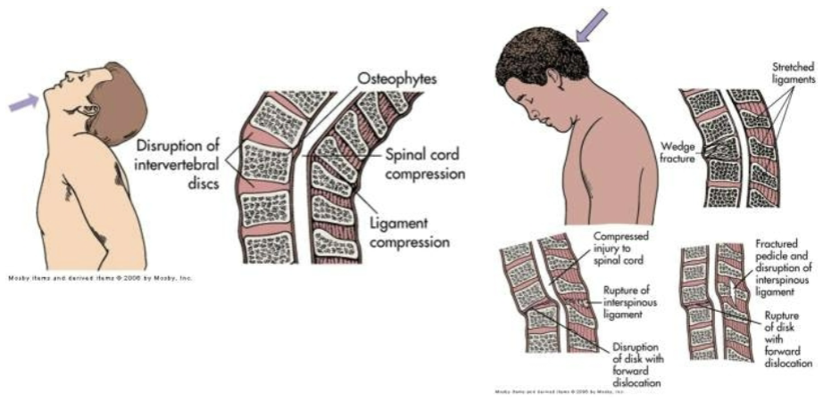
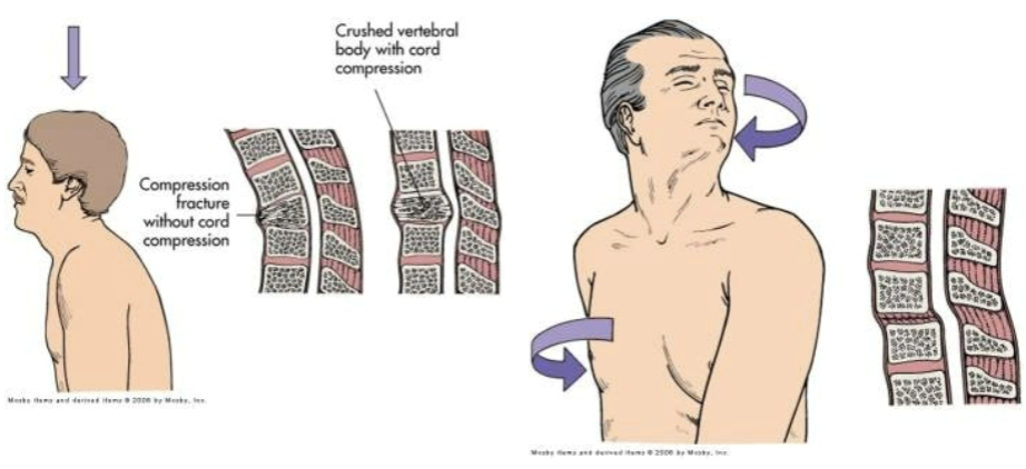
    • spinal shock (may last 7-20 days or more)

      • normal activity of the spinal cord ceases at and below the level of injury. sites lack continuous nervous discharges from the brain

      • complete loss of reflex function (skeletal, bladder, bowel, sexual function, thermal control, and autonomic control)

      • loss of motor and sensory function depends on level of injury
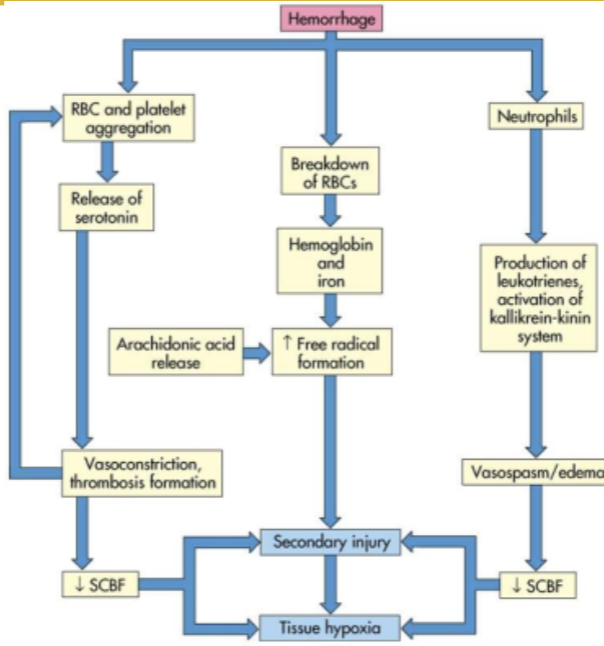
    • paraplegia

    • quadriplegia

    • autonomic hyperreflexia (dysreflexia)

      • massive, uncompensated cardiovascular response to stimulation of the sympathetic nervous system

      • stimulation of the sensory receptors below the level of the cord lesion

      • there are pictures of normal and dysreflexia pathways but they’re super blurry

  • degenerative disorders of the spine:

    • occur in the vertebral disk

    • degenerative disk disease (ddd)

      • common in individuals 30 and older

      • normally no signs functional incapacity

      • occurs due to biochemical and biomechanical alterations of intervertebral disk tissue

        • spondylolysis- degenerative process of vertebral column, associated with soft tissue

        • spondylolisthesis- slip of vertebra due to stress

        • spinal stenosis- narrowing of spinal canal ex. due to annulus

          • annulus fibrosis is the tough, outer layer of the intervertebral discs and if its degenerated or damaged, the inner pulpous part of the intervertebral disc can bulge or herniate out into the spinal canal (which narrows it)

        • (these 3 things can result from ddd)

    • low back pain

    • herniated intervertebral disk caused by trauma or degenerative disk disease

  • cerebrovascular disorders:

    • the common clinical manifestation of cerebrovascular disease is CVAs

    • cerebrovascular accidents (cvas) (commonly called a stroke, refers to a sudden interruption of blood flow in the brain, ‘accident’ bc it is sudden and unexpected)

      • thrombotic stroke

        • arterial occlusions caused by thrombi formed in arteries supplying the brain or in the intracranial vessels

        • transient ischemic attacks

      • embolic stroke

        • fragments that break from a thrombus formed outside the brain

      • (for both of these) i don’t think the location the clot formed is important to the classification, just whether it was the thrombus or an embolus

      • hemorrhagic stroke- ex. caused by hypertension

      • lacunar stroke- a type of ischemic stroke that happens when blood flow to a small area of the brain is blocked ex. caused by thickening of arterial wall

      • cerebral infarction- area of brain loses blood supply bc of vascular occlusion

      • cerebral hemorrhage- ex. caused by hypertension

      • intracranial aneurysm

        • may result from

          • arteriosclerosis

            • hardening/thickening of the arterial wall, can cause an aneurysm bc arteriosclerosis weakens the arterial wall bc it makes it stiffer and bulge under pressure (when it should be able to expand and contract based on blood flow), also bc it causes structural changes which can cause an imbalance of forces acting on the wall which can cause weak spots, and also bc the added layers increase the blood pressure in that area (bc narrower = increased pressure) which can add more stress to weak areas

          • congenital abnormalities

          • trauma

          • inflammation

          • infection

        • it is asymptomatic, but clinical manifestation may arise from cranial nerve compression

          • the signs vary, depending on location

        • diagnosis: mri, ct scan

        • treatment: surgery

          • under cva bc it can lead to an actual cva (so considered a type of cva once it leads to the stroke)

      • vascular malformations

        • symptoms include

          • nondescriptive chronic headache

          • 50% experience seizure disorders caused by compression

          • 50% experience hemorrhage (subarachnoid, subdural, or intracerebral)

        • diagnosis: ct scan, mri, transcranial doppler, and mra (magnetic resonance angiography)

        • treatment: surgery

          • under cva bc it can lead to an actual cva (so considered a type of cva once it leads to the stroke)

      • subarachnoid hemorrhage

        • blood escapes from defective or injured vasculature into the subarachnoid space

        • manifestations

          • headache, nausea, neck stiffness, photophobia (light sensitivity)

          • kernig sign- straightening the knee w the hip and knee in a flexed position produces pain in the neck and back

          • brudzinski sign- passive flexion of the neck produces neck pain and increase rigidity

  • headache:

    • migrane headache

      • trigger factors ex. stress, hunger, weather change, noise

    • cluster headache

      • several attacks can occur during the day for a periods of days followed by a long period of spontaneous remission

    • chronic paroxysmal hemicrania

      • cluster type headache that occurs with more daily frequency but w shorter duration

    • tension type headache

      • mild to moderate bilateral headache w a sensation of a tight band or pressure around the head

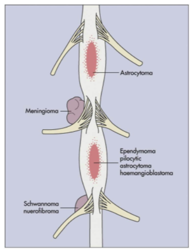
  • central nervous system tumors:

    • cranial tumors

      • primary intracerebral tumors (gliomas)

        • astrocytoma

        • oligodendroglioma

        • ependymoma

      • primary extracerebral tumors (extracerebral is outside brain but inside cranium)

        • meningioma

        • nerve sheath tumors

        • metastatic carcinoma

          • i dont think this should be under primary extracerebral tumors bc it wouldn’t really originate as extracerebral….

        • for both of the above primary means it starts there (so primary intracerebral originated in the brain)

    • spinal cord tumors

      • intramedullary tumors (within the spinal cord itself)

      • extramedullary tumors

        • intradural

        • extradural

      • manifestations

        • compressive syndrome (spinal cord or surrounding structures is compressed)

        • irritative syndrome (spinal cord or nerve roots is irritated either by chemicals released, local inflammation caused by the tumor, or direct contact)

        • syringomyelic syndrome- inflammation of spinal cord
  • infection and inflammation of the cns:

    • cns may be affected directly by bacteria, virus, fungi, protozoa, and rickettsiae (rickettsiae are a type of bacteria but they have unique characteristics and modes of transportation)

    • meningitis

      • bacterial meningitis primarily (an) infection of (the) pia mater and (pia) archnoid

      • aseptic (viral, nonpurulent (no pus), (and) lymphocytic (elevated number of lymphocytes)) meningitis (aseptic here means non bacterial)

      • fungal meningitis

      • tubercular (tb) meningitis (also caused by a bacteria but bacterial meningitis listed above is probably from a different bacteria)

    • encephalitis

      • inflammation of brain tissue

      • acute febrile illness, usually of viral origin w nervous system involvement

      • most common forms of encephalitis are caused by arthropod borne viruses and herpes simplex virus

  • neurologic complications of aids:

    • human immunodeficiency associated cognitive dysfunction

    • hiv myelopathy (disease of the spinal cord)

    • hiv neuropathy

    • aseptic viral meningitis

    • opportunistic infections

    • cytomegalovirus infections (usually very mild symptoms unless immunocompromised)

    • parasitic infection

    • cns neoplasms (abnormal growth of tissue)

  • peripheral nervous system disorders:

    • acons traveling to and from cns can be injured

    • neuropathies (damage to nerves)

      • generalized symmetric polyneuropathies

        • symmetric involvement of sensory, motor, or autonomic fibers

      • generalized neuropathies

        • affect the cell body of only one type of peripheral neuron

      • i think polyneuropathy is a type of neuropathy that specifically affects multiple nerve types)

      • symptoms: muscle strength and muscle tone are affected

      • many neuropathies can be reversed by eliminating the cause if possible

    • plexus injuries

      • involves the nerve plexus distal to the spinal roots but proximal to the formation of the peripheral nerves

  • neuromuscular junction disorders:

    • nerve impulse at neuromuscular junction are disrupted due to

      • nutritional deficit, certain drugs (ex. reserpine)

      • certain disorders that disrupt the synthesis or packaging of neurotransmitters or release of the neurotransmitter

  • alterations in arousal: (as in awakeness)

    • can be:

      • strutural

        • divided by location above or below tentorial plate (tentorial plate separates the cerebrum and cerebellum)

      • metabolic

      • psychogenic

    • coma is produced by either

      • bilateral hemisphere damage or suppression

      • brain stem lesions or metabolic derangement that damages or suppresses the reticular activating system

    • clinical manifestations:

      • level of consciousness changes

      • pattern of breathing

        • posthyperventilation apnea (phva)

        • cheyne-stoke respirations (csr) (cyles of deep, rapid breathing followed by periods of apnea)

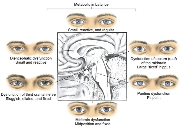
      • pupillary changes

      • oculomotor responses (movement by the ocular muscles aka movement of the eyeball)

      • motor responses

      • vomiting, yawning, hiccups

  • brain death (total brain death):

    • body can no longer maintain internal homeostasis

    • brain death criteria:

      • completion of all appropriate and therapeutic procedures

      • unresponsive coma (absence of motor and reflex responses)

      • no spontaneous respirations (apnea)

  • brain death (brain stem death):

    • brain death criteria:

      • no ocular responses

      • isoelectric eeg (shows no detectable brain activity)

      • persistence 6-12 hrs after onset

  • cerebral death:

    • cerebral death (irreversible coma) is death of the cerebral hemispheres exclusive of the brain stem and cerebellum

    • no behavioral or environmental responses

    • the brain can continue to maintain internal homeostasis

    • survivors of cerebral death: (one of the below, not all)

      • remain in a coma

      • emerge into a persistent vegetative state

      • progress into a minimal conscious state (mcs)

      • locked in syndrome (pretty sure this can’t happen with cerebral death bc thats paralysis but still cognitively intact, at least partially; chatgpt agrees)

  • alterations in awareness:

    • selective attention

      • ability to select from available, competing environmental and internal stimuli

      • sensory inattentiveness

        • extinction

          • basically, if a person has specific damage on one side of their brain, it will be much harder to register stimulus on the physical side associated with that side of the brain. they wont be able to register (sudden) stimuli on both sides at the same time (the unaffected side will take precedence often even if the affected sides stimulus came first). They will still register one but not the other. if there is no stimulus on the unaffected side, they generally will have no issue registering stimuli on the affected side. (ex. left brain damage, if theres a loud horn honking unexpectedly on the physical left side, they wont register anything (even constant stimuli) on the physical right side)

        • neglect syndrome

          • generally associated with damage to the right hemisphere (so affecting the physical left side); both sudden and constant stimuli will be ignored (and totally unaware of its presence) on the physical side associated w the side of the brain thats damaged. this might even apply to themselves (they might neglect personal grooming on that side or even not recognize their own limbs on that side [even if they can see it])

      • selective attention deficit

    • memory

    • amnesia

      • retrograde amnesia

      • anterograde amnesia

      • executive attention deficits

        • adhd

      • image processing

  • seizures:

    • syndrome vs disease

    • sudden, transient alteration of brain function caused by an abrupt explosive, disorderly discharge of cerebral neurons

    • motor, sensory, autonomic, or psychic signs

    • convulsion

      • tonic clonic (jerky, contract relax) movements associated with some seizures

    • idiopathic epilepsy- no underlying cause can be found

      • some forms of epilepsy do have known causes

    • etiologic factors

      • cerebral lesions

      • biochemical disorders

      • cerebral trauma

      • epilepsy

    • partial seizures

      • simple, complex, secondary generalized

    • generalized seizures

      • absent, myoclonic (brief, shock like jerks), clonic (rhythmic jerking), tonic clonic (tonic == body stiffens), atonic (sudden loss of muscle tone which results in falling/collapsing)

    • unclassified epileptic seizure

    • aura (warning signs immediately before a seizure)

    • prodoma (warning signs that can occur hours or even days before a seizure)

    • tonic phase

      • contraction

    • clonic phase

      • relaxation

    • postictal phase

  • data processing deficits:

    • agnosia (registers sensory info but cant recognize or interpret it)

      • tactile, visual, auditory, etc.

    • dysphasia

      • expressive dysphasia (difficult to speak or write coherently, words typically are said properly but either not right words or not right order. they usually know what they want to say but cant make a coherent sentence. might also struggle w word retrieval)

      • receptive dysphasia (difficulty understanding spoken and written language)

      • transcortical dysphasia (difficulty w both)

    • aphasia (generally a more severe or complete version of dysphasia)

  • acute confusional states (acs):

    • transient disorders of awareness that result from cerebral dysfunction

      • secondary to drug intoxication, metabolic disorder, or nervous system disease

      • delirium

        • hyperkinetic (excessive or abnormal movement)

        • hypokinetic (reduced or slower movement)

        • these 2 are under delirium bc delirium can cause these in specific form (ex. restlessness or lethargy)

  • dementia:

    • progressive failure of cerebral functions that is not caused by an impaired level of consciousness

    • losses:

      • orientation

      • memory

      • language

      • judgement

      • decision making

  • alzheimer disease (ad):

    • familial, early and late onset

    • nonhereditary (sporadic, late onset)

    • theories

      • mutation for encoding amyloid precursor protein

      • alteration in apolipoprotein e

      • loss of neurotransmitter stimulation of choline acetyltransferase

    • neurofibrillary tangles (abnormal aggregates of a specific protein found inside neurons; accumulation of these tangles disrupts the normal functioning of neurons)

    • senile plaques (aka amyloid plaques, abnormal clumps of protein that accumulate btwn neurons in the brain, disrupt cell communication and trigger inflammatory responses which leads to neuronal damage and cognitive decline)

    • clinical manifestations:

      • forgetfulness

      • emotional upset

      • disorientation

      • confusion

      • lack of concentration

      • decline in abstraction, problem solving, and judgement

    • diagnosis is made by ruling out other causes of dementia

  • cerebral hemodynamics:

    • the dynamic of blood flow in the brain, including the regulation, distribution, and alterations in cbf

    • cbf (cerebral blood flow)

    • cpp (cerebral perfusion pressure)

    • cbv (cerebral blood volume)

    • cerebral oxygenation

  • increased intracranial pressure (iicp):

    • normal 5-15mmHg

    • caused by an increase in intracranial content

      • tumor growth, edema, excessive csf, or hemorrhage

    • stage 1

      • the body can compensate for iicp, may not have noticeable symptoms but may experience slight changes in consciousness or subtle neurological signs. cerebral perfusion is maintained

    • stage 2

      • compensatory mechanisms start to fail, symptoms: slight changes in mental status (ex. confusion or lethargy), decrease in the ability to maintain normal blood flow to the brain etc. may be increased blood pressure (to try to maintain normal brain perfusion)

    • stage 3

      • significant elevation of icp leading to reduced cerebral perfusion. symptoms: altered level of consciousness, severe headache, pupillary changes, abnormal posturing (body positioning) etc. risk of brain herniation increases

    • stage 4

      • most critical stage, often referred to as herniation, there is a life threatening increase in icp that leads to brain herniation and severe neurological impairment. symptoms: coma, loss of reflexes, cardiovascular instability etc.

  • herniation syndromes:

    • supratentorial herniation:

      • uncal

        • uncus (small structure on the medial surface of the temporal lobe) or hippocampal gyrus (or both) shifts from the middle fossa through the tentorial notch into the posterior fossa

      • central

        • downward shift of the diencephalon (region of the brain btwn the cerebral hemispheres and the brainstem) through the tentorial notch

      • cingulate

        • congulate gyrus (a curved fold of brain tissue above the corpus callosum) shifts under the falx cerebri (a fold of dura mater)

    • infratentorial herniation:

      • cerebellar tonsil shifts through foramen magnum

  • cerebral edema:

    • increase in the fluid (intracellular or extracellular) within the brain

    • types

      • vasogenic (increased permeability of the blood brain barrier)

      • cytotoxic (swelling of brain cells from an influx of water bc of cellular injury or metabolic dysfunction)

      • interstitial (accumulation of csf)

  • hydrocephalus:

    • excess fluid within the cranial vault, subarachnoid space, or both

    • caused by interference in csf flow

      • decreased reabsorption

      • increased fluid production

      • obstruction within the ventricular system

    • noncommunicating hydrocephalus (aka obstructive hydrocephalus; happens when there is a blockage in the flow of csf in the ventricles)

      • internal (blockage is in the ventricles, cant flow btwn ventricles)

      • intraventricular (accumulation in the ventricles)

    • communicating (extraventricular) hydrocephalus (csf can flow btwn ventricles but cant be properly reabsorbed into the bloodstream)

    • acute hydrocephalus

    • normal pressure hydrocephalus

  • alterations in neuromuscular function:

    • muscle tone

      • hypotonia (decreased muscle tone, floppiness or reduced resistance to passive movement)

      • hypertonia

        • spasticity (tightness and resistance to movement)

        • gegenhalten (paratonia) (resistance to passive movement that occurs in response to the external force, the muscle tone increases once the force is applied)

        • dystonia (involuntary muscle contractions)

        • rigidity

  • alterations in movement:

    • paresis (partial weakness or reduced strength) and paralysis

      • upper motor neuron syndromes

        • hemiparesis or hemiplegia

        • diplegia

        • paraparesis or paraplegia

        • quadriparesis or quadriplegia

        • pyramidal motor syndromes (results from damage to the pyramidal tracts [pathways in the brain that control voluntary movement])

        • spinal shock (temporary paresis/paralysis)
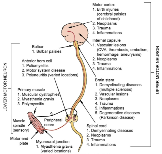
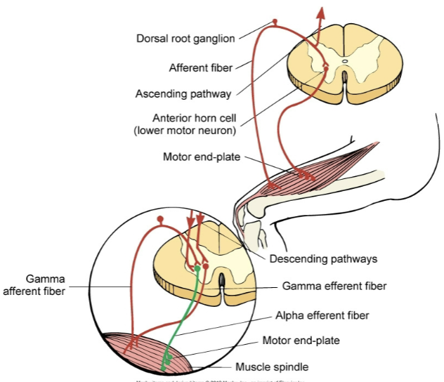
    • lower motor neuron syndromes

      • flaccid paresis or flaccid paralysis

      • hyporeflexia (reduced or diminished reflex responses) or areflexia (no reflex responses)

      • fibrillation (spontaneous, irregular contractions of individual muscle fibers)

      • amyotrophies (degeneration and atrophy of muscle tissue from loss of motor neurons)

        • paralytic poliomyelitis

          • infectious disease caused by polio virus, leads to inflammation of spinal cord and motor neurons, can result in paralysis and muscle weakness

        • nuclear palsies

          • disorders that result from damage to the cranial nerve nuclei in the brainstem (leading to deficits in the functions controlled by those nerves)

        • guillain barre syndrome

          • autoimmune disorder attacking the peripheral nervous system

        • progressive bulbar palsy

          • causes degeneration of the bulbar region (part of the brain stem) which leads to progressive weakness and atrophy of the muscles innervated by cranial nerves

        • bulbar palsy

          • like progressive bulbar palsy but results from conditions like stroke or trauma, symptoms either stabilize or improve rather than progressively worsen

    • hyperkinesia

      • excessive movement

      • chorea (involuntary, irregular, unpredictable muscle movements), wandering, tremor at rest

      • postural tremor etc.

    • paroxysmal dyskinesias

      • dyskinesia- sudden, involuntary, abnormal movements

      • paroxysm- a sudden, intense episode or attack of a condition or symptom

    • tardive dyskinesia

      • tardive- delayed or late onset of symptoms; in this specific context- dyskinesia that develops after prolonged exposure to certain meds

    • huntington disease

      • also known as chorea

      • autosomal dominant hereditary degenerative disorder

      • severe degeneration of the basal ganglia (caudate nucleus [a part of the basal ganglia]) and frontal cerebral atrophy

        • depletion of gamma-aminobutyric acid (gaba)

    • hypokinesia

      • decreased movement

      • akinesia

      • bradykinesia (slowed movement)

      • loss of associated movement

  • parkinson disease:

    • severe degeneration of the basal ganglia (corpus striatum) involving the dopaminergic nigrostriatal pathway

      • parkinsonian tremor, rigidity, bradykinesia

      • postural disturbances

      • autonomic and neuroendocrine symptoms

      • congitive affective symptoms

    • secondary parkinsonism (parkinsons symptoms from something else thats not parkinsons)

  • disorders of posture (stance):

    • dystonia (involuntary muscle contractions resulting in abnormal postures, twisting movements, or repetitive motions)

      • dystonic postures and movements

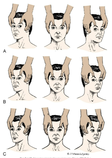
      • decorticate posture (A; also legs are turned in)

      • decerebrate posture (B, also back is arched)

      • basal ganglion posture (stiff/rigid limbs, flexed posture of limbs, stooped/leaning forward trunk)

      • senile posture (forward flexed trunk, rounded shoulders, slight flexion of hips and knees)

  • disorders of gait:

    • spastic gait (stiff/jerky movements, tendency to drag 1/both legs, reduced arm swing on affected side, difficulty w balance/coordination)

    • scissors gait (legs cross/scissor when walking, narrow base of support, short steps, hypertonia in legs)

      • base of support: the area beneath a person that supports their weight, determined by the distance btwn their feet when standing/walking

    • cerebellar gait (unsteady/wide based walking, difficulty w balance, lurching/swaying movements, frequent stumbling/falling)

    • basal ganglion gait (reduced arm swing, shuffling steps w a short stride length, difficulty initiating movements [freezing], stooped posture)

    • senile gait (slow/shuffling steps, broad base of support, reduced arm swing, difficulty w balance/coordination)

  • disorders of expression:

    • hypermimesis (exaggerated expressions of emotions)

    • hypomimesis (reduced expressions of emotions)

    • dyspraxias and apraxias (difficulty [dys]/inability [a] planning and executing coordinated movements)

  • extrapyramidal motor syndromes:

    • dysfunction of the extrapyramidal system which is part of the motor control pathway in the brain

    • basal ganglia motor syndromes (caused by dysfunction of the basal ganglia, results in bradykinesia, hypertonia, tremors [specifically resting tremors which happen when not moving the limb], dystonia, chorea etc.)

    • cerebellar motor syndromes (caused by dysfunction of the cerebellum, results in ataxia, dysmetria, tremors [specifically intention tremors which happen when trying to move the limb], hypotonia, difficulty w balance/posture etc.)

      • ataxia- lack of coordination and unsteady movements

      • dysmetria- inability to the control the distance/range of movements

Lecture 5: Alteration in Neurological Function

  • Brain Trauma:

    • traumatic brain injury- a traumatic insult to the brain

      • resulting physical, intellectual, emotional, social, and vocational changes (idk if that means it does or it can result in)

      • caused 50% by motor vehicle accidents, 21% falls, 29% by violence and sports

    • head injuries are broadly categorized into blunt and open trauma

      • blunt trauma (closed, nonmissile)

        • head strikes hard surface or a rapidly moving object strikes the head

        • the dura remains intact and brain tissues are not exposed to the environment

        • the trauma causes focal (local) or diffuse (general) brain injuries

      • open trauma (penetrating, missile)

        • injury breaks the dura and exposes the cranial contents to the environment

        • the trauma causes primarily focal injuries ex. weapon (idk what weapon is doing here)

    • coup injury

      • injury directly below (i think below as in below the cranium) the point of impact

      • objects such as a baseball bat striking the front of the head usually produce only coup injuries

    • contrecoup injuries

      • injury on the pole opposite the site of impact

      • objects striking the side of the head produces contrecoup injury

      • the injury is caused by the skull on the other side. the brain moves and collides with the hard surface of the cranium

        • i think for a fall or impact, the initial impact puts energy into the skull in the opposite direction (for every action there is an equal and opposite reaction and also maybe rebounding?) and then it moves and collides w the other side of the cranium). thats how i understand it at least

    • compound fractures

      • compound fractures are caused by objects striking the head with great force or by the head striking an object forcefully

  • focal brain injury:

    • focal brain injury is specific observable brain lesion

      • epidural hemorrhage, subdural hematoma, and intracerebral hematoma

        • hemorrhage- active bleeding, hematoma- accumulated blood post (or during) active bleeding (usually once it stops or slows down)

        • epidural being the only hemorrhage is probably random

    • force of impact typically produces contusions (bruise produced by bleeding into the skin)

    • contusions can cause

      • extradural (epidural) hemorrhages or hematomas (collection of blood in soft tissue)

      • subdural hematomas

      • intracerebral hematomas

extradural/epidural hematoma: skull fracture w tear of the middle meningeal artery and veinsubdural hematoma
  • diffuse brain injury:

    • diffuse brain injury or diffuse axonal injury (dai) results from

      • shaking, inertial effort

      • acceleration/deceleration (ex. rotational acceleration can cause shearing, tearing, or stretching of nerve fibers that can lead to axonal damage)

        • axonal damage- shearing, tearing, or stretching of nerve fibers

      • severity of damage corresponds to the amount of shearing force applied to the brain and brain stem

    • dai categories:

      • mild concussion

      • classical concussion

      • diffuse axonal injuries (dai): mild, moderate, and severe

        • first 2 are categories of diffuse brain injuries (dai seems to be used a little more broadly in these slides) while the last is specifically categories of dai

  • mild concussion:

    • temporary axonal disturbances causing attention and memory deficits but no loss of consciousness

    • 3 forms of mild concussion:

      • 1- confusion, disorientation, and momentary amnesia (partial or total loss of memory)

      • 2- momentary confusion and retrograde amnesia develops after 5-10 min

      • 3- confusion w retrograde and anterograde amnesia develops after impact

  • classic cerebral concussion:

    • grade 4 (might be connected to 1,2 & 3 from the last slide bc i think theyre all grades of concussions just 4 is classic while 1-3 is mild)

      • disconnection of cerebral systems from the brain stem and reticular activating system

      • physiologic and neurologic dysfunction without substantial anatomic disruption

      • loss of consciousness (<6 hrs)

      • anterograde and retrograde amnesia

  • diffuse axonal injury:

    • produces a traumatic coma lasting more than 6 hrs bc of axonal disruption

    • 3 forms of dai

      • mild- post traumatic coma hasts 6-24 hrs

      • moderate- actual tearing of some axons in both hemispheres

      • severe- involves severe mechanical disruption of many axons (extending to diencephalon)

        • diencephalon- part of the brain right above the brain stem, contains thalamus, hypothalamus etc.

  • clinical manifestation of focal brain injury:

    • loss of consciousness (no longer than 5 min)

    • loss of reflexes

      • results the individual to fall to the ground

      • cessation of respiration

      • bradycardia and low blood pressure

    • increasing in csf pressure

    • ecg and eec (electroencephalographic) changes

    • returning to full alert- minutes to days (full level of consciousness may never return)

    • for diagnoses ct scan and mri should be done

  • spinal cord trauma:

    • most commonly occurs due to vertebral injuries

    • vertebral injuries can be classified as

      • simple fracture ex. fracture of transverse or spinous process

      • compressed fracture ex. fracture that leads to compression of vertebral body

        • the vertebral body fractures which lessens its structural strength and causes it to be compressed which can put pressure on the spinal cord and surrounding nerves

      • comminuted (burst) fracture- vertebral body is shattered into fragments

      • dislocation

    • traumatic injury of vertebral and neural tissues are caused due to compressing, pulling, or shearing forces

    • most common locations: cervical (1,2 4-7) and t1-l2 lumbar vertebrae

      • locations reflect most mobile portions of vertebral column and the locations where the spinal cord occupies most of the vertebral canal

    • spinal shock (may last 7-20 days or more)

      • normal activity of the spinal cord ceases at and below the level of injury. sites lack continuous nervous discharges from the brain

      • complete loss of reflex function (skeletal, bladder, bowel, sexual function, thermal control, and autonomic control)

      • loss of motor and sensory function depends on level of injury

    • paraplegia

    • quadriplegia

    • autonomic hyperreflexia (dysreflexia)

      • massive, uncompensated cardiovascular response to stimulation of the sympathetic nervous system

      • stimulation of the sensory receptors below the level of the cord lesion

      • there are pictures of normal and dysreflexia pathways but they’re super blurry

  • degenerative disorders of the spine:

    • occur in the vertebral disk

    • degenerative disk disease (ddd)

      • common in individuals 30 and older

      • normally no signs functional incapacity

      • occurs due to biochemical and biomechanical alterations of intervertebral disk tissue

        • spondylolysis- degenerative process of vertebral column, associated with soft tissue

        • spondylolisthesis- slip of vertebra due to stress

        • spinal stenosis- narrowing of spinal canal ex. due to annulus

          • annulus fibrosis is the tough, outer layer of the intervertebral discs and if its degenerated or damaged, the inner pulpous part of the intervertebral disc can bulge or herniate out into the spinal canal (which narrows it)

        • (these 3 things can result from ddd)

    • low back pain

    • herniated intervertebral disk caused by trauma or degenerative disk disease

  • cerebrovascular disorders:

    • the common clinical manifestation of cerebrovascular disease is CVAs

    • cerebrovascular accidents (cvas) (commonly called a stroke, refers to a sudden interruption of blood flow in the brain, ‘accident’ bc it is sudden and unexpected)

      • thrombotic stroke

        • arterial occlusions caused by thrombi formed in arteries supplying the brain or in the intracranial vessels

        • transient ischemic attacks

      • embolic stroke

        • fragments that break from a thrombus formed outside the brain

      • (for both of these) i don’t think the location the clot formed is important to the classification, just whether it was the thrombus or an embolus

      • hemorrhagic stroke- ex. caused by hypertension

      • lacunar stroke- a type of ischemic stroke that happens when blood flow to a small area of the brain is blocked ex. caused by thickening of arterial wall

      • cerebral infarction- area of brain loses blood supply bc of vascular occlusion

      • cerebral hemorrhage- ex. caused by hypertension

      • intracranial aneurysm

        • may result from

          • arteriosclerosis

            • hardening/thickening of the arterial wall, can cause an aneurysm bc arteriosclerosis weakens the arterial wall bc it makes it stiffer and bulge under pressure (when it should be able to expand and contract based on blood flow), also bc it causes structural changes which can cause an imbalance of forces acting on the wall which can cause weak spots, and also bc the added layers increase the blood pressure in that area (bc narrower = increased pressure) which can add more stress to weak areas

          • congenital abnormalities

          • trauma

          • inflammation

          • infection

        • it is asymptomatic, but clinical manifestation may arise from cranial nerve compression

          • the signs vary, depending on location

        • diagnosis: mri, ct scan

        • treatment: surgery

          • under cva bc it can lead to an actual cva (so considered a type of cva once it leads to the stroke)

      • vascular malformations

        • symptoms include

          • nondescriptive chronic headache

          • 50% experience seizure disorders caused by compression

          • 50% experience hemorrhage (subarachnoid, subdural, or intracerebral)

        • diagnosis: ct scan, mri, transcranial doppler, and mra (magnetic resonance angiography)

        • treatment: surgery

          • under cva bc it can lead to an actual cva (so considered a type of cva once it leads to the stroke)

      • subarachnoid hemorrhage

        • blood escapes from defective or injured vasculature into the subarachnoid space

        • manifestations

          • headache, nausea, neck stiffness, photophobia (light sensitivity)

          • kernig sign- straightening the knee w the hip and knee in a flexed position produces pain in the neck and back

          • brudzinski sign- passive flexion of the neck produces neck pain and increase rigidity

  • headache:

    • migrane headache

      • trigger factors ex. stress, hunger, weather change, noise

    • cluster headache

      • several attacks can occur during the day for a periods of days followed by a long period of spontaneous remission

    • chronic paroxysmal hemicrania

      • cluster type headache that occurs with more daily frequency but w shorter duration

    • tension type headache

      • mild to moderate bilateral headache w a sensation of a tight band or pressure around the head

  • central nervous system tumors:

    • cranial tumors

      • primary intracerebral tumors (gliomas)

        • astrocytoma

        • oligodendroglioma

        • ependymoma

      • primary extracerebral tumors (extracerebral is outside brain but inside cranium)

        • meningioma

        • nerve sheath tumors

        • metastatic carcinoma

          • i dont think this should be under primary extracerebral tumors bc it wouldn’t really originate as extracerebral….

        • for both of the above primary means it starts there (so primary intracerebral originated in the brain)

    • spinal cord tumors

      • intramedullary tumors (within the spinal cord itself)

      • extramedullary tumors

        • intradural

        • extradural

      • manifestations

        • compressive syndrome (spinal cord or surrounding structures is compressed)

        • irritative syndrome (spinal cord or nerve roots is irritated either by chemicals released, local inflammation caused by the tumor, or direct contact)

        • syringomyelic syndrome- inflammation of spinal cord

  • infection and inflammation of the cns:

    • cns may be affected directly by bacteria, virus, fungi, protozoa, and rickettsiae (rickettsiae are a type of bacteria but they have unique characteristics and modes of transportation)

    • meningitis

      • bacterial meningitis primarily (an) infection of (the) pia mater and (pia) archnoid

      • aseptic (viral, nonpurulent (no pus), (and) lymphocytic (elevated number of lymphocytes)) meningitis (aseptic here means non bacterial)

      • fungal meningitis

      • tubercular (tb) meningitis (also caused by a bacteria but bacterial meningitis listed above is probably from a different bacteria)

    • encephalitis

      • inflammation of brain tissue

      • acute febrile illness, usually of viral origin w nervous system involvement

      • most common forms of encephalitis are caused by arthropod borne viruses and herpes simplex virus

  • neurologic complications of aids:

    • human immunodeficiency associated cognitive dysfunction

    • hiv myelopathy (disease of the spinal cord)

    • hiv neuropathy

    • aseptic viral meningitis

    • opportunistic infections

    • cytomegalovirus infections (usually very mild symptoms unless immunocompromised)

    • parasitic infection

    • cns neoplasms (abnormal growth of tissue)

  • peripheral nervous system disorders:

    • acons traveling to and from cns can be injured

    • neuropathies (damage to nerves)

      • generalized symmetric polyneuropathies

        • symmetric involvement of sensory, motor, or autonomic fibers

      • generalized neuropathies

        • affect the cell body of only one type of peripheral neuron

      • i think polyneuropathy is a type of neuropathy that specifically affects multiple nerve types)

      • symptoms: muscle strength and muscle tone are affected

      • many neuropathies can be reversed by eliminating the cause if possible

    • plexus injuries

      • involves the nerve plexus distal to the spinal roots but proximal to the formation of the peripheral nerves

  • neuromuscular junction disorders:

    • nerve impulse at neuromuscular junction are disrupted due to

      • nutritional deficit, certain drugs (ex. reserpine)

      • certain disorders that disrupt the synthesis or packaging of neurotransmitters or release of the neurotransmitter

  • alterations in arousal: (as in awakeness)

    • can be:

      • strutural

        • divided by location above or below tentorial plate (tentorial plate separates the cerebrum and cerebellum)

      • metabolic

      • psychogenic

    • coma is produced by either

      • bilateral hemisphere damage or suppression

      • brain stem lesions or metabolic derangement that damages or suppresses the reticular activating system

    • clinical manifestations:

      • level of consciousness changes

      • pattern of breathing

        • posthyperventilation apnea (phva)

        • cheyne-stoke respirations (csr) (cyles of deep, rapid breathing followed by periods of apnea)

      • pupillary changes

      • oculomotor responses (movement by the ocular muscles aka movement of the eyeball)

      • motor responses

      • vomiting, yawning, hiccups

  • brain death (total brain death):

    • body can no longer maintain internal homeostasis

    • brain death criteria:

      • completion of all appropriate and therapeutic procedures

      • unresponsive coma (absence of motor and reflex responses)

      • no spontaneous respirations (apnea)

  • brain death (brain stem death):

    • brain death criteria:

      • no ocular responses

      • isoelectric eeg (shows no detectable brain activity)

      • persistence 6-12 hrs after onset

  • cerebral death:

    • cerebral death (irreversible coma) is death of the cerebral hemispheres exclusive of the brain stem and cerebellum

    • no behavioral or environmental responses

    • the brain can continue to maintain internal homeostasis

    • survivors of cerebral death: (one of the below, not all)

      • remain in a coma

      • emerge into a persistent vegetative state

      • progress into a minimal conscious state (mcs)

      • locked in syndrome (pretty sure this can’t happen with cerebral death bc thats paralysis but still cognitively intact, at least partially; chatgpt agrees)

  • alterations in awareness:

    • selective attention

      • ability to select from available, competing environmental and internal stimuli

      • sensory inattentiveness

        • extinction

          • basically, if a person has specific damage on one side of their brain, it will be much harder to register stimulus on the physical side associated with that side of the brain. they wont be able to register (sudden) stimuli on both sides at the same time (the unaffected side will take precedence often even if the affected sides stimulus came first). They will still register one but not the other. if there is no stimulus on the unaffected side, they generally will have no issue registering stimuli on the affected side. (ex. left brain damage, if theres a loud horn honking unexpectedly on the physical left side, they wont register anything (even constant stimuli) on the physical right side)

        • neglect syndrome

          • generally associated with damage to the right hemisphere (so affecting the physical left side); both sudden and constant stimuli will be ignored (and totally unaware of its presence) on the physical side associated w the side of the brain thats damaged. this might even apply to themselves (they might neglect personal grooming on that side or even not recognize their own limbs on that side [even if they can see it])

      • selective attention deficit

    • memory

    • amnesia

      • retrograde amnesia

      • anterograde amnesia

      • executive attention deficits

        • adhd

      • image processing

  • seizures:

    • syndrome vs disease

    • sudden, transient alteration of brain function caused by an abrupt explosive, disorderly discharge of cerebral neurons

    • motor, sensory, autonomic, or psychic signs

    • convulsion

      • tonic clonic (jerky, contract relax) movements associated with some seizures

    • idiopathic epilepsy- no underlying cause can be found

      • some forms of epilepsy do have known causes

    • etiologic factors

      • cerebral lesions

      • biochemical disorders

      • cerebral trauma

      • epilepsy

    • partial seizures

      • simple, complex, secondary generalized

    • generalized seizures

      • absent, myoclonic (brief, shock like jerks), clonic (rhythmic jerking), tonic clonic (tonic == body stiffens), atonic (sudden loss of muscle tone which results in falling/collapsing)

    • unclassified epileptic seizure

    • aura (warning signs immediately before a seizure)

    • prodoma (warning signs that can occur hours or even days before a seizure)

    • tonic phase

      • contraction

    • clonic phase

      • relaxation

    • postictal phase

  • data processing deficits:

    • agnosia (registers sensory info but cant recognize or interpret it)

      • tactile, visual, auditory, etc.

    • dysphasia

      • expressive dysphasia (difficult to speak or write coherently, words typically are said properly but either not right words or not right order. they usually know what they want to say but cant make a coherent sentence. might also struggle w word retrieval)

      • receptive dysphasia (difficulty understanding spoken and written language)

      • transcortical dysphasia (difficulty w both)

    • aphasia (generally a more severe or complete version of dysphasia)

  • acute confusional states (acs):

    • transient disorders of awareness that result from cerebral dysfunction

      • secondary to drug intoxication, metabolic disorder, or nervous system disease

      • delirium

        • hyperkinetic (excessive or abnormal movement)

        • hypokinetic (reduced or slower movement)

        • these 2 are under delirium bc delirium can cause these in specific form (ex. restlessness or lethargy)

  • dementia:

    • progressive failure of cerebral functions that is not caused by an impaired level of consciousness

    • losses:

      • orientation

      • memory

      • language

      • judgement

      • decision making

  • alzheimer disease (ad):

    • familial, early and late onset

    • nonhereditary (sporadic, late onset)

    • theories

      • mutation for encoding amyloid precursor protein

      • alteration in apolipoprotein e

      • loss of neurotransmitter stimulation of choline acetyltransferase

    • neurofibrillary tangles (abnormal aggregates of a specific protein found inside neurons; accumulation of these tangles disrupts the normal functioning of neurons)

    • senile plaques (aka amyloid plaques, abnormal clumps of protein that accumulate btwn neurons in the brain, disrupt cell communication and trigger inflammatory responses which leads to neuronal damage and cognitive decline)

    • clinical manifestations:

      • forgetfulness

      • emotional upset

      • disorientation

      • confusion

      • lack of concentration

      • decline in abstraction, problem solving, and judgement

    • diagnosis is made by ruling out other causes of dementia

  • cerebral hemodynamics:

    • the dynamic of blood flow in the brain, including the regulation, distribution, and alterations in cbf

    • cbf (cerebral blood flow)

    • cpp (cerebral perfusion pressure)

    • cbv (cerebral blood volume)

    • cerebral oxygenation

  • increased intracranial pressure (iicp):

    • normal 5-15mmHg

    • caused by an increase in intracranial content

      • tumor growth, edema, excessive csf, or hemorrhage

    • stage 1

      • the body can compensate for iicp, may not have noticeable symptoms but may experience slight changes in consciousness or subtle neurological signs. cerebral perfusion is maintained

    • stage 2

      • compensatory mechanisms start to fail, symptoms: slight changes in mental status (ex. confusion or lethargy), decrease in the ability to maintain normal blood flow to the brain etc. may be increased blood pressure (to try to maintain normal brain perfusion)

    • stage 3

      • significant elevation of icp leading to reduced cerebral perfusion. symptoms: altered level of consciousness, severe headache, pupillary changes, abnormal posturing (body positioning) etc. risk of brain herniation increases

    • stage 4

      • most critical stage, often referred to as herniation, there is a life threatening increase in icp that leads to brain herniation and severe neurological impairment. symptoms: coma, loss of reflexes, cardiovascular instability etc.

  • herniation syndromes:

    • supratentorial herniation:

      • uncal

        • uncus (small structure on the medial surface of the temporal lobe) or hippocampal gyrus (or both) shifts from the middle fossa through the tentorial notch into the posterior fossa

      • central

        • downward shift of the diencephalon (region of the brain btwn the cerebral hemispheres and the brainstem) through the tentorial notch

      • cingulate

        • congulate gyrus (a curved fold of brain tissue above the corpus callosum) shifts under the falx cerebri (a fold of dura mater)

    • infratentorial herniation:

      • cerebellar tonsil shifts through foramen magnum

  • cerebral edema:

    • increase in the fluid (intracellular or extracellular) within the brain

    • types

      • vasogenic (increased permeability of the blood brain barrier)

      • cytotoxic (swelling of brain cells from an influx of water bc of cellular injury or metabolic dysfunction)

      • interstitial (accumulation of csf)

  • hydrocephalus:

    • excess fluid within the cranial vault, subarachnoid space, or both

    • caused by interference in csf flow

      • decreased reabsorption

      • increased fluid production

      • obstruction within the ventricular system

    • noncommunicating hydrocephalus (aka obstructive hydrocephalus; happens when there is a blockage in the flow of csf in the ventricles)

      • internal (blockage is in the ventricles, cant flow btwn ventricles)

      • intraventricular (accumulation in the ventricles)

    • communicating (extraventricular) hydrocephalus (csf can flow btwn ventricles but cant be properly reabsorbed into the bloodstream)

    • acute hydrocephalus

    • normal pressure hydrocephalus

  • alterations in neuromuscular function:

    • muscle tone

      • hypotonia (decreased muscle tone, floppiness or reduced resistance to passive movement)

      • hypertonia

        • spasticity (tightness and resistance to movement)

        • gegenhalten (paratonia) (resistance to passive movement that occurs in response to the external force, the muscle tone increases once the force is applied)

        • dystonia (involuntary muscle contractions)

        • rigidity

  • alterations in movement:

    • paresis (partial weakness or reduced strength) and paralysis

      • upper motor neuron syndromes

        • hemiparesis or hemiplegia

        • diplegia

        • paraparesis or paraplegia

        • quadriparesis or quadriplegia

        • pyramidal motor syndromes (results from damage to the pyramidal tracts [pathways in the brain that control voluntary movement])

        • spinal shock (temporary paresis/paralysis)
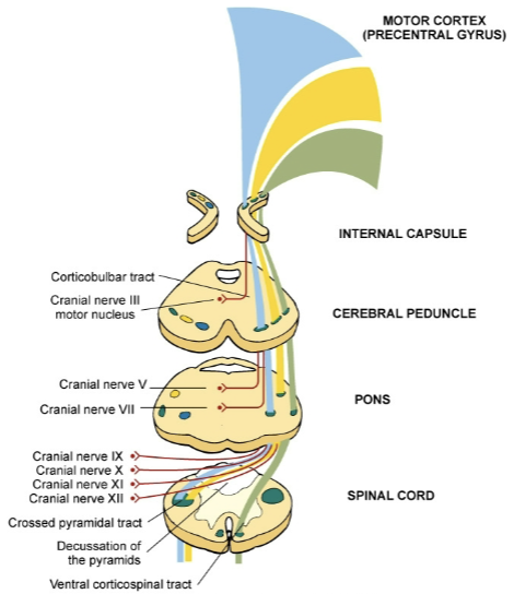
    • lower motor neuron syndromes

      • flaccid paresis or flaccid paralysis

      • hyporeflexia (reduced or diminished reflex responses) or areflexia (no reflex responses)

      • fibrillation (spontaneous, irregular contractions of individual muscle fibers)
      • amyotrophies (degeneration and atrophy of muscle tissue from loss of motor neurons)

        • paralytic poliomyelitis

          • infectious disease caused by polio virus, leads to inflammation of spinal cord and motor neurons, can result in paralysis and muscle weakness

        • nuclear palsies

          • disorders that result from damage to the cranial nerve nuclei in the brainstem (leading to deficits in the functions controlled by those nerves)

        • guillain barre syndrome

          • autoimmune disorder attacking the peripheral nervous system

        • progressive bulbar palsy

          • causes degeneration of the bulbar region (part of the brain stem) which leads to progressive weakness and atrophy of the muscles innervated by cranial nerves

        • bulbar palsy

          • like progressive bulbar palsy but results from conditions like stroke or trauma, symptoms either stabilize or improve rather than progressively worsen

    • hyperkinesia

      • excessive movement

      • chorea (involuntary, irregular, unpredictable muscle movements), wandering, tremor at rest

      • postural tremor etc.

    • paroxysmal dyskinesias

      • dyskinesia- sudden, involuntary, abnormal movements

      • paroxysm- a sudden, intense episode or attack of a condition or symptom

    • tardive dyskinesia

      • tardive- delayed or late onset of symptoms; in this specific context- dyskinesia that develops after prolonged exposure to certain meds

    • huntington disease

      • also known as chorea

      • autosomal dominant hereditary degenerative disorder

      • severe degeneration of the basal ganglia (caudate nucleus [a part of the basal ganglia]) and frontal cerebral atrophy

        • depletion of gamma-aminobutyric acid (gaba)

    • hypokinesia

      • decreased movement

      • akinesia

      • bradykinesia (slowed movement)

      • loss of associated movement

  • parkinson disease:

    • severe degeneration of the basal ganglia (corpus striatum) involving the dopaminergic nigrostriatal pathway

      • parkinsonian tremor, rigidity, bradykinesia

      • postural disturbances

      • autonomic and neuroendocrine symptoms

      • congitive affective symptoms

    • secondary parkinsonism (parkinsons symptoms from something else thats not parkinsons)

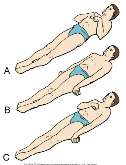
  • disorders of posture (stance):

    • dystonia (involuntary muscle contractions resulting in abnormal postures, twisting movements, or repetitive motions)

      • dystonic postures and movements

      • decorticate posture (A; also legs are turned in)

      • decerebrate posture (B, also back is arched)

      • basal ganglion posture (stiff/rigid limbs, flexed posture of limbs, stooped/leaning forward trunk)

      • senile posture (forward flexed trunk, rounded shoulders, slight flexion of hips and knees)
  • disorders of gait:

    • spastic gait (stiff/jerky movements, tendency to drag 1/both legs, reduced arm swing on affected side, difficulty w balance/coordination)

    • scissors gait (legs cross/scissor when walking, narrow base of support, short steps, hypertonia in legs)

      • base of support: the area beneath a person that supports their weight, determined by the distance btwn their feet when standing/walking

    • cerebellar gait (unsteady/wide based walking, difficulty w balance, lurching/swaying movements, frequent stumbling/falling)

    • basal ganglion gait (reduced arm swing, shuffling steps w a short stride length, difficulty initiating movements [freezing], stooped posture)

    • senile gait (slow/shuffling steps, broad base of support, reduced arm swing, difficulty w balance/coordination)

  • disorders of expression:

    • hypermimesis (exaggerated expressions of emotions)

    • hypomimesis (reduced expressions of emotions)

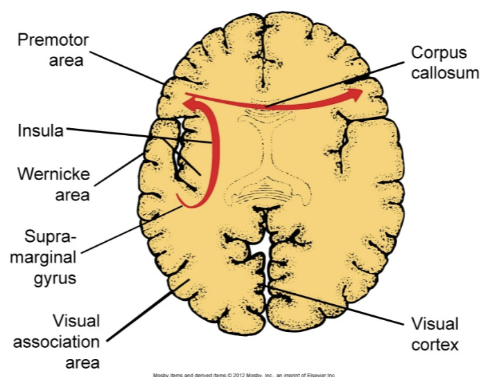
    • dyspraxias and apraxias (difficulty [dys]/inability [a] planning and executing coordinated movements)
  • extrapyramidal motor syndromes:

    • dysfunction of the extrapyramidal system which is part of the motor control pathway in the brain

    • basal ganglia motor syndromes (caused by dysfunction of the basal ganglia, results in bradykinesia, hypertonia, tremors [specifically resting tremors which happen when not moving the limb], dystonia, chorea etc.)

    • cerebellar motor syndromes (caused by dysfunction of the cerebellum, results in ataxia, dysmetria, tremors [specifically intention tremors which happen when trying to move the limb], hypotonia, difficulty w balance/posture etc.)

      • ataxia- lack of coordination and unsteady movements

      • dysmetria- inability to the control the distance/range of movements

robot