Interactivity: DNA Structure
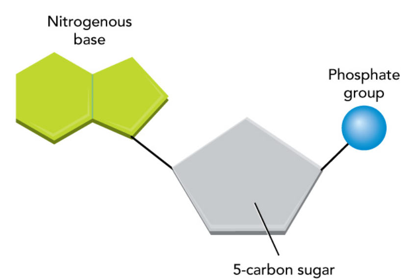
DNA is a large macromolecule classified as a nucleic acid. DNA is a polymer consisting of many smaller monomer units called nucleotides. Each nucleotide has a 5-carbon sugar ring, a phosphate group, and a nitrogenous base.
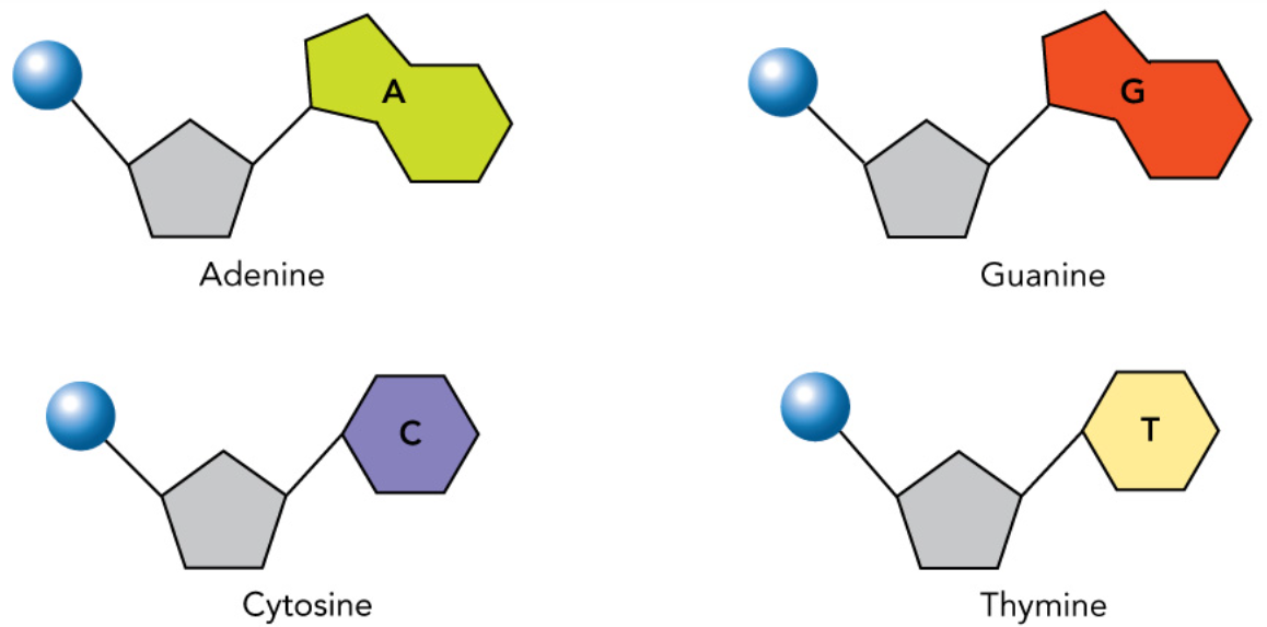
DNA contains four different types of nucleotides, each containing a different nitrogenous base: guanine, adenine, thymine, and cytosine.
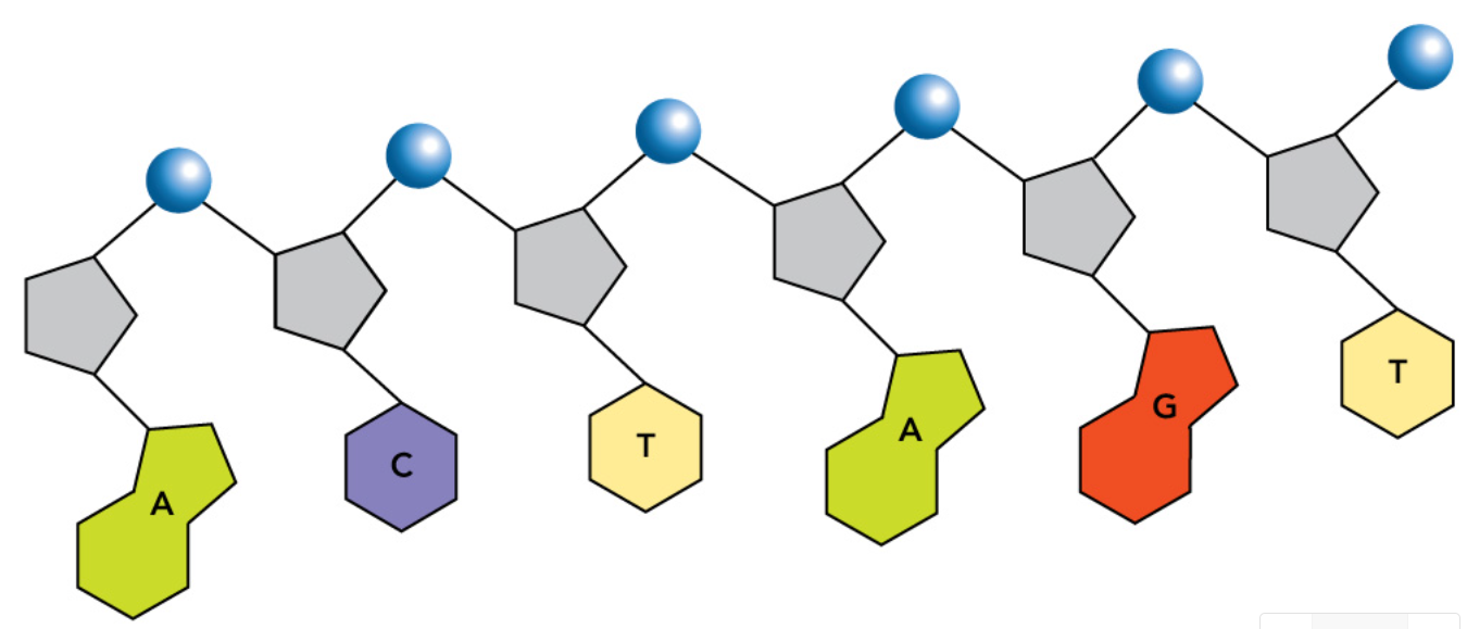
In each strand of DNA, nucleotides are connected by covalent bonds between the sugar of one nucleotide and the phosphate group of the next nucleotide. Some chromosomes can contain a few hundred million pairs of nucleotides.
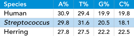
After the bases of DNA were identified, Erwin Chargaff analyzed the percentages of adenine, thymine, guanine, and cytosine in the DNA of many different organisms. This table shows the percentages of A, T, G, and C for a human, a bacterium, and a fish. Chargaff observed an approximate rule about these percentages in all of his samples: What is the rule?
A = T; G = C
Rosalind Franklin’s Contribution
Rosalind Franklin and Maurice Wilkins of King's College, London, tried to deduce the structure of the DNA molecule through the technique of X-ray diffraction. In this method, an isolated DNA molecule is bombarded with X rays and the pattern of reflected rays is shown on a photographic film. By observing parts of the pattern, you can deduce what some of the molecular structure must be.
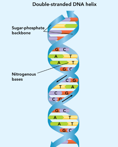
In 1952, Franklin produced an X-ray diffraction photograph that revealed two important clues about the structure of DNA. First is that the sugar-phosphate strands of DNA are twisted around each other in a double helix structure. Second is that the nitrogenous bases appear to be stacked at regular intervals near the center of the molecule.
Double Helix
In 1951, James Watson and Francis Crick began working together at Cambridge University to build a model of DNA. By this time Linus Pauling had showed that proteins often had a helical structure, so Watson and Crick tried using variations on a helix for their model, without success. However, after viewing Franklin's X-ray diffraction image, Watson and Crick were able to complete their model of DNA.
You can think of DNA as two strands of nucleotides, with each strand formed by a backbone of sugar and phosphate bonded together. First, the two strands are connected by hydrogen bonds between the nitrogenous bases, which are located between the two strands, forming the rungs of a ladder. Then, the entire ladder is twisted to form a double-stranded helix.
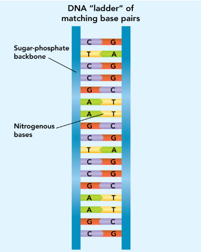
Base Pairing
A key part of the Watson-Crick model came when Watson realized that adenine could form hydrogen bonds with thymine and guanine could form hydrogen bonds with cytosine. This explains why A = T and G = C in Chargaff's rules. Also, these two hydrogen-bonded nucleotide pairs had the exact same width, so they could form the rungs of the DNA ladder.
The fact that these pairs could match up only in this way meant that the sequence of bases in one strand could determine the sequence of bases in a second strand created from the first. The second strand is said to be complementary to the first strand. Individual bases are paired so that the identity of any base determines the identity of the base paired with it; that is, the complementary base.