Chemical Signalling by Neurotransmitters
Communication Between Neurons
Chemical
Presynaptic cell -releasing neurotransmitter
Snaptic cleft
synaptic vesicles
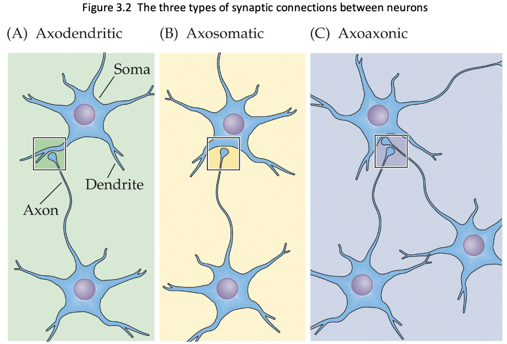
Usually, the communication between neurons is axodendritic communication
Neurons can contain more than one type of neurotransmitter
Classical neuropeptides (growth hormone)
Types of snapses:
Acodendritic - axon to dendrite
Axosomatic - axon to cell body
Axononic - axon to axon
Neuromuscular junction - axon to muscle
Electrical - electric current flows along specialized proteins
Mixed - both chemical and electrical transmission
Criteria for Status as a Neurotransmitter
presynaptic cell contains a substance and a method of making it.
There is a way to inactivate the substance.
Stimulation of a neuron leads to the release of substances.
A postsynaptic cell contains receptors for a substance
Application of a substance or an agonist to postsynaptic receptors should have a similar effect to stimulation of the presynaptic neuron.
Antagonistic drug should block the effects of both introducing the substance to the cleft and sitmulating the presynaptic neuron.
( All criteria not required)
Neurotransmitters
Classical
Acetylcholine (ACh)
Monoamines (biogenic amines)
Catecholamines
Norepinephrine (NE)
Dopamine (DA)
Epinephrine (E) (aka adrenaline)
Indoleamine
Serotonin (5-HT)
Others
Histamine (HA)
Amino acids
Gamma-aminobutyric acid (GABA)
Glutamate (excitatory)
Glycine (inhibitory, anti-inflammatory)
Purines -energy metabolism
Adenosine triphosphate (ATP)
Adenosine
Nonclassical
Neurppeptides
Endogenous opiates (morphinelike substances)
Enkephalins, Endorphins
Vairous hormones
Somatostatin, Vasopressin, Corticotropin-Releasing Factor (CRF)
Substance P
Orexin/hypocretin
Neurotrophic Factors
brain-derived neurotrophic factors (BDNF), nerve growth factor (NGF)
Many others
Lipids
Anadamide
2-arachidonoglycerol
Gases
Nitric oxide (NO)
Carbon Monoxide (CO)
Hydrogen Sulphide (H2S)
Neurotrophic Factors: Neuron growth factors that influence growth, cell differentiation and survival, and maintenance of synaptic connections determine which dendrites grow and which react
NGF (nerve growth factor) is secreted by peripheral organs; guides the growth of axons to establish synapses with the target organ.
BDNF (brain-derived neurotrophic factor) is released from skeletal muscle; guides motor neuron development and survival; supports survival of neurons and brain cells, promotes synaptic connections between neurons, and is important for learning and LTM storage; also plays a role in adult neurogenesis.
Neurtrophin-3 -3 (NT3)
Nerotoprpin- 4/5
Neuromodulator: affects the activity of NTs by enhancing, reducing, or prolonging the transmitter’s effectiveness (all neurotransmitters) -in textbook
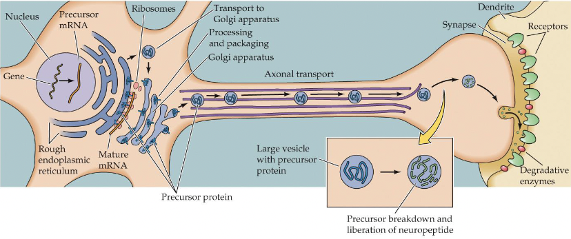
All NTs (except neuropeptides) can be produced anywhere in the cell, but are usually made in the axon terminals (lots of enzymes).
Neuropeptides are manufactured in the cell body.
Implication: Takes longer to replenish the supply of neuropeptides.
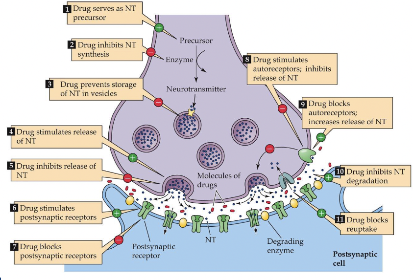
Release of Neurotransmitters
Exocytosis at active zones-
Synaptobrevin and synaptagmin-1 (proteins) help vesicles fuse with the membrane
Endocytosis (vesicles recycling only occurs with small vesicles) (clathrin is a protein that helps)
Three mechanisms control the release of NTs:
Rate of firing
Probability of NT release
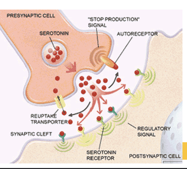
The presence of autoreceptors (feedback mechanism) helps a neuron determine whether or not the release neurotransmitter.
Inactivation of Neurotransmitters
Enzymatic breakdown in the cleft (e.g., acetylcholinesterase (AChE) breaks down ACh).
Reuptake by transporters on the axon terminal
Uptake by transporters on the post-synaptic cell or nearby glial cells
Drugs can interfere with inactivation
- e.g., Cocaine exerts effects by blocking reuptake of DA, 5-HT, and NE
Types of Neurotransmitter Receptors
Ionotropic (fast receptors)
4 or 5 protein subunits
ion channel opens when NT binds (ligand-gated channel receptors)
desensitization: channel is closed even though ligands are bound to the receptor.
Metaborophic (slower but longer lasting)
a.k.a. - G protein-coupled receptors (GPCRs)
1 protein subunit that crosses the membrane 7 times
no channels/pores; instead, activate G proteins (membrane), which either
(a) open or close ion channels on the membrane
(b) stimulate or inhibit effector enzymes in the cell membrane, which create or break down second messengers. The increase or decrease in second messengers causes changes in the cell.
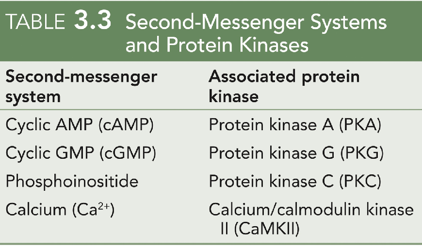
Second Messenger Systems
Molecules inside the cell that activate protein kinases to cause phosphorylation of substrate proteins
The added phosphate groups alter the function of the protein
Phosphorylation of nuclear proteins can turn gene expression on or off
Tyrosine Kinase (trk) Receptors
mediate the action of neurotrophic factors:
NGF (nerve growth factor), BDNF (brain-derived neurotrophic factor), NT -3 and NT-4 (neurotrophins).
Nerotropic factors bring two trk receptors together, resulting in reciprocal phosphorylation of tyrosine residues and activation of other protein kinases.
TrkA - NGF
trkB -BDNF & NT-4
trkC - NT-3
Neurotransmitters Outside of the CNS
Many of the same transmitters occur both inside and outside the CNS
Some neurotransmitters are synthesized by gut bacteria; drugs may alter the human microbiome and influence health.
The gut-brain axis refers to signalling back and forth between the gut (including the gut microbiome) and the brain.
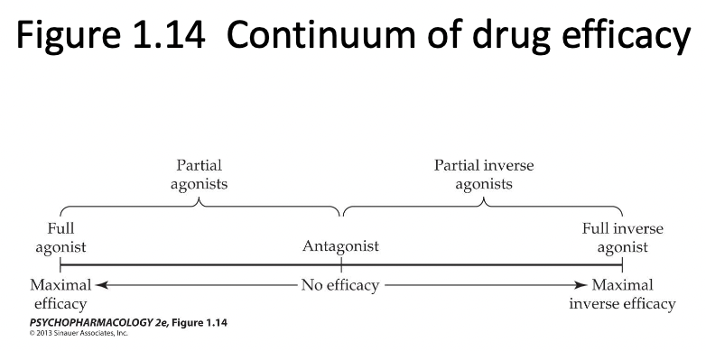
Drugs can have at least 4 actions when they bind to a receptor.
agonistic or partial agonistic action: bind to the receptor site that NT normally binds to and initiate a cellular response to the NT
allosteric action: bind to a receptor site near the normal site of a NT, which can then facilitate NT binding.
antagonistic action: binds to the receptor site that NT normally binds to, but NO initiation of cellular response and blocks NT from binding (antagonistic action).
2 types of antagonists.
(a) Competitive antagonists: drugs that compete with agonists to bind receptors but do not initiate intracellular effects, reducing the effect of the agonist (e.g., Naloxone)
(b) Noncompetitive antagonists reduce the effect of agonists by:
interfering with cell processes that were initiated by the agonist
Bindng to the receptor at a site other than the agonist binding site.
Disturbing the cell membrane supporting the receptor
Inverse agonistic action: initiates a biological action that is opposite to that produced by an agonist (and more than the basal activity - receptor needs to have some basal activity). H1 antihistomine
Allosteric Modulators
Many ionotropic and metabolic receptors have additional binding sites, called allosteric sites
Allosteric modulators: Molecules, such as drugs, that bind to such sites and alter the functioning of the receptor.
It can have a positive or a negative effect on receptor signalling.
How Drugs Alter Synaptic Transmission
The Endocrine System
Endocrine Glands and Their Respective Hormones.
Hormons are secreted by endocrine glands; circulated in the bloodstream
Target cells have hormone-specific receptors.
The same substance sometimes acts as both a neurotransmitter and a hormone.
Adrenal Glands
Adrenal medulla secretes epinephrine (EPI) and norepinephrine (NE) (monoamines).
Adrenal cortex secretes glucocorticoids (steroid hormones), Dehydroepiandrosterone (DHEA) and its metabolite DHEA sulphate (DHEAS) (both of these are transformed into androgens and estrogens)
Gonads:
Ovaries secrete estrogens and progesterone.
Testes secrete androgens (e.g., testosterone).
Islets of Langerhans, in the pancreas, secrete insulin and glucagon (peptide hormones) to regulate glucose.
Thyroid gland -secretes thyroxine (T4) and triiodothyronine (T3) - regulates energy metabolism.
Pineal gland - secretes melatonin - control of sleep and other rhythms
Pituitary gland - secretes hormones that control other glands.
Anterior pituitary - secretes stimulatory hormones: TSH, ACTH, FSH, LH, GH, and PRL.
Posterior pituitary - secretes vasopressin and oxytocin.
The hypothalamus secretes releasing hormones to trigger the secretion of stimulating hormones by the anterior pituitary
Hypothalamic releasing hormones: released by neurons in the median eminence.
Blood vessels in pituitary stalk carry them to the anterior pituitary.
Tytotropin-releasing hormone (TRH) stimulates TSH, corticotropin
Hormonal and Neurotransmitter Functions of Oxytocin and Vasopressin
Vasopressin (VP) and oxytocin (OT): synthesized in the hypothalamus by neurons whose axons reach the posterior pituitary gland.
VP (antidiuretic hormone) regulates water retention in the kidneys.
OT stimulates uterine contraction during childbirth and triggers milk letdown from the breasts.
VP and OT neurons form the paraventricular nucleus, connected to many other brain regions.
The brain areas receiving VP and/or OT
Oxytocin research: OT can influence a variety of social behaviours, including empathy, altruism, trust, and social memory.
Mechanism of Hormone Action
Most peptide hormones act through membrane metabotropic receptors.
Insulin uses tyrosine kinase receptors
Steroid and thyroid hormones operate mostly through intracellular receptors in the cell nucleus; they function as transcription factors.
Thus, it can have slower but long-lasting effects.
The Endocrine System is Important to Pharmacologists
Dugs can adversely alter endocrine function.
Hormones may alter behavioural responses to drugs.
Hormones sometimes have psychoactive properties
Because pituitary hormones are controlled by neurotransmitters in the brain, the endocrine system can tell us if a neurotransmitter system has been altered.