Chapter 7 - Axial Skeleton
The Skeleton
Consists of:
Bones, cartilage, joints, & ligaments
Composed of 206 named bones grouped into 2 divisions
Axial skeleton (80 bones)
Appendicular skeleton (126 bones)
The Axial Skeleton
Consists of skull, vertebral column, & thoracic cage (ribs & sternum)
Forms the long axis of the body
Helps support the head, neck, and trunk
The Skull
Formed by cranial and facial bones
The Cranium
Considered the body’s most complex bony structure
Serves to surround and protect the brain
Provides attachment for head & neck muscles
Facial bones serve to
Form framework of the face
Form cavities for the sense organs of sight, taste, and smell
Provide openings for the passage of air & food
Hold the teeth in place
Anchor muscles of the face
Cranial Bones
Formed from eight large bones
Paired bones include
Temporal bones
Parietal bones
Unpaired bones include
Frontal bone
Occipital bone
Sphenoid bone
Ethmoid bone
Parietal Bones & Sutures
Parietal bones form superior & lateral parts of the skull
Sutures - interlocking, immovable joint that will unite the skull bones firmly together; rigid zig-zaggy appearance
Major sutures of the cranium
Coronal suture - Articulates the parietal and frontal bones together
Runs in the coronal plane
Squamous suture - Articulates the parietal and temporal bones together
Lambdoid suture - Articulates the parietal and occipital bones together
Sagittal suture - Articulates the parietal bones together
Lies right on the midline
Occipitomastoid suture - Between the occipital bone and mastoid process
ONLY suture that doesn’t concern the parietal bone
Frontal Bone (unpaired)
Forms the forehead and roofs of orbits
Supraorbital margin - superior margin of orbits
Glabella - smooth part of frontal bone between superciliary arches (structures deep to the eyebrows)
Borders the frontal sinuses
Located near the nasal cavities
Occipital Bone (unpaired)
Forms the posterior portion of the cranium & cranial base
Part of the skull that forms walls holding the cerebellum
Foramen magnum located at its base
“Magnum” = meaning large
Marks the connection of the brain to the spinal cord
Flanked by a pair of structures called “occipital condyles”
Temporal Bones (paired)
Lie inferior to parietal bones
Specific regions of temporal bone
Squamous - plate shaped portion of the bone
Tymphanic - surrounding the external acoustic meatus
Contains styloid process which attaches to stylohyoid ligament
Mastoid - contains the mastoid process, a lump right behind the ear
Site for neck attachment
Contains air sinuses
Petrous - harbor cavities that belong to the middle & inner ear
Can only be seen from the inferior aspect
Sphenoid bone (unpaired)
“Keystone” cranial bone because it forms a central wedge that articulates w/ every other cranial bone
Spans the width of the cranial floor
Resembles a butterfly or bat w/ spread wings
Consists of a body and three pairs of processes
Greater wings
Lesser wings
Pterygoid process
Sella turcica - “turkish saddle”
Contains a depression on it called the “hypophyseal fossa”
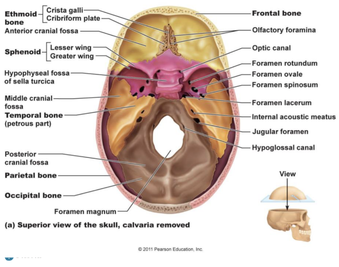
Ethmoid bone (unpaired)
Bone that is most deeply situated into the skull
Delicate, thin bone
Lies between nasal and sphenoid bones
Forms most of the medial bony region between the nasal cavity and orbits
Cribriform plate - superior surface of the ethmoid bone
Forms the roof of the nasal cavity
Contain olfactory foramina (tiny holes that olfactory nerves pass through)
Crista galli - divides the cribriform plate into 2 sections
Attachment site for a fibrous membrane called the falx cerebri
Perpendicular plate - forms superior part of nasal septum
Flanked by lateral masses (L and R lateral mass)
Filled w/ ethmoidal air cells (a.k.a ethmoid sinuses)
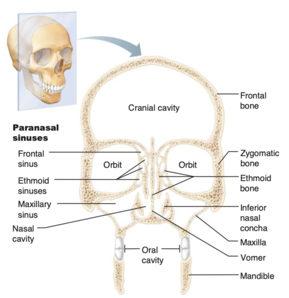
Facial Bones
Unpaired
Mandible
Vomer
Paired
Maxillae
Zygomatic bones
Nasal bones
Lacrimal bones
Tear drop shape
Palatine bones
Inferior nasal conchae
Shell shaped
Mandible (unpaired)
ONLY bone of the skull that moves
Lower jaw bone is largest and strongest facial bone
Composed of 2 major parts
Horizontal body
Alveolar margin
Superior border of the horizontal body
Contain “alveoli” (tooth sockets) that secure the teeth in place
Ramus of mandible
Meets the body at the mandibular angle
Temporomandibular joint (TMJ) - formed by the the articulation of the mandibular condyle to the temporal bone
Maxillary Bones (paired)
Form the upper jaw
Central portion of the facial skeleton
Also contains an alveolar margin
Inferior border of the maxilla
Contain alveoli
Contains maxillary sinuses
Make up ¼ of the paranasal sinuses
Zygomatic process that meets w/ the zygomatic bone
Palatine process
Forms the anterior part of the hard palate
Only visible from the anterior aspect
Articulates w/ every other facial bone except for the mandible
Zygomatic bones (paired)
Also known as the cheekbones
Joins w/ 3 different zygomatic processes
Zygomatic process belonging to the temporal bone
Zygomatic process belonging to the frontal bone
Zygomatic process belonging to the maxilla
Zygomatic arch is held together by articulating bones
Other Bones of the Face
Nasal Bones (paired)
Form the bridge of the nose
Very delicate
Palatine bone (paired)
Horizontal plate of this bone forms the posterior aspect of the hard palate
Perpendicular plate forms part of the lateral wall of the nasal cavity
Vomer (unpaired)
Forms the inferior region of the nasal septum
Completion of the septum is aided by septal (hyaline) cartilage
Inferior nasal conchae (unpaired)
Projecting medially from the lateral portion of the nasal cavity
Superior to the hard palate
Lacrimal bones (paired)
Nasal Cavity
Superior to the hard palate
Harbor sensory receptors that help in sense of smell
Contains the following:
Roof (floor of the nasal cavity)
Floor (roof of the oral cavity)
Lateral walls - formed by the superior nasal concha and middle nasal concha
Contains canal-like passageways called “meatuses” in between, w/ purpose of increasing air turbulence
Paranasal Sinuses
Air-filled sinuses located within
Frontal
Ethmoid
Sphenoid
Maxillary
Lined w/ mucus membrane that warms and moistens inhaled air
Help lighten the skull
The Hyoid Bone
Connects to temporal bone via stylohyoid ligament
Special because it is the only bone w/ no direct articulation with any other bone
Not considered part of the skull, but still associated with it
Structures:
Centrally positioned body
Greater horns
Lesser horns (location of connection to stylohyoid ligament)
Acts as a movable base for the tongue
The Vertebral Column (Spine)
Formed from 26 bones in the adult
Acts as the main support of the body axis
Transmits weight of trunk to the lower limbs’
Vertebral cavity harbors and protects the spinal cord
Articulates w/ the ribs
Get larger as you move down inferiorly (adjusts to the increasing weight in each section)
Held in place by ligaments of dense regular CT
Anterior and posterior longitudinal ligaments - run straight, attach to the vertebral bodies
Ligamentum flavum (a.k.a. adjacent vertebrae)
5 major regions
Cervical; C1-C7
Thoracic; T1-T12
Lumbar; L1-L5
Sacral; 5 fused vertebrae
Coccyx; (usually) 4 fused vertebrae
Intervertebral discs
Cushion-like pads between vertebrae that absorb shock
Found between all vertebrae EXCEPT between C1 and C2
Composed of
Nucleus pulposus forming the inner sphere
Annulus fibrosus forming concentric rings that limit the expansion of nucleus pulposus
Herniated disc - Nucleus pulposus squeezes out of its usual position and compresses spinal nerve fluid, causing excruciating pain
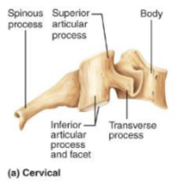
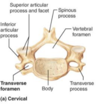
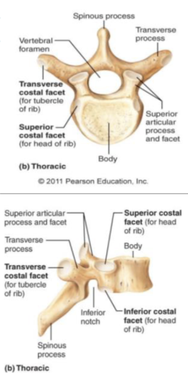
General Structure of Vertebrae
Common structures include
Body
Vertebral arch
Vertebral foramen → forms vertebral canal w/ all 26 vertebrae
Spinous process
Transverse process
Superior & inferior articular process
Superior & inferior articular facet → on the surface of the process
Differentiating Between Vertebrae
Cervical vertebrae - smallest and lightest
C1 (atlas)
Only vertebrae that lacks a body and spinous process
Vertebral foramen is ring-shaped
Supports the skull
Superior articular facets receive the occipital condyles
Allows flexion & extension of neck
C2 (axis)
Has a normal spinous process (not bifid)
Dens (odontoid process) projects superiorly through C1
Result of the body of the atlas fusing with the axis
Participates in side to side head movement (indicating no)
C3-C6 are typical cervical
vertebrae
Body is wider laterally
Spinous processes are
short and bifid
Vertebral foramen are
large and triangular
Transverse processes
contain transverse foramina
C7
“Vertebra prominens” - very large & long spinous process (not bifid)
Thoracic vertebrae
All articulate w/ ribs
Have heart-shaped bodies from the superior
view
Spinous processes are long and point
inferiorly
Vertebral foramen is circular
Transverse processes will form the letter “W”
Lumbar vertebrae
Bodies are very thick and robust
Transverse processes are thin/flat and
tapered
Spinous processes are thick, blunt, and point
posterior
“Hatchet” shaped
Vertebral foramina are triangular
Sacrum
Shapes the posterior wall of pelvis
Superior surface articulates w/ L5
Inferiorly articulates w/ coccyx
Sacral promontory (only visible from anterior aspect)
Sacral foramina - allow passage of sacral nerves
Coccyx
The “tailbone”
Offers only slight support to pelvic organs
The Thoracic Cage
Forms the framework of the chest
Components
Thoracic vertebrae - posteriorly
Ribs - laterally
Sternum and costal cartilage - anteriorly
Protects thoracic organs
Supports shoulder girdle & upper limbs
Attachment sites for skeletal muscles at the back, neck, shoulder, and chest
Sternum
Shaped like a dagger
Manubrium - handle of the “dagger”
Jugular notch - indentation at the superior portion of manubrium
Clavicular notch - indentation for association w/ the clavicle
Attached to rib #1
Major portion is made up of the body
Contains notches to receive ribs 2-7
Xiphoid process - inferiorly lying
Made up of costal cartilage
Does Not completely become bone until we're 40
The Ribs
All ribs attach to vertebral column posteriorly
True ribs (pairs 1-7)
“True” because they directly connect to the sternum via costal cartilage
False ribs (pairs 8-12)
“False” because they are indirectly connected to the sternum, and
Floating ribs (pairs 11 & 12)
No anterior connection to the sternum AT ALL, literally floating