Radiology Chapters 38-42
Key terms Chapter 38:
ALARA Concept: As low as reasonably achievable
Ampere: A unit of electric current
Anode: Positive electrode in the x-ray tube
Atom: Basic unit of matter
Bremsstrahlung radiation: “braking radiation”; the sudden deceleration of electrons as they interact with highly positively changed nuclei
Cathode: Negative electrode in the x-ray tube
Central ray: X-rays at the center of the beam
Contrast: Differences in degrees of blackness on an image
Control panel: Portion of the x-ray unit that contains the master switch, the indicator light, the selector buttons, and the exposure button
Density: Overall darkness or blackness of an image
Dental radiography: Process of recording images of the teeth and adjacent structures to x-radiation
Dental radiograph: A photographic image produced on a receptor by the passage of x-ray through teeth and related structures
Dental radiographer: Any person who positions, exposes, and produces dental x-ray image receptors
Digital imaging: Filmless method of capturing an image and displaying it by using an image receptor. and electronic signal, and a computer to process and store the image
Image: A picture or likeness of an object
Image Receptor: A recording medium; examples include x-ray film, phosphor plate, or digital sensor
Imaging, dental: The creation of digital, print, or film representations of anatomic structures for the purpose of diagnosis
Kilovoltage: Highest voltage of x-ray tube used during and exposure
Latent: Time between exposure to ionizing radiation and appearance of symptoms
Lead apron: Device used to protect the reproductive and blood-forming tissues from scatter radiation
Magnification: Proportional enlargement of an image
Master switch, Indicator light, Selector buttons, and Exposure button: Components of control panel
Matter: Anything that occupies space and has form or shape
Milliampere (MA): One one-thousandth (1/1000) of an ampere; a unit of measurement used to describe the intensity of an electrical current
Penumbra: Blurred or indistinct area that surrounds an image
Personal radiation monitoring badge: Device that measures exposure of personnel to ionizing radiation by measuring the intensity of visible light emitted from a crystal in the detector when heated; the intensity of light emitted depends on the radiation exposure
Photon: Minute (tiny) bundle of pure energy that has no weight or mass
Primary beam: Most penetrating beam produced at the target of the anode
Primary radiation: Same as a primary beam
Quality (of x-ray beam): Mean energy or penetrating ability of the x-ray beam
Quantity (of x-ray beam): Number of x-rays produced in the dental unit; the quantity of x-rays produced is controlled by mA
Radiation: Forms of waves of energy emission through space or material
Radiograph: Image produced on photosensitive film by exposing the film to radiation and then processing it
Radiology: The science of study of radiation as used in medicine; a branch of medical science that deals with the therapeutic use of x-rays, radioactive substances, and other forms of radiant energy
Radiography: The art and science of making radiographs by the exposure of a receptor to x-rays
Scatter radiation: Form of secondary radiation that occurs when an x-ray beam has been deflected from its path by interaction with matter
Sensor: Solid-state image receptor that contains a silicon chip with an electric circuit
Sharpness: Measure of how well an image reproduces the fine details or outline of an object
Somatic effects: Effects of radiation that cause illness and are responsible for poor health (such as cancer, leukemia, and cataracts) but are not passed on to offspring
Thyroid collar: Flexible lead shield that is placed securely around the neck
Tubehead: Part of the x-ray unit that contains the x-ray tube, the high-voltage and low-voltage transformers, and insulating oil
Tungsten target: Focal spot in the anode
X-radiation: High-energy ionizing electromagnetic radiation
X-ray: A beam of energy that has the power to penetrate substances and record image shadows on receptors (Photographic film or digital sensors)
Dental imaging enables the dentist to see conditions that are not visible in the oral cavity and to identify many conditions that might otherwise remain undetected.
Uses of dental images
Dental caries in the early stages
identify bone loss in the early stages
Locate abnormalities in surrounding hard and soft tissues
Evaluate growth and development
Provide information during dental procedures (root canals therapy)
Document a patient’s condition at a specific time
Radiation, which is used to produce all types of dental images, has the ability to cause damage to all types of living tissues.
Any exposure to radiation, no matter how small, has the potential to cause harmful biological changes in the operator and the patient
Wilhelm Conrad Roentgen discovered x-ray in 1895
The shorter the wavelength the greater the energy is
higher energy, short wavelength- can penetrate matter more easily
Lower energy, longer wavelength
All machines have 3 primary components: tubehead, extension arm, and control panel
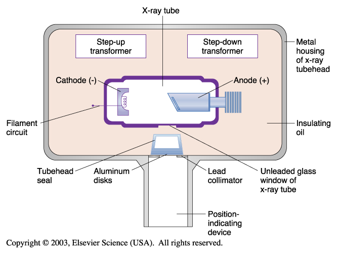
Components of the tubehead
The housing is the metal body that contains the x-ray tube
Insulating oils fills the housing and surrounds the x-ray tube. Oil prevents overheating by absorbing the heat created by the production of x-rays
The tubehead seal, made of leaded glass or aluminum, keeps the oil in the tubehead and acts as a filter for the x-ray beam
The x-ray tube is where x-rays are produced
The transformer alters the voltage of incoming electrical current
The collimator limits the size of the x-ray beam to a circular 2-inch opening at the PID
When the shape and size of the beam changes to a rectangle only slight larger than the film, the amount of tissue exposed to radiation can be reduced by more than half
The aluminum filter is a 0.5mm thick sheet located inside the PID
X-ray Tube
X-ray tube is the heart of the x-ray generating system
Dental Radiography Principles and Techniques Sixth Edition:
Chapter 1
Discovery of X-Radiation
Wilhelm Conrad Roentgen discovered the X-ray on November 8, 1895
Earlier Experiments
A sealed glass tube from which most of the air had been evacuated. The original vacuum was aka Geissler tube
Johann Wilhelm Hittorf- Used the vacuum tube to study fluorescence
In 1870 he observed that the discharges emitted from the negative electrode of the tube traveled in straight lines, produced heat, and resulted in a greenish fluorescence. He called the discharges cathode rays
In 1894, Phillip Lenard discovered that cathode rays could penetrate thin window of aluminum foil built into the walls of the glass tubes and cause fluorescent screens to glow
Pioneers In Dental X-Radiation
Otto Walkhoff made the first dental radiograph in 1895
That same year W.J. Morton, a New York physician, made the first practical use of radiographs in the United States using a skull
C. Edmund Kells was the first credited doctor with the first practical use of radiographs in dentistry in 1896
William H. Rollins is a Boston dentist who developed the first dental x-ray unit
Frank Van Woert is a dentist from New York City who was the first to use film in intraoral radiography
History of Dental Radiographic Techniques
Weston Price introduced the bisecting technique in 1904
Howard Riley Raper Redefined the original bisecting technique and introduced the bite-wing technique in 1925
The paralleling technique was introduced by C. Edmund Kells in 1896
in 1933 Hisatugu Numata of japan was the first to expose a panoramic radiograph
History of Dental Digital Imaging
Digital imaging allows for instant and easy transmission of images and electronic storage
History of Three-Dimensional Digital Imaging
In 1999, cone-beam computed tomography was introduced to dentistry
Chapter 2
Atomic and Molecular Structure
The world is composed of matter and energy
Matter- Anything that occupies space and has mass
When matter is altered, energy results
The fundamental unit of matter is the atom
Nucleus- dense core of the atom, is composed of particles known as protons and neutrons
Proton- Carry positive electrical charges
Neutron- Carry no electrical charge
The number of protons and neutrons determines its mass number or atomic weight
Atomic Weight- The number of protons inside the nucleus equals the number of electrons outside the nucleus
Elements- Substances made up of only one type of atom
Electrons- Tiny, negatively charged particles that have very little mass
Electrons travel around the nucleus in well defined paths known as orbits or shells
Ionization
An atom that gains or loses and electron and becomes electrically unbalanced is known as an ion
Ionization- The production of ions, or the process of converting an atom into ions
Radiation and Radioactivity
Radiation- The emission and propagation of energy through space or a substance in the form of waves or particles
Radioactivity- The process by which certain unstable atoms or elements undergo spontaneous disintegration, or decay, in an effort to attain a more balanced nuclear state
Ionizing Radiation
Ionizing Radiation- Radiation that is capable of producing ions by removing or adding an electron to an atom
It can be classified into two groups:
Particle radiation
Electromagnetic Radiation
Particulate Radiation
Particulate Radiation- Tiny particles of matter that posses mass and travel in straight lines and at high speeds
Cathode Rays- Streams of high-speed electrons that originate in the x-ray tube
Electromagnetic Radiation
Electromagnetic Radiation- The propagation of wavelike energy (without mass) through space or matter
Electromagnetic Spectrum- Electromagnetic radiations are arranged according to their energies
Principle Concept: Characterizing electromagnetic radiations as discrete bundles of energy called photons or quanta
Photons- Bundles of energy with no mass or weight that travel as waves at the speed of light and move through space in a straight line, “carrying the energy” of electromagnetic radiation
Wave Concept: Characterizing electromagnetic radiations as waves and focuses on the properties of velocity, wavelength, and frequency
Velocity- The speed of the wave
Wavelength- The distance between the crest of one wave and the crest of the next
Frequency- The number of wavelengths that pass a given point in a certain amount of time
If the frequency of the wave is high, the wavelength will be short
If the Frequency of the wave is low, the wavelength will be long
Long wavelength, low frequency
Short wavelength, high frequency
X-Radiation
X-rays- Can be defined as weightless bundles of energy (photons) without an electrical charge that travel in waves with a specific frequency at the speed of light
X-ray photons interact with the materials they penetrate and cause ionization
X-ray Machine
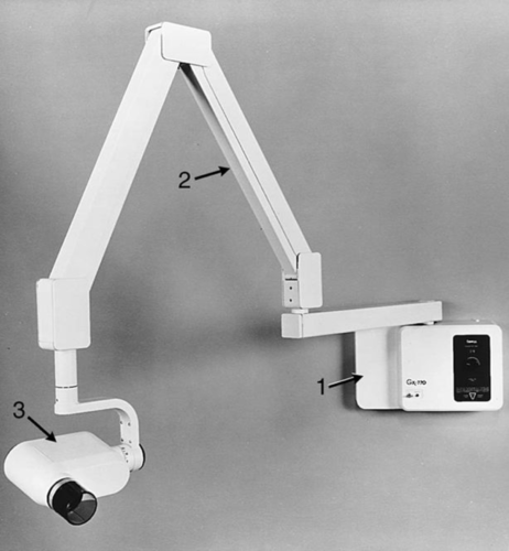 1. Control Panel
Extension Arm
Tubehead
Control Panel- Contains an on-off switch and indicator light, an exposure button and indicator light, and control devices (time, Kvp, mA) to regulate the x-ray beam
Extension Arm- Suspends the x-ray tubehead and houses the electrical wires that extend from the control panel to the tubehead. The extension arm allows for movement and positioning of the tubehead
Tubehead- Is a tightly sealed, heavy metal housing that contains the x-ray that produces dental x-rays.
Components of the tube head
Metal Housing- The metal body of the tubehead that surrounds the x-ray tube and transformers and is filled with oil- protects the x-ray tube and grounds the high-voltage components
Insulating Oil- The oil that surrounds the x-ray tube and transformers inside the tubehead- prevents overheating by absorbing the heat created by the production of x-rays
Tubehead Seal- The aluminum or leaded-glass covering of the tubehead that permits the exit of x-rays from the tubehead- seals the oil in the tubehead and acts as a filter to the x-ray beam
X-ray Tube- The heart of the x-ray generating system
Transformer- A device that alters the voltage of incoming electricity
Aluminum Disk- Sheets of 0.5-mm thick aluminum placed in the path of the x-ray beam, filter out the non-penetrating, longer wavelength x-rays.
Lead Collimator- A lead plate with central hole that fits directly over the opening of the metal housing, where the x-rays exit restricts the size of the x-ray beam
Position-indicating Device (PID)- Open-ended, lead-lined cylinder that extends from the opening of the metal housing of the tubehead- aims and shapes the x-ray beam. The PID is sometimes referred to as the cone
X-ray Tube
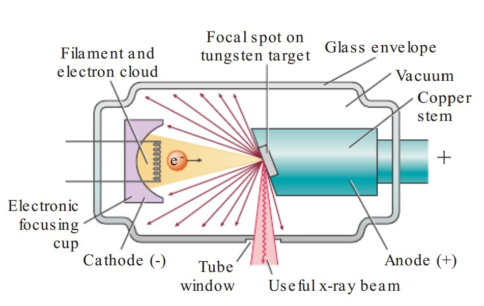
The X-ray tube is the heart of the x-ray generating system; It is a critical to the production of x-rays
Leaded-Glass Housing- A leaded-glass vacuum tube that prevents x-rays from escaping in all directions. One central area of the leaded-glass tube has a “window” that permits the x-ray beam to exit the tube and directs the x-ray beam toward the aluminum disks, lead collimator, and PID
Cathode- Negatively electrode, consists of a tungsten wire filament in a cup-shaped holder made of molybdenum. The purpose of the cathode is to supply the electrons necessary to generate x-ray. In the x-ray tube, the electrons produced in the negative cathode are accelerated toward the positive anode
Tungsten Filament- Coiled wire made of tungsten, which produces electrons when heated
Molybdenum Cup- Focuses the electrons into a narrow beam and directs the beam across the tube toward the tungsten target of the anode