DNA Structure
Cast of Characters
Francis Crick
X-ray diffraction on polypeptides and proteins
James Watson
Created the structural model
~Asshole~
Maurice Wilkins
Worked with X-ray crystallography
First work on X-ray diffraction of DNA
Rosalind Franklin
Used x-ray diffraction to obtain pictures of the DNA molecule
INDEPENDENTLY determined structure
PERIOD I LOVE HERRRRR
Where Did They Start?
Biochemistry- Some biological molecules, like proteins can form helical structures. Discovered by Linus Pauling, Robert Corey, and Herman Branson.
Transmission Genetics-
Must be able to replicate
Must contain information
Must be variable (alleles)
Must be relatively stable
Using the molecular information obtained by chemist and the data of Rosalind Franklin:
Previous Discoveries + Physical “Blueprint” = Model of DNA (Sugar phosphate outside, Nitrogenous bases on the inside in pairs)
The 3D Structure of DNA
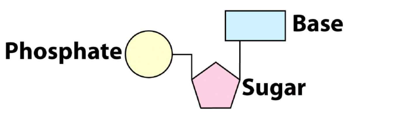
Nucleotide: 3 Basic Components
Deoxyribonucleic Acid
Deoxyribose- sugar
Nitrogen base- pyrimidine or purine (5 types)
Phosphoric acide- phosphate
Nucleotide = all three components
Nucleotide: Nitrogenous Base
DNA Base Names have Y in them and so does the Pyrimidine- Pyrimidines have the simple structure of single ringPurines have the more complicated structure of the double ring
Nucleotide: Phosphate Groups
Can have 1, 2, 3 phosphate groups
the nucleotides loose in the cell have 3 phosphate bases and is used for chemical reaction in the cell: ATP (Adenine Triphosphate)
Single DNA Strands
DNA is a polymer: A chain of nucleotide monomers
5’ End (5 prime end)- the end of the DNA strand thatr has the phosphate group
3’ end (3 prime end)- has the three sugars at the end, nucelotides are always added to the 3’ end
DNA always read 5’ to 3’
Nucleotide triphosphate is added to a growing DNA strand by an enzyme called DNA polymerase
the different ends (5’ and 3’) are called Polarity/ Directionality = chemical polarity with respect to the numbers carbons in the sugar
DNA Duplex
DNA is two antiparallel and Complementary DNA strands
2 stands come together due to complimentary base pairing
Built by Phosphodiester bonds
Complementary Base Pairs
Always a pyrimidine (one ring) and a purine (two rings)
A-T (or A-U) and C-G (C-G is harder to separate bc three Hydrogen Bonds)
Complementary Base Pairing by Hydrogen Bonds (Weak bonds)
EX: 5’ end- TGTA
The reverse complementary- ACAT
The Double Helix
Two strands in a helix
One helical turn is 3.4 nm
Each turn has ~10 base pairs
The diameter is 2nm in width
3D Structure
DNA is NOT perfectly Symmetrical
The minor and the major grooves are important for interaction between DNA and proteins
Major groove has more interaction
DNA exists as packaged form as chromosomes inside a cell
There are 6 billion bases in our genome which equates to 6 feet of Linear DNA that needs to fit into a nucleus ( ~5-8 micron) (equivalent to fitting 24 mi of string into a tennis ball) ⬇
Happens through DNA Organization
DNA In the Cell
Supercoiling-
DNA can be supercoiled (over or under winding of DNA)
Supercoiled can either be positive (over) or negative (under -making the DNA helix more like the ladder)
Most DNA in most organisms is negatively supercoiled
REMEMBER: When writing the complimentary sequence, you read/write it from 5’ to 3’ (bottom to top)
DNA Organization
Organization starts with…
Supercoiling
DNA can be supercoiled
Supercoiled can be either negative or positive
Most DNA in most organisms is negatively supercoiled during important molecular processes
Nucleosomes and Chromatosomes
Nucleosomes are made up of a group of eight histones that act as the core- term nucleosome refers to both the histone core and the DNA wrapped around it
Histones are highly conserved proteins
Protein “tails” are stretches of amino acid that are often places where chemical modification can change uniform protein function.
DNA backbone is negatively charged while the histones are positively charged- allows the DNA to cling to the histone core and wrap around it
Chromatosomes- has the properties of the nucleosomes but now it has Histone 1
A bound H1 protein locks DNA into place, there are now two full turns of DNA around the octamer (166 base pairs) forming a chromatosomes
H1 acts like scotch tape and keeps DNA in place on the chromatosome
DNA: approx 3 BP per nm length→ “beads on a string”: approx 20 BP per nm
Solenoid Model (30nm Fiber)
Where the “beads on a string” continue to wind and coil to form…
Now at 100 BP per nm
Loop and Scaffold Model
Loops form on a protein scaffold of non- histone proteins
More condensation of DNA
The loops/fibers are then coiled around each other
The chromosome scaffold is a filamentous framework made up of a large
number of distinct nonhistone scaffold proteins
Steps Laid out:
Supercoiling
Nucleosomes and chromatosomes
Beads on a string
Solenoid
Loops
Coils
Loop and Scaffold model