8.1 the cardiovascular and respiratory system
the cardiovascular system
- heart
- blood vessels
- blood
function
- delivery of O2, fuel and nutrients to the tissues of the body
- removal of CO2 and waste products from the tissues
- maintenance of a constant body temperature - thermoregulation
- prevention of infection (immune function)
the heart
- formed from myocardium, a specialized muscle tissue
- surrounded by percardium (tough protective sac that fits loosely over the heart) which allows the heart to expland and contract
- epicardium - lines the outside of the heart
- endocardium - lines the inside of the heart
- made up of 4 separate chambers:
- atria and ventricles
- considered a double pump and is divided into the right and left heart separated by the interventricular septum
- right heart: pumps deoxygenated blood to the lungs (pulmonary circulation)
- left heart: pumps oxygenated blood to the rest of the body (systemic circulation)
- working out the heart increases the myocardium
- working out your cardio will increase the size of the ventricles to pump more blood
- healthy hearts have a lower BPM but pump more blood per pump
proper blood flow through the heart
- deoxygenated blood enters the right atrium of the heart through the superior and inferior vena cava
- deoxygenated blood travels from the right atrium through the tricuspid valve into the right ventricle
- deoxygenated blood is pumped from the right ventricle through the pulmonary valve into the pulmonary artery
- the pulmonary artery carries the blood to the lungs to be oxygenated
- blood from the lungs travels through the pulmonary veins into the left atrium of the heart
- oxygenated blood travels from the left atrium through the mitral valve into the left ventricle
- oxygenated blood is pumped from the left ventricle through the aortic valve into the aorta
- the aorta carries the blood to other arteries that bring the blood to the rest of the body
excitation of the heart
- the sinoatrial node (SA node)
- specialized region of tissue found in the wall of the right atrium
- the location where electrical signals are initiated (pacemaker)
- atrioventricular node (AV node)
- passes electrical signal from atria into ventricles
- passes electrical signal to the bundle of His (atrioventricular bundle)
- bundle of His pass electrical signal to the purkinje fibers
- purkinje fibers pass electrical signals to the myocardium
- the myocardium contracts
- leads to contraction of the heart
- leads to the pumping of blood
electrical activity of the heart
- measured using an electrocardiogram (ECG)
- graphical representation of electrical sequence of events occurring with each contraction of the heart
- each wave generated during contraction is named
- P wave - represents contraction of the atria
- QRS complex - represents contraction of the ventricles
- T wave - represents the filling of the ventricles
- missing - filling of the atria (masked by the QRS complex)
cardiac cycle
- the cardiac cycle is a series of events occurring through one heartbeat
- diastole phase - relaxation, heart fills with blood
- systole phase - contraction, heart contracts and ejects blood
the vascular system and blood
- the vascular system is a network of vessels the transport blood throughout the body, vessels are divided into 4 categories
- arteries - carry oxygenated blood away from the heart to different organs (elastic and do not have valves)
- arterioles - smaller arteries
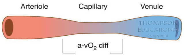
- capillaries - responsible for the exchange of gases and nutrients with the tissue
- venules - smaller veins
- veins - returns deoxygenated blood back to the heart (elastic and do have valves)
the return of blood from the veins
- 4 tools that assist the body in the return of the blood in the veins to the heart
- the skeletal muscle pump
- upon contraction of skeletal muscle, blood is pushed back to the heart
- the thoracic pump
- pressure in the veins in the chest decrease while pressure in the abdominal cavity increase upon intake of breath
- blood flows from high pressure to low pressure, pushing the blood from the abdomen to the thoracic cavity
- the nervous system
- sends signals to the veins allowing the veins to constrict and allow more blood to go back to the heart
- valves in the veins
properties of blood
- plasma (55% of blood)
- fluid component of blood (mostly water)
- blood cells
- red blood cells (erythrocytes) (45%)
- made in bone marrow
- transports O2 and CO2 in the blood
- transports nutrients and waste
- contains hemoglobin
- white blood cells (leukocytes) (<1%)
- destroys foreign elements
- critical in the function of the immune system
- platelets
- regulate blood clotting
blood pressure
- blood pressure is the force exerted by the blood against the walls of the arteries
- measuring blood pressure - systolic pressure over diastolic pressure
- systolic BP - pressure observed in the arteries during contraction phase
- diastolic BP - pressure observed during relaxation phase
normal BP
- normal bp is 120mmHg over 80mmHg
- hypertension
- BP greater than 140 over 90
- factors affecting BP that are controllable
- diet (sodium, saturated fat, cholesterol)
- aerobic exercise
- stress
- factors we do not have control over
- age
- genetics
BP and health
- blood pressure is a commonly used indicator of health
- elevated BP (hypertension) is a major risk factor for cardiovascular disease
- aerobic exercise training leads to improvements in resting blood pressure within three weeks to three months of starting exercise
- further improvements are made when exercise is coupled with improvements in diet (low fats, and cholesterol and high in fiber and complex carbohydrates)
- cardiovascular disease: atherosclerosis - associated with the narrowing of the coronary arteries resulting from the accumulation of hard deposits of cholesterol called plaque in the lumen of the arteries
- arteriosclerosis - the hardening of the arteries
- myocardial infarction: heart attack - blockage involving the death of some of the cardiac muscle
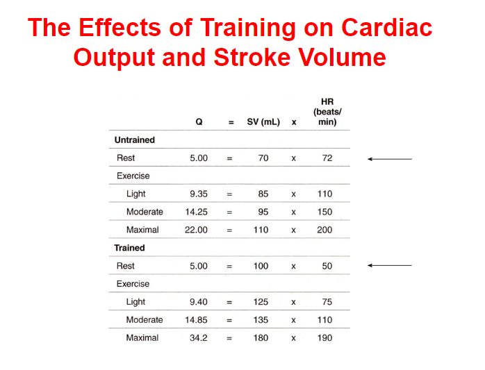
cardiac output and stroke volume
- cardiac output (q) - the amount of blood pumped out of the left ventricle in one minute (L/min)
- stroke volume (SV) - the amount of blood pumped out of the left ventricle with each beat (mL)
- Q = HR x SV
- average cardiac output during exercise
- 25-30 L/min
- average cardiac output at rest
- 5-6 L/min
the effects of aerobic training on the cardiovascular system
- Increase in venous return
- Increase in ventricular volume and thickness of ventricle walls (myocardium) = increase in SV
- Increase in Q
- Increase in the number of capillaries that deliver oxygen to the myocardium
- Increase in diameter of coronary arteries
- Increase in blood volume
- Increase in red blood cells
- at rest: increase in SV and decrease in HR
- during exercise: blood flow is redirected to the muscles from the organs (except the brain where blood flow is unaltered)
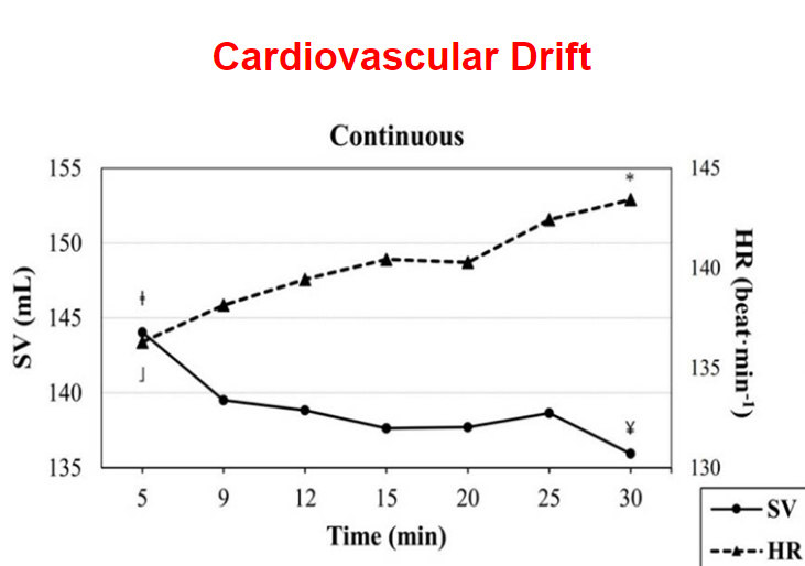
cardiovascular drift
- cardiovascular drift is a phenomenon that occurs when during an easy steady state aerobic activity (run) there is an increase in HR and decrease in SV to maintain Q
- begins after 10-20 minutes (without an increase in effort)
- heightened during warm temperatures
- caused by:
- increase in body temperature and redistribution of blood flow to the skin for cooling
- decrease in plasma volume due to dehydration
how endurance and resistance exercise affect blood pressure
- during endurance exercise
- systolic blood pressure increases (but diastolic does not due to vasodilation of the arteries)
- during resistance exercise
- systolic and diastolic blood pressure increases
- post exercise hypotension
- blood pressure drops below normal resting values persisting for about 24 hrs after exercise due to pooling of blood in vasodilated muscle beds
the respiratory system
- composed of structures that allow
- passage of air from outside the body to the lungs
- gas exchange to occur
- 3 main functions
- supply O2 to the blood
- remove CO2 from the blood
- regulate blood pH
- divided into 2 zones
- conductive zone
- respiratory zone
the conductive zone
- composed of structures that transport air to the lungs
- mouth and nose
- pharynx
- larynx
- trachea
- primary and secondary bronchi
- tertiary and terminal bronchioles
- warms and humidified air
- filters air (hair and mucous)
the respiratory zone
- composed of structures involved with the exchange of gases
- respiratory bronchioles
- alveolar sacs (ab. 300 million)
mechanisms of breathing
- inspiration
- contraction of diaphragm (lowers)
- thoracic cavity expands
- air enters
- expiration
- relaxation of diaphragm (goes up)
- air is expelled
ventilation and control of ventilation
- ventilation (Ve) - the volume of air that is moved by the lungs in one minute
- can increase up to 100/200 L of air/minute
- tidal volume (Vt) - volume of air in each breath
- respiratory frequency (f) - the number of breaths per minute
- at rest - 12 breaths/min
- during exercise - 30/40 breaths/min
- central chemoreceptors are found in the brain stem and they detect changes in brain CO2 and pH
- peripheral chemoreceptors are found in places like the aorta
adaptations to training
- regular aerobic training leads to very few adaptations in the respiratory system at rest
- increase in Vt
- decrease in f
- no changes in Ve
- during sub-maximal exercise
- increase in Vt
- decrease in f
- increase in Ve
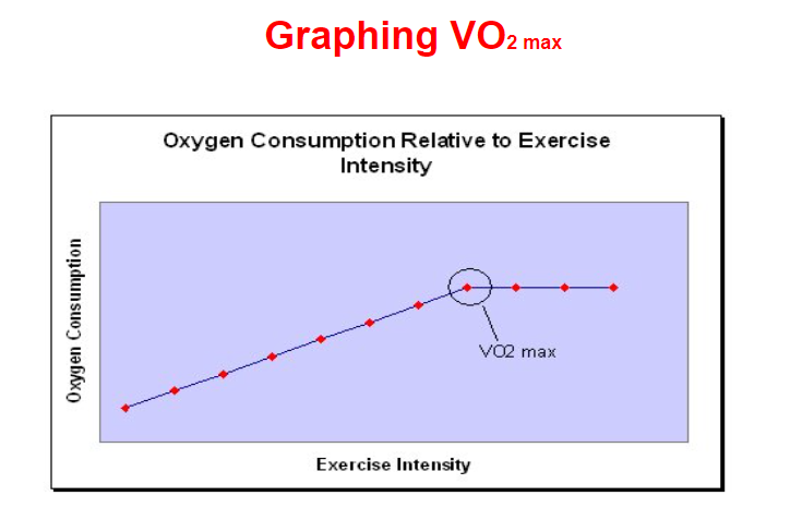
maximal oxygen consumption (VO2 max)
- maximum rate of oxygen consumption (VO2 max) - maximal amount of oxygen that can be taken in and used for metabolic production of ATP in 1 minute/kg of body weight during maximal exercise
- measured while participant performs incremental exercise to exhaustion
- used as a measure of aerobic fitness
- the limiting factor in healthy people is the cardiovascular system, when this system is unable to meet the demands of the working muscle and deliver adequate amounts of oxygen
- limitations due to:
- inadequate Q
- inadequate hemoglobin concentration
how is VO2 max calculated
- VO2 max - mL/kg/min
- VO2 max = (volume of air inspired x %O2 inspired) - (volume of air expired x %O2 expired)
- average female in the good range is 40-43 mL/kg/min
- average male in the good range is 46-50 mL/kg/min
- the most effective way to improve VO2 max is to train using high intensity interval training where the athlete maximizes the amount of time he/she is at or close to VO2 max
- VO2 max is largely based on genetics, specifically hemoglobin concentrations and types of muscle fibers
- VO2 max peaks at age 18 and decreases by 1% per year
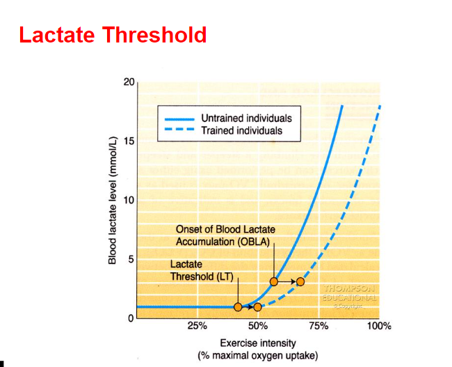
ventilatory and lactate thresholds
- ventilatory threshold - point when ventilation increases much more rapidly than workload
- usually occurs at 65-85% of VO2 max depending of level of fitness
- due to increase in lactic acid and decrease in blood pH (due to increase in CO2)
- lactate threshold - point where blood lactate exceeds the body’s ability to clear it
- the best predictor of performance in endurance events
- onset of blood lactate accumulation - point when blood lactate levels begin to accumulate rapidly
- both LT and OBLA can be shifted to the right with aerobic training
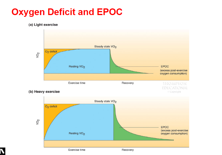
oxygen deficit
- ultimately the delivery of oxygen is matched to the demand of oxygen but there is a lag since the physiological mechanisms are not instantaneous
- oxygen deficit - the difference between the oxygen required to perform a task and the oxygen actually consumed prior to reaching a new steady state
- steady state - submaximal exercise levels where oxygen uptake and heart rate level off, where oxygen demands and energy production are evenly balanced and where the body maintains a steady level of exertion for a fairly extended period of time
- the trained person will reach this plateau quicker than an untrained person and will have a smaller oxygen deficit for an exercise of a given duration
excess post-exercise oxygen consumption (EPOC)
- excess post-exercise oxygen consumption - the additional oxygen taken in during this recovery period in order to restore balance
- the additional oxygen requirements are used for
- replenish oxygen to the various body systems that were taxed during the exercise which include
- refilling phosphocreatine reserves in muscles
- replenishing oxygen in the blood and tissue
- lowering elevated heart rate and breathing
- lowering body temperature
- increased blood lactate removal
amount of oxygen delivered to the skeletal muscle
- a-VO2 difference - one way to measure the amount of oxygen that has been delivered to a skeletal muscle by measuring the amount of arterial blood before it arrives and venous blood after it leaves indication how much oxygen is removed from the blood in the capillaries
- at rest: 4.5 mL/100L/min
- during exercise: 16mL/100L/min (80-85% extraction)