Chapter 8- Special Senses
Special Senses
- Special senses include:
- Smell
- Taste
- Sight
- Hearing
- Equilibrium
- Special sense receptors
- Large, complex sensory organs
- Localized clusters of receptors
The Eye and Vision
- 70% of all sensory receptors are in the eyes
- Each eye has over 1 million nerve fibers carrying information to the brain
Anatomy of the Eye
- Accessory structures include the:
- Extrinsic eye muscles (operating from the outside)
- Eyelids
- Conjunctiva
- Lacrimal apparatus
External and Accessory Structures
- Eyelids
- Meet at the medial and lateral commissure (canthus)
- Eyelashes
- Tarsal glands produce an oily secretion that lubricates the eye
- Ciliary glands are located between the eyelashes
- Conjunctiva
- Membranes that lines the eyelids and eyeball
- Connects with the transparent cornea
- Secretes to lubricate the eye and keep it moist
- Lacrimal apparatus = lacrimal gland + ducts
- Lacrimal gland—produces lacrimal fluid (tears); situated on lateral end of each eye
- Tears drain across the eye into the lacrimal canaliculi, then the lacrimal sac, and into the nasolacrimal duct-, which empties into the nasal cavity
- Tears contain:
- Dilute salt solution (saline)
- Mucus
- Antibodies
- Lysozyme (enzyme that destroys bacteria)
- Function of tears
- Cleanse, protect, moisten, lubricate the eye
- Extrinsic eye muscles
- 6 muscle attach attach to the outer surface of the eye
- Produce gross eye movements
Internal Structures: The Eyeball
- Three layers, or tunics, form the wall of the eyeball
- Fibrous layer: outside layer
- Vascular layer: middle layer
- Sensory layer: inside layer
- Humors are fluids that fill the interior of the eyeball
- Lens divides the eye into two chambers
- Fibrous layer = sclera + cornea
- Sclera
- White connective tissue layer ”white of the eye”
- Cornea
- Transparent, central anterior portion
- Allows for light to pass through
- Repairs itself easily
- The only human tissue that can be transplanted without fear of rejection
- Vascular layer
- Choroid is a blood-rich nutritive layer that contains a pigment(prevents light from scattering) & is modified anteriorly into two smooth muscle structures
- Ciliary body
- Iris -—regulates amount of light entering eye
- Pigmented layer—gives eye color
- Pupil—rounded opening in the iris
- Sensory layer
- Retina contains two layers
- Outer pigmented layer absorbs light and prevents it from scattering
- Inner neural layer contains receptor cells (photoreceptors)
- Rods
- Cones
- Electrical signals pass from photoreceptors via a two-neuron chain
- Bipolar neuronsGanglion cells
- Signals leave the retina toward the brain through the optic nerve
- Optic disc- (blind spot) is where the optic nerve leaves the eyeball
- Cannot see images focused on the optic disc
- Rods
- Most are found toward the edges of the retina
- Allow vision in dim light and peripheral vision
- All perception is in gray tones
- Cones
- Allow for detailed color vision
- Densest in the center of the retina
- Fovea centralis–lateral to blind spot
- Area of the retina with only cones
- Visual acuity(sharpest vision) is here
- No photoreceptor cells are at the optic disc, or blind spot
- Cone sensitivity
- Three types of cones
- Each cone type is sensitive to different wavelengths of visible light
- Lens
- Flexible, biconvex (convex on both sides) crystal-like structure
- Held in place by a suspensory ligament attached to the ciliary body
- Lens divides the eye into two chambers
- Anterior (aqueous) segment
- Anterior to the lens
- Contains aqueous humor, a clear, watery fluid
- Posterior (vitreous) segment
- Posterior to the lens
- Contains vitreous humor, a gel-like substance
- Aqueous humor
- Watery fluid found between lens and cornea
- Similar to blood plasma
- Helps maintain intraocular pressure
- Provides nutrients for the lens and cornea
- Reabsorbed into venous blood through the scleral venous sinus, or canal of Schlemm
- Vitreous humor
- Gel- like substance posterior to the lens
- Prevents the eye from collapsing
- Helps maintain intraocular pressure
- Ophthalmoscope
- Instrument used to illuminate the interior of the eyeball and fundus (posterior wall)
- Can detect diabetes, arteriosclerosis, degeneration of the optic nerve and retina
Physiology of Vision
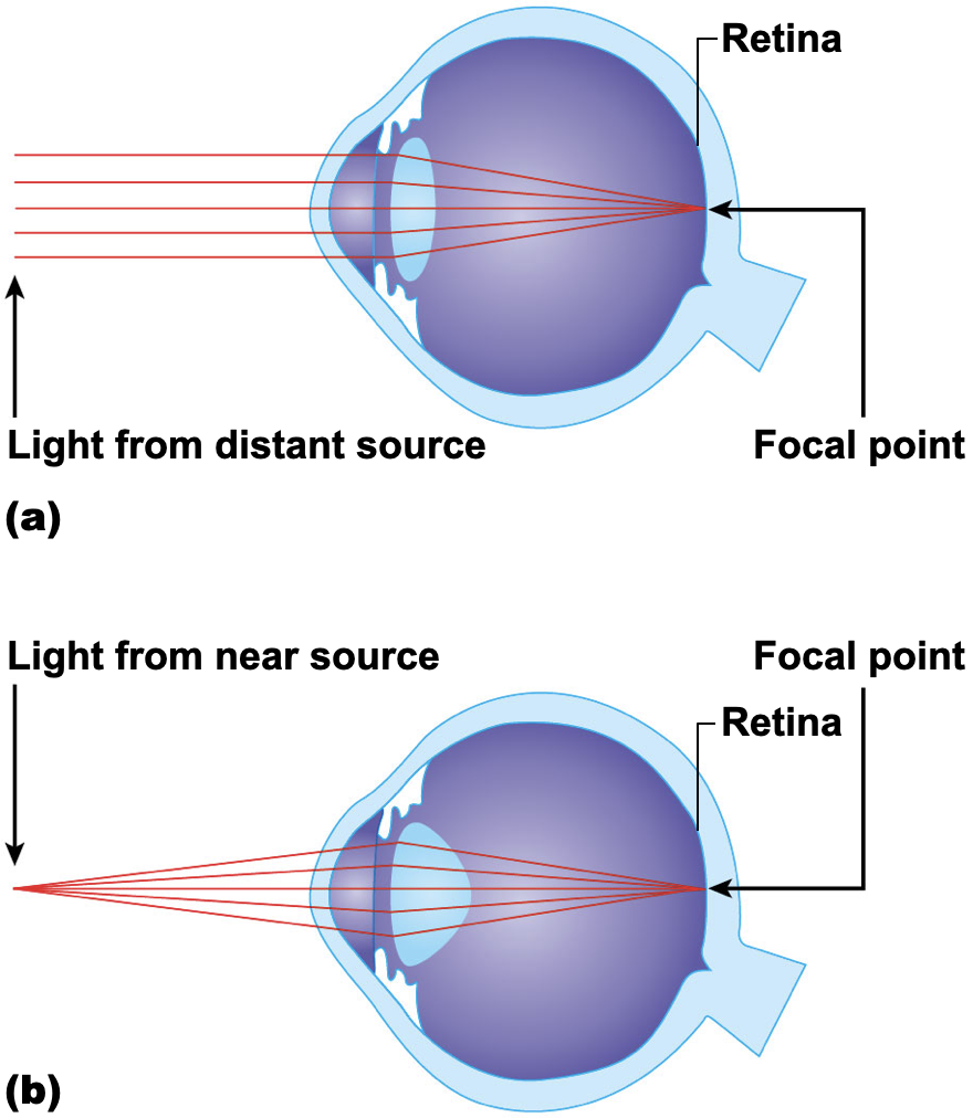
- Path of light through eye & light refraction
- Light must be focused to a point on the retina for optimal vision
- Light is bent, or refracted, by the cornea, aqueous humor, lens, and vitreous humor
- The eye is set for distant vision (over 20 feet away)
- Accommodation—the lens must change shape to focus on closer objects (less than 20 feet away)
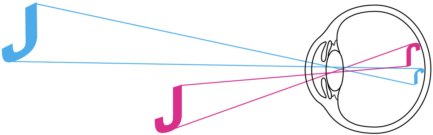
- Pathway of light through the eye and light refraction (continued)
- Image formed on the retina is a real image
- Real images are:
- Reversed from left to right
- Upside down
- Smaller than the object
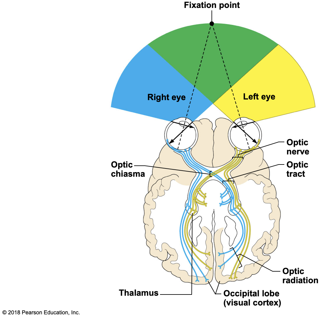
- Visual fields and visual pathways to brain
- Optic nerve
- Bundle of axons that exit the back of the eye carrying impulses from the retina
- Optic chiasma
- Location where the optic nerves cross
- Fibers from the medial side of each eye cross over to the opposite side of the brain
- Visual fields & visual pathways to the brain
- Optic tracts
- Contain fibers from the lateral side of the eye on the same side and the medial side of the opposite eye
- Synapse with neurons in the thalamus- (relaying of sensory signals, including motor signals, to the cerebral cortex, and the regulation of consciousness, sleep, and alertness)
- Optic radiation
- Axons from the thalamus run to the occipital lobe
- Synapse with cortical cells, and vision interpretation (seeing) occurs
- Summary of the pathway of impulses from the retina to the point of visual interpretation
- Optic nerve
- Optic chiasma
- Optic tract
- Thalamus
- Optic radiation
- Optic cortex in occipital lobe of brain
- Visual fields
- Each eye “sees” a slightly different view
- Field of view overlaps for each eye
- Binocular vision results and provides:
- Depth perception (three-dimensional vision)
A Closer Look
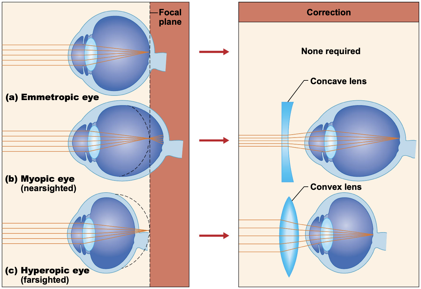
- Emmetropia—eye focuses images correctly on the retina
- Myopia (nearsightedness)
- Distant objects appear blurry
- Light from those objects fail to reach the retina and are focused in front of it
- Results from an eyeball that is too long
- Hyperopia (farsightedness)
- Near -objects are blurry, whereas distant objects are clear
- Distant objects are focused behind the retina
- Results from an eyeball that is too short or from a “lazy lens”
- Astigmatism
- Images are blurry
- Results from light focusing as lines, not points, on the retina because of unequal curvatures of the cornea or lens
- Convergence: reflexive movement of the eyes medially when we focus on a close object
- Photopupillary reflex: bright light causes pupils to constrict
- Accommodation pupillary reflex: viewing close objects causes pupils to constrict
The Ear: Hearing and Balance
- Ear houses two senses
- Hearing
- Equilibrium (balance)
- Receptors are mechanoreceptors (respond to touch or feel)
- Different organs house receptors for each sense
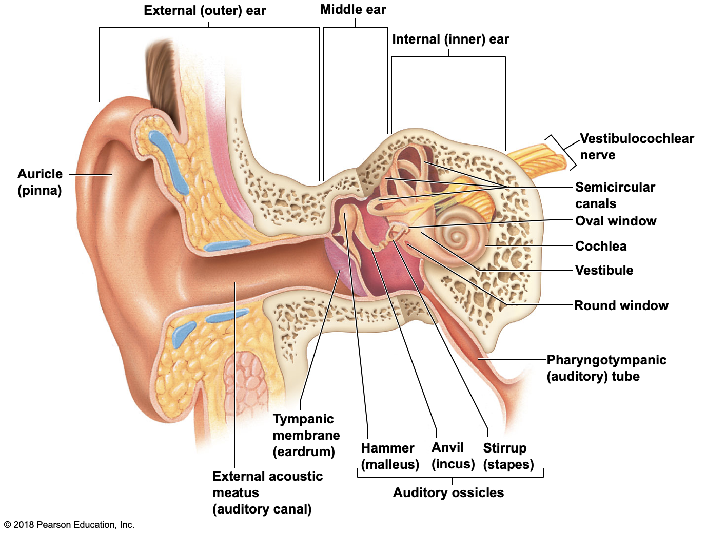
Anatomy of the Ear
- The ear is divided into three areas
- External (outer) ear
- Middle ear
- Internal (inner) ear
- External (outer) ear
- Auricle (pinna)
- Ext. acoustic meatus (auditory canal)
- Narrow chamber in the temporal bone
- Lined with skin and ceruminous (earwax) glands
- Ends at the tympanic membrane (eardrum)
- External ear is involved only in collecting sound waves
- Middle ear cavity (tympanic cavity)
- Air filled, mucosa-lined cavity within the temporal bone
- Involved only in the sense of hearing
- Located between tympanic membrane and oval window and round window
- Pharyngotympanic tube (auditory tube)
- Links middle ear cavity with the throat
- Equalizes pressure in the middle ear cavity so the eardrum can vibrate
- Middle ear cavity (tympanic cavity)
- Three bones (ossicles) span the cavity
- Malleus(hammer), Incus(anvil), Stapes(stirrup)
- Function
- Transmit vibration from tympanic membrane to the fluids of the inner ear
- Vibrations travel: hammer -> anvil -> stirrup -> oval window of inner ear
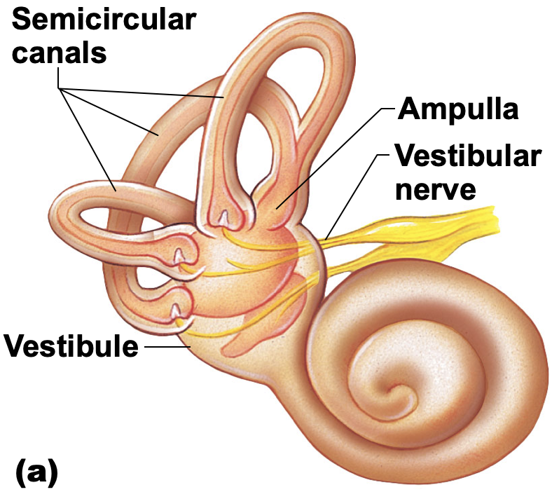
- Internal (inner) ear
- Sense organs for hearing and balance
- Bony labyrinth (osseous labryrinth) consists of:
- Cochlea, vestibule, semicircular canals
- Bony labyrinth is filled with perilymph
- Membranous labyrinth is suspended in perilymph and contains endolymph
Equilibrium
- Equilibrium receptors of the inner ear are called the vestibular apparatus
- Vestibular apparatus has two functional parts
- Static equilibrium
- Dynamic equilibrium
Static Equilibrium
- Maculae—receptors in the vestibule
- Report on the position of the head
- Help us keep our head erect
- Send information via the vestibular nerve (division of cranial nerve VIII) to the cerebellum of the brain
- Anatomy of the maculae
- Hair cells are embedded in the otolithic membrane
- Otoliths (tiny stones) float in a gel around hair cells
- Movements cause otoliths to roll and bend hair cells
Dynamic Equilibrium
- Crista ampullaris
- Responds to angular or rotational of the head
- In ampulla of each semicircular canal
- Tuft of hair cells covered with cupula (gelatinous cap)
- If the head moves, the cupola drags against the endolymph
- Hair cells are stimulated, impulse travels vestibular n. to the cerebellum
Hearing
- Spiral organ of Corti
- Located within the cochlear duct
- Receptors = hair cells on the basilar membrane
- Gel-like tectorial membrane is capable of bending hair cells
- Cochlear nerve attached to hair cells transmits nerve impulses to auditory cortex on temporal lobe
- Pathway of vibrations from sound waves
- Ear drumossiclesoval window
- Sound is amplified by the ossicles
- Pressure waves cause vibrations in the basilar membrane in the organ of Corti
- Hair cells of the tectorial membrane are bent when the basilar membrane vibrates against it
- An action potential starts in the cochlear nerve (cranial nerve VIII), and the impulse travels to the temporal lobe
- High pitched sounds disturb the short, stiff fibers of the basilar membrane
- Receptor cells close to the oval window are stimulated
- Low pitched sounds disturb the long, floppy fibers of the basilar membrane
- Specific hair cells further along the cochlea are affected
Hearing and Equilibrium Deficits
- Deafness is any degree of hearing loss
- Conduction deafness results when the transmission of sound vibrations through the external and middle ears is hindered
- Sensorineural deafness results from damage to the nervous system structures involved in hearing
- Meniere’s affects inner ear and causes progressive deafness and perhaps vertigo (sensation of spinning)
Chemical Senses: Smell & Taste
- Chemoreceptors
- Stimulated by chemicals in solution
- Taste has five types of receptors
- Smell can differentiate a wider range of chemicals
- Both senses complement each other and respond to many of the same stimuli
Olfactory Receptors/Sense of Smell
- Olfactory receptors in roof of nasal cavity
- Olfactory receptor cells (neurons) with long cilia (olfactory hairs) detect chemicals
- Chemicals must be dissolved in mucus for detection by chemoreceptors called olfactory receptors
- Impulses are transmitted via the olfactory filaments to the olfactory nerve (I)
- Smells interpreted in the olfactory cortex
Taste Buds and Sense of Taste
- Taste buds house the receptor organs
- Locations of taste buds
- Most are on the tongue
- Soft palate
- Superior part of the pharynx
- Cheeks
- The tongue is covered with projections called papillae that contain taste buds
- Vallate (circumvallate) papillae
- Fungiform papillae
- Filiform papillae
- Gustatory cells are the taste receptors
- Possess gustatory hairs (long microvilli)
- Gustatory hairs protrude through a taste pore
- Hairs are stimulated by chemicals dissolved in saliva
- Impulses are carried to the gustatory complex by several cranial nerves because taste buds are found in different areas
- Facial nerve (cranial nerve VII)
- Glossopharyngeal nerve (cranial nerve IX)
- Vagus nerve (cranial nerve X)
- Taste buds are replaced frequently by basal cells
- Five basic taste sensations
- Sweet receptors respond to sugars, saccharine, some amino acids
- Sour receptors respond to H+ ions or acids
- Bitter receptors respond to alkaloids
- Salty receptors respond to metal ions
- Umami receptors respond to the amino acid glutamate or the beefy taste of meat
Developmental Aspects of the Special Senses
- Special sense organs are formed early in embryonic development
- Maternal infections during the first 5 or 6 weeks of pregnancy may cause visual abnormalities as well as sensorineural deafness in the developing child
- Vision requires the most learning
- The infant has poor visual acuity (is farsighted) and lacks color vision and depth perception at birth
- The eye continues to grow and mature until age 8 or 9
- Age-related eye issues
- Presbyopia—“old vision” results from decreasing lens elasticity that accompanies aging
- Difficulty to focus for close vision
- Lacrimal glands become less active
- Lens becomes discolored
- Dilator muscles of iris become less efficient, pupils remain constricted
- The newborn infant can hear sounds, but initial responses are reflexive
- By the toddler stage, the child is listening critically and beginning to imitate sounds as language development begins
- Age-related ear problems
- Presbycusis—type of sensorineural deafness that may result from otosclerosis (ear ossicles fuse)
- Congenital ear problems usually result from missing pinnas and closed or missing external acoustic meatuses
- Taste and smell are most acute at birth and decrease in sensitivity after age 40 as the number of olfactory and gustatory receptors decreases