

Chapter 14: Coordination and Response: II Animal Receptor Organs
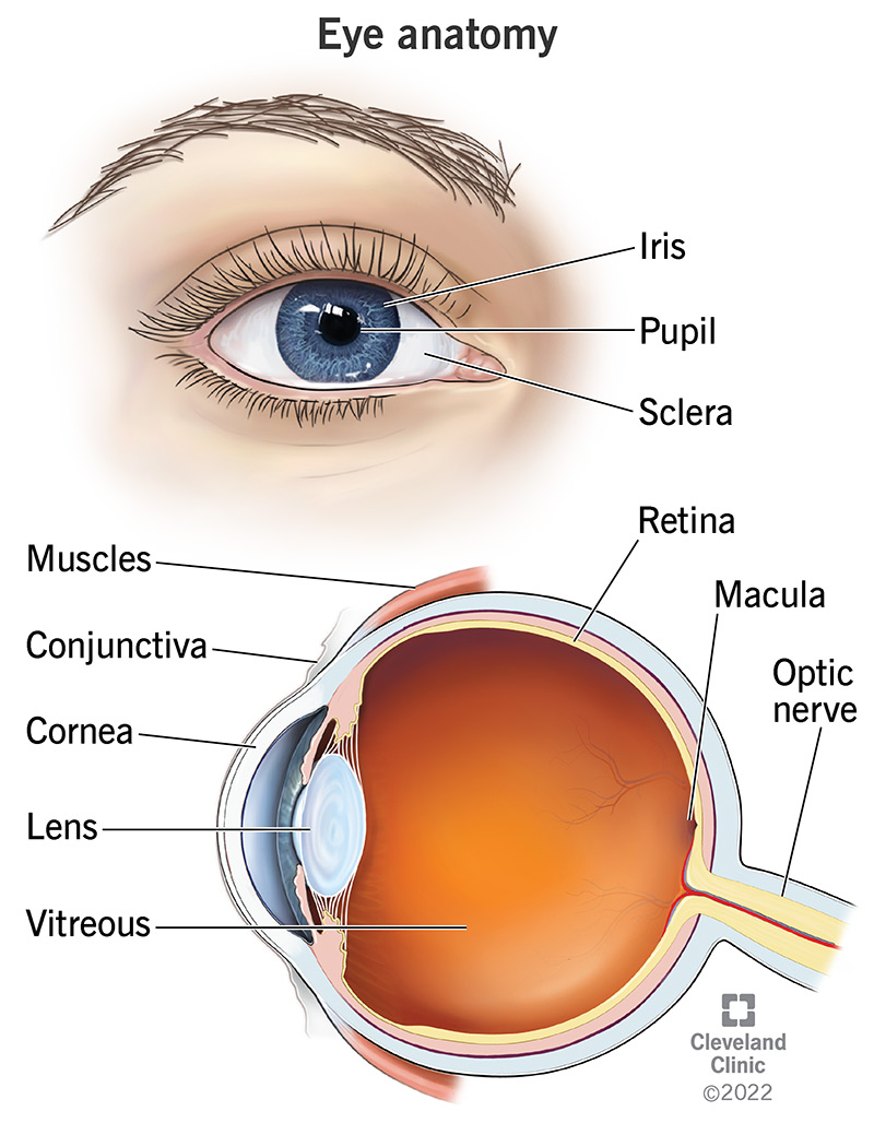
The Mammalian Eye
Cornea
A clear layer coating iris.
Refracts light in the eye.
Iris
Colored section of eye.
Controls amount of light entering by contracting and dilating pupil.
Pupil
Allows entrance of light.
Lens
Behind iris.
Focuses image on retina.
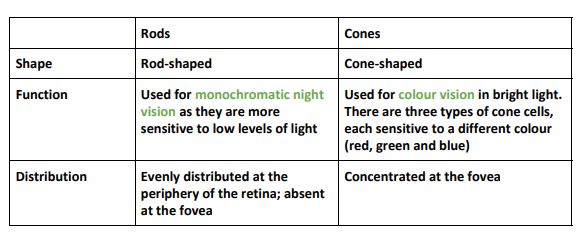
Retina
Contain photoreceptors; rods and cons.
Optic Nerve
Each photoreceptor cell is attached to neuron.
Carries nerve impulses to brain.
Fovea
Section in middle of retina.
Provides clear images.
Ciliary Muscles
Alters the thickness of muscles.
Choroid Coat
Contains number of blood capillaries which nourish eye.
Painted black (due to supply of blood vessels) to prevent internal reflection of light.
Aqueous Humor
Watery fluid.
Vitreous Humor
Transparent jelly like fluid.
NOTE: both aqueous humor and vitreous humor serve to keep eyeball form and refract light.
Rods and Cons
Controlling the entry of light in eye
Achieved by altering diameter of pupil.
Size of pupil controlled by iris muscles - circular and radial muscles.
When circular muscle contract, radial muscles relax, and pupil becomes smaller.
When radial contract, circular muscle relax , and pupil enlarges (diltes).
Pupil enlarges when surrounding light intensity is low, and smaller when the light intensity is high.
Vision
Focusing (Accomodation)
It is the adjustment of lens of the eye so that clear images of objects at different distances are formed on retina.
Focusing on distant object
Circular ciliary muscles relax.
Suspensory ligaments are pulled tight - lens become flatter and less convex.
Focusing on nearer object
Circular ciliary muscles contract.
Suspensory ligaments are released loosely - lens becomes thicker and more convex.