4.0 Overview

Chapter 4: Host Basics
An Introduction to Bacteria
Bacteria are prokaryotes and among the oldest organisms on the planet. They are also among the simplest free-living life-forms known. Like all prokaryotes, they are single-cell organisms and lack a nuclear membrane and other internal membrane-bound organelles. Bacteria are small, usually in the range of 1 to 5 microns (μm) across (a human red blood cell is 8 μm).
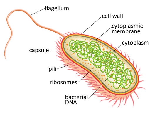
All bacteria have a cytoplasmic plasma membrane made of lipids that enclose the inner workings of the bacterial cell. A cell wall constructed of sugars and proteins surrounds the plasma membrane and gives the bacteria structural strength. Bacteria can have an additional lipid membrane and a slimy capsule that protects them from harsh environments or performs other functions. Some bacteria have appendages called “pili” and “flagella” (singular are “pilus” and “flagellum,” respectively). These hair-like structures protrude from the bacteria and perform specialized functions. Inside the plasma membrane all bacteria contain a DNA genome (typically 2–10 megabase pairs Mbp). But unlike the genome in eukaryotic cells, the bacterial genome is not enclosed by a lipid membrane (Figure 4.0-1). Genes are transcribed by RNA polymerase to make messenger RNAs (mRNA), which are used by ribosomes as substrates for protein synthesis. Because bacteria lack a nuclear membrane, the processes of transcription (mRNA synthesis) and translation (protein synthesis) are tightly coupled events.
|
|---|
Figure 4.0-1. Bacterial structure. |
The Bacterial World
Bacteria are extremely abundant and by mass outweigh all plants and animals on the planet, combined. They are present in almost all habitats and naturally reside in and on you. Bacteria are also tremendously diverse, and different bacteria thrive in different environments. In each environment, bacteria have rich interactions with other forms of life. For example, the many different bacteria that live within the human gastrointestinal tract contribute to our health by helping us digest food and build our immune system. There are, however, bacteria that cause devastating human diseases like cholera (Vibrio cholerae) and tuberculosis (Mycobacterium tuberculosis). Wherever bacteria live, their environment is hardly ever theirs alone and can be a hostile place. They must deal with toxins and antibiotics naturally produced by fungi and other
bacteria, as well as bacteriophages that can infect them. Over time, bacteria have evolved a variety of strategies to combat these assaults, making them incredibly successful organisms despite their simplicity.
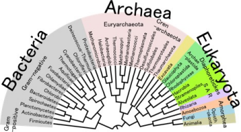
All free-living organisms contain ribosomes for protein synthesis, and these ribosomes contain large RNA molecules that contribute to catalytic function. Different bacterial species have slightly different ribosomal RNA (rRNA) sequences, and these can be used to construct models—or a phylogeny—of their evolutionary relationships (Figure 4.0-2).
|
|---|
Fig. 4.0-2. Phylogeny showing evolutionary relationships between the three phyla. |
The Bacterial Life Cycle
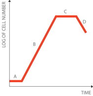
It is common for bacteria to reproduce asexually by dividing in half through a process known as binary fission. Bacterial growth is thus exponential, with one cell dividing to produce two cells, each of these dividing to produce four cells, then each of those dividing to produce eight cells, and so on. Fortunately, various influences, including resource limitations, lead to growth cessation. Otherwise, the world soon would be engulfed in bacteria! In the lab, when bacteria are growing in a liquid medium, this stage of growth is referred to as “stationary phase” (Figure 4.0-3) and the culture is sometimes referred to as being “saturated.” The number of bacterial cells in a saturated cultured is typically 2–4 x 10⁹ cells/ml.
|
|---|
Figure 4.0-3. Bacterial growth over time. When inoculated into fresh media, bacteria will take some time recover without growth. This is called the “lag phase” (A). Bacteria will then enter a logarithmic (or exponential) growth phase (B), where the number of bacteria doubles once every time period (the period depends on the bacterial species). Once nutrients are used up, the bacterial number will remain constant in the stationary phase (C), and eventually decline in the death phase (D). |
The growth rate of bacteria can be measured as the “doubling time,” that is, the time it takes for the number of bacterial cells to double. The doubling time varies greatly among different bacteria, and it also fluctuates widely depending on environmental aspects such as temperature and nutrient availability. Some bacteria can grow with doubling times as fast as 15 –20 minutes, whereas others may take days or even weeks to double.
In the lab we normally study a bacterial strain in isolation from other types of bacteria. We provide the bacteria with growth media, either in the form of a solid (agar) or a liquid, that is rich in nutrients to encourage their growth. This is called “culturing bacteria.” When a bacterial sample is streaked onto solid media, we can observe individual bacterial colonies. All the bacteria in a colony are genetically identical (apart from any in which rare mutations have occurred) because they arose from a single bacterium. The number of cells in a bacterial colony varies enormously depending on how large the colony is, but it is generally in range of 107–108 cells.
The morphology of bacterial colonies—that is, their color, size, and shape—differs greatly from species to species, ranging from yellow to purple and from smooth to rough. If many bacterial cells (e.g., 105) are added to solid media, individual colonies do not form. Instead, the bacteria will grow as a lawn that encompasses the entire surface area of the plate. When bacteria are cultured in liquid, they diffuse in the solution, and as they accumulate, the liquid becomes increasingly cloudy as it forms a saturated culture.