Chapter 3: Animal Architecture
]]Animal Body Plans]]
@@Symmetry@@
Balanced proportions – correspondence in size and shape of parts on opposite sides of a median plane
- Spherical Symmetry:
- Any plane passing through the center divides the body into equivalent, or mirrored, halves
- Occurs chiefly among some unicellular eukaryote groups
- Rare in animals
- Best suited for floating and rolling
- Radial Symmetry:
- Body can be divided into similar halves by more than 2 planes passing through the longitudinal axis
- One end of the longitudinal axis is usually the mouth
- Biradial Symmetry:
- Only 1 or 2 planes passing through the longitudinal axis produce mirrored halves
- Single or paired parts limit symmetrical planes
- E.g., ctenophores
- Bilateral Symmetry:
- Applies to animals divided along a sagittal plane into 2 mirrored portions
- Right and left halves
- Major innovation
- Much better fitted for directional movement
- Strongly associated with cephalization:
- Differentiation of a head end
- Accompanied by the concentration of nervous tissue and sense organs
@@Asymmetry@@
- Not balanced
- No plane through which they are divided into identical halves
]]Development of Animal Body Plans]]
An animal’s body plan forms through an inherited developmental sequence.
Begins with a zygote
- Single cell
- Will divide into a larger number of smaller cells
Blastomeres
Process = cleavage
- Orderly sequence of cell divisions
- Occurs in several different ways
Sponges and cnidarians lack a distinct pattern
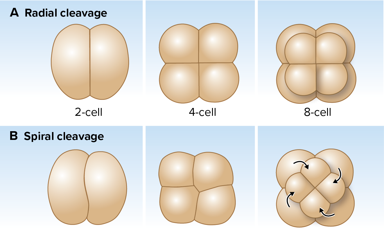
Bilaterians typically exhibit 2 types:
@@Radial Cleavage@@
- Tiers or layers of cells on top of one another
- Typically occurs with regulative development
- If blastomere is separated from others, can adjust or “regulate” its development
- Result: complete embryo
@@Spiral Cleavage@@
- Cleavage planes diagonal to the polar axis
- Unequal cells are produced by alternate clockwise and counterclockwise cleavage around the axis of polarity
- Typically occurs with a form of mosaic development
Organ-forming determinants in egg cytoplasm are positioned within egg
- Before first cleavage division
- Result: separated blastomeres still develop as if part of the whole
- Defective, partial embryo
@@Cleavage@@
Proceeds until the zygote is divided into many small cells
- Surround a blastocoel: Fluid-filled cavity.
Blastula
- Hollow ball of cells
- Becomes a gastrula
- Except in sponges
- Process: gastrulation
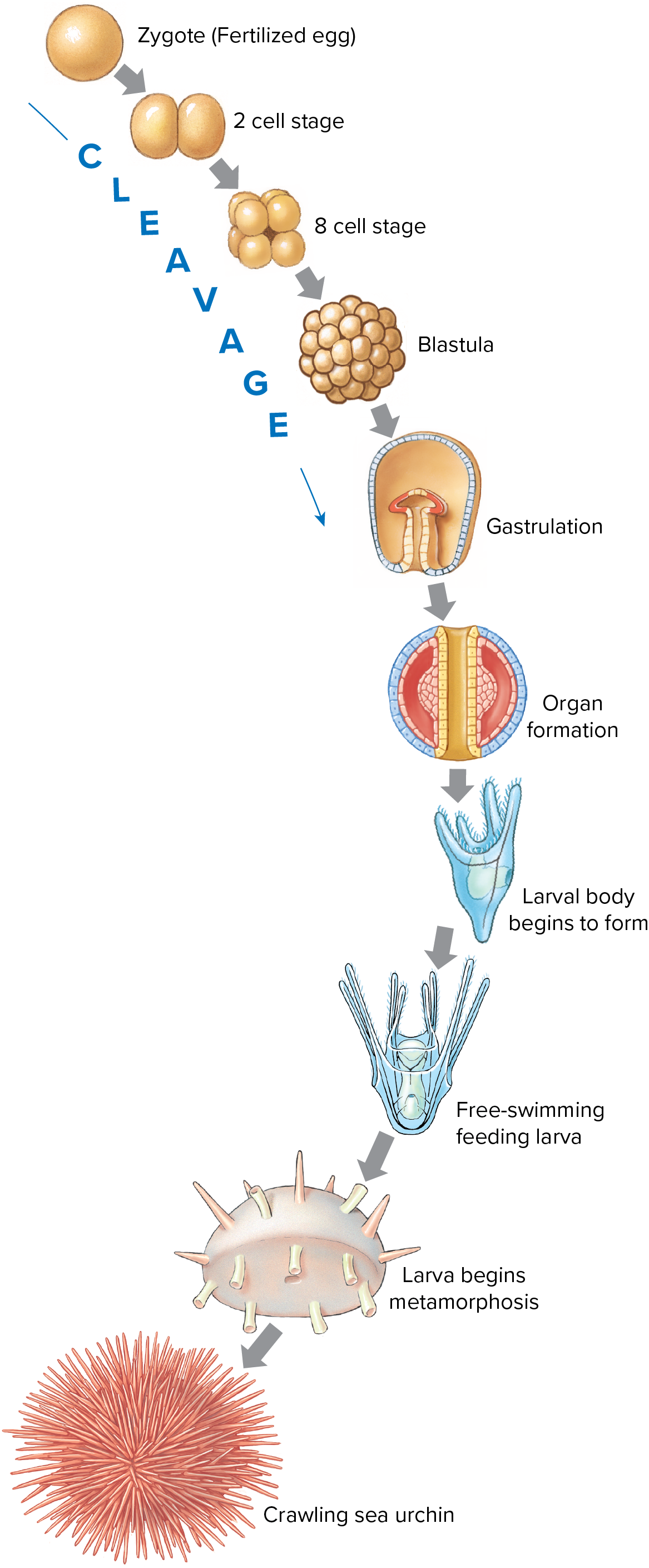
@@Gastrulation@@
- 2-3 germ layers develop
- Primary cell layers
- Some of the first lineage-specific stem cells
- Ectoderm
- Outer layer
- Differentiates into epidermis and nervous system cells
- Endoderm
- Innermost layer
- Surrounds and defines inner body cavity = Gastrocoel
- Will become gut cavity
- Mesoderm
- Middle germ layer
- Gives rise to connective tissues, muscle, urogenital and vascular systems, and peritoneum
@@Body Cavities@@
Gut cavity
- Development from gastrocoel
- Always has at least 1 opening
- Blastophore
One opening: blind or incomplete gut
Most animals develop 2nd opening to gut
- Creates a tube
- Complete gut
- Typically surrounded by a fluid-filled cavity
- Coelom if lined with mesoderm
Coelom
- Body cavity in triploblastic animals
- Lined with mesodermal peritoneum
- In some animals, mesoderm only lines the outer edge of the blastocoel
- Lies next to ectoderm
- Pseudocoelom
- In some animals, mesoderm completely fills blastocoel
- Acoelomate animals
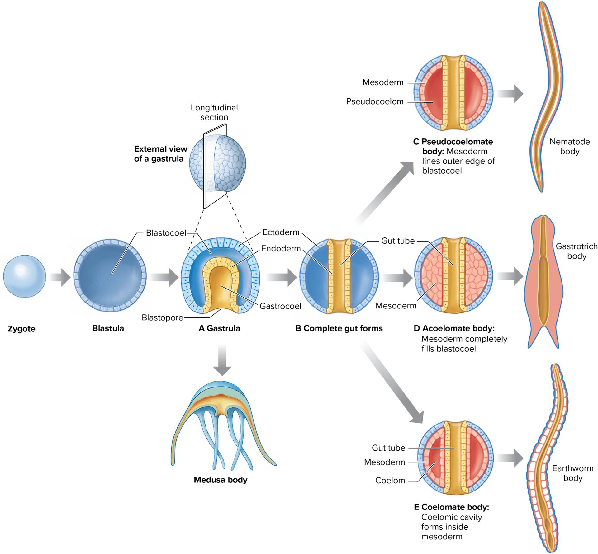
Evolution was a major development for bilaterians
Advantages:
- Tube-within-a-tube
- Space for viscera, cushioning, protection, hydrostatic skeleton (aids movement)
]]How Many Body Plans Are There?]]
Animals comprise 32 phyla.
Sponges only have a cellular level of organization
All others:
- Diploblastic: 2 germ layers. (E.g., Phylum Cnidaria)
- Triploblastic: 3 germ layers
- Bilateria: 2 groups that differ in various developmental characteristics
- Protostomia
- Deuterostomia
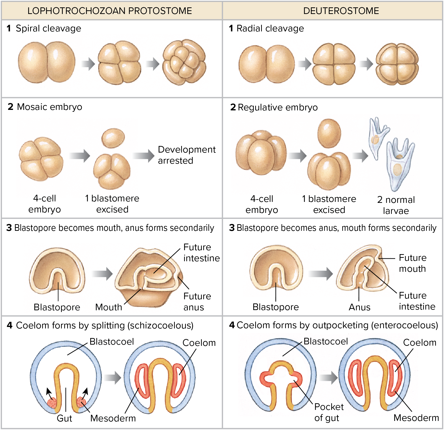
@@Deuterostome Body Plans:@@
Blastopore becomes anus
- “Second mouth”
- Refers to formation of mouth from a second opening in embryo
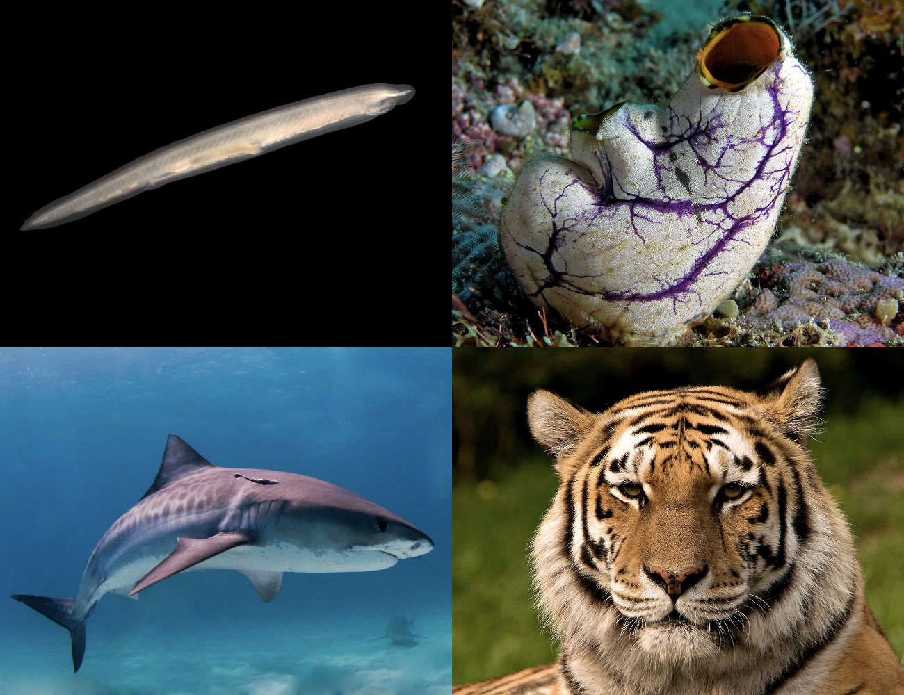
3 Deuterostome phyla:
1. Echinodermata (Sea stars and relatives)
2. Hemichordata
3. Chordata
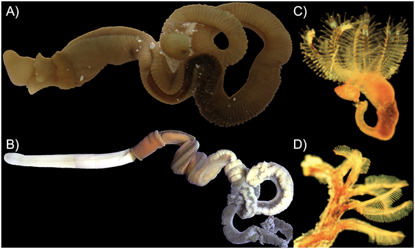
@@Protostome Body Plans:@@
- First embryonic opening (blastopore) becomes the mouth
- 2 subgroups:
- Ecdysozoa
- Molting animals
- Arthropods, nematodes, and 6 other phyla
- Lophotrochozoa
- Very diverse group
- 17 phyla
]]Components of Animal Bodies]]
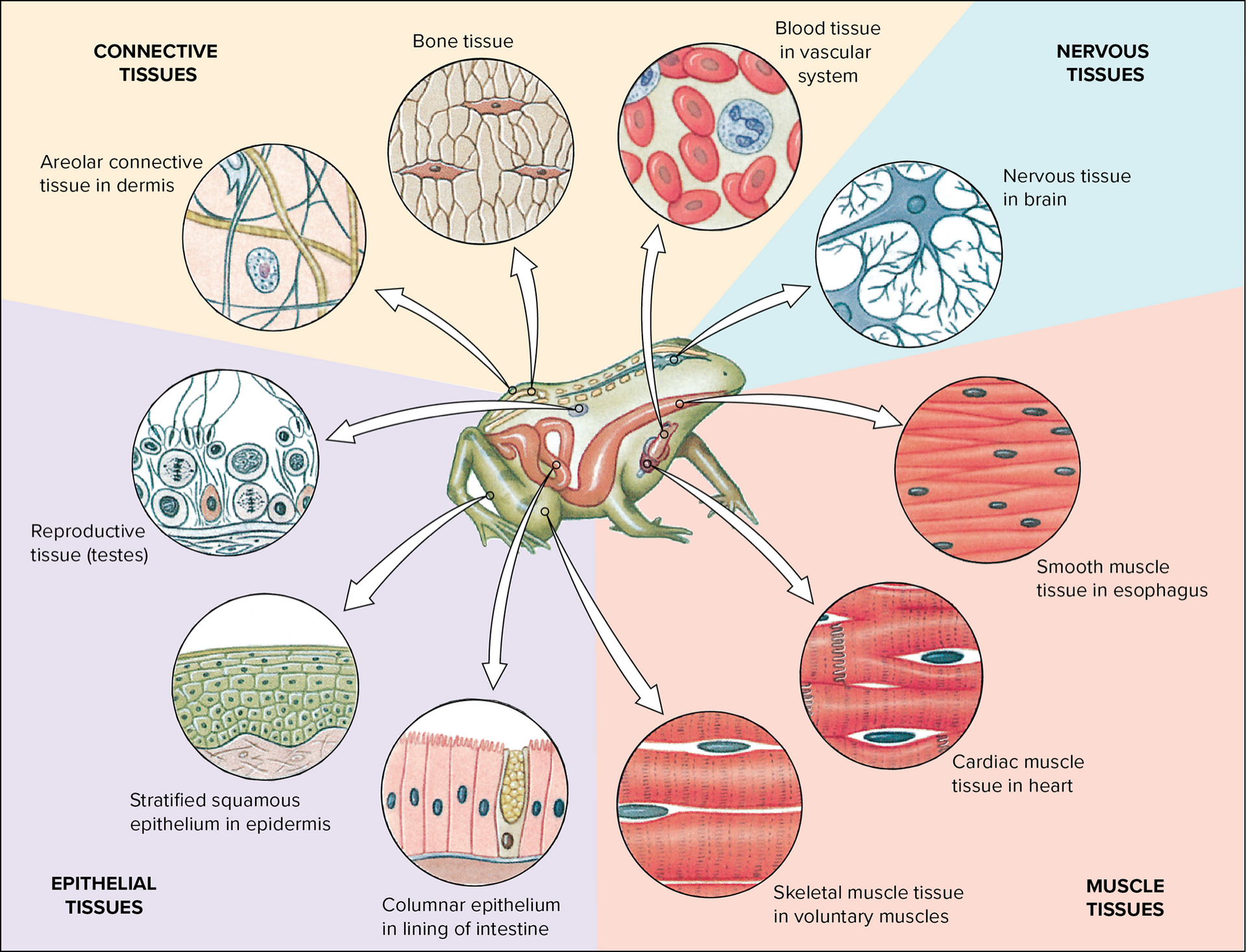
All animal bodies consist of:
- @@Cellular Components@@
- Tissues and organs derived from embryonic germ layers
- A group of cells specialized for performing a common function
- Study of tissues = histology
- 4 kinds:
- Epithelial
- Connective
- Muscular
- Nervous
- @@Extracellular Components@@
- Fluids and structures that cells deposit outside their cell membranes
@@Epithelial Tissue@@
- Sheets of cells
- Cover external or internal surfaces
- Often modified into glands that produce mucus, hormones, or enzymes
@@Connective Tissue@@
- Diverse group
- Various binding and supportive functions
- Widespread throughout body
@@Muscle Tissue@@
- Most common tissue of most animals
- Originates from mesoderm
- Specialized for contraction
- Types: Skeletal, Cardiac, Smooth
@@Nervous Tissue@@
Specialized for receiving stimuli and conducting impulses from one region to another
Types of cells:
- Neurons
- Neuroglia
