X910_ALBiology_S2T2
Learning Objectives
Living cells replicate for growth, repair, and replenishment.
Eukaryotic cells divide through mitosis for genetically identical daughter cells.
Cell cycle stages include interphase, mitosis, and cytokinesis.
Cancer arises from loss of cell division control.
Prokaryotic cells divide through binary fission.
Viruses replicate using cellular hosts.
Mitosis
Mitosis results in genetically identical daughter cells, known as diploids.
Meiosis involves the splitting of parent cells to form four daughter cells each with half of the original parent genetics; these cells are haploid.
Cells spend time in interphase before active mitosis.
Mitosis stages: prophase, late prophase, metaphase, anaphase, telophase, cytokinesis.
Prophase
In the first phase of mitotic division, we first see the chromosomes forming. They start as long threads of DNA molecules before becoming shorter and thicker. The spindle fibres continue to pull the chromosomes until they line up across the centre of the cell, also known as the metaphase plate.
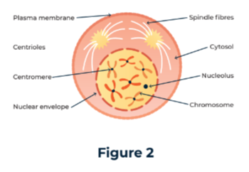
In Figure 2, centrioles move to opposite sides of the cell, forming spindle fibers that stretch across. The nuclear envelope breaks down as the nucleolus also disappears from the cell, releasing chromosomes into the cytoplasm. Spindle fibers attach to chromosomes and pull them towards the cell center.
Metaphase
The spindle fibres continue to pull the chromosomes until they line up across the centre of the cell, also known as the metaphase plate.
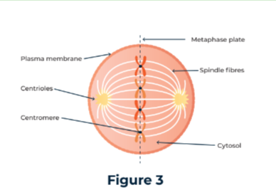
The chromosomes are now made up of two chromatids which are an exact copy of the DNA from the parent. The central part of a chromosome that joins the chromatids is the centromere, and this is where the spindle fibres are attached.
Anaphase
The centromeres now split, dividing the chromatids too, and the spindle fibres continue to pull which results in half the chromatids moving to one pole of the cell, the other half to the opposite pole.
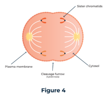
During this phase, the spindle fibres are surrounded by mitochondria that provide the energy required for this process. The cleavage furrow, where the cell will finally split, starts to form.
Telophase
This phase is characterised by the disappearance of the chromosomes to form much less defined chromatin.
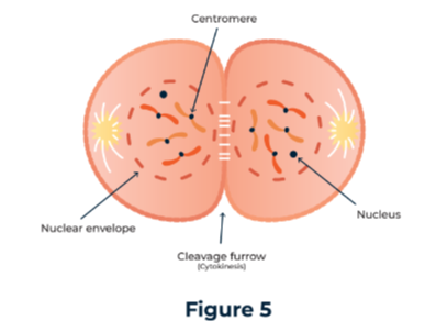
The spindle fibres disintegrate too as the nuclear envelope and nucleolus reform in both halves of the cell. Eventually, the cell finally splits in a process called cytokinesis, along where the cleavage furrow had started to form, through the centre of the cell.
Binary Fission
Prokaryotic cells divide through binary fission.
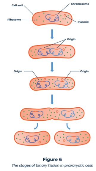
Stages of binary fission:
Stage 1: Parent cell ready to divide and replicate
Stage 2: The DNA and plasmids replicate
Stage 3: DNA and plasmids move apart attaching to the cell membrane
Stage 4: Cell membrane begins to lengthen, starting to pinch inwards in the centre
Stage 5: Two separate cells are formed, as pinching continues and splits the now elongated cell. A new cell wall forms between the two identical daughter cells.
Replication of Viruses
Viruses cannot divide and replicate on their own.
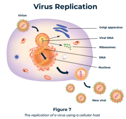
Viruses all have proteins on their protein coat (the capsid) that allow them to attach to the host cell, use cellular hosts to replicate by injecting viral nucleic acid .
Viral components are produced, reformed, and egested to continue the replication process, able to continue the process and continue to proliferate.
The Cell Cycle
Cell cycle includes stages of cell growth and division.
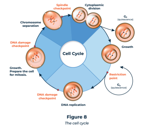
Stages: interphase (longest phase where no cell division occurs), nuclear division (commences division as a whole and involves the division of the nucleus within the cell), cytokinesis (sees the cytoplasm divide, creating two new cells).
Can be mitotic (produce identical daughter cells) or meiotic (produce four cells, with half the genetics).
Cell Division & Cancer
Cancer is caused by loss of control over cell division.
Mutated genes (Damage to important genes that regulate the cell cycle and mitosis and their products become damaged and this leads to) lead to unchecked cell growth and tumour formation.
Mitosis: it is regulated by specific genes that can mutate and therefore lead to uncontrolled mitosis which, as we know, leads to copies of parent cells and, in the case of cancer, proliferation of the cancer causing cells explaining the rapid growth of tumours as the cancer cells grow in number exponentially.
Cancer treatments disrupt cell division process to prevent replication or disrupt spindle
Drawbacks of cancer treatments: so the drawback of most chemotherapy drugs is that they adversely affect many healthy cells in the process, because it is not just cancer cells that replicate their DNA or form spindle fibres, these are fundamental parts of the division of any cell in the human body.
The distinction between healthy cells and cancerous cells is the speed at which they divide, with cancerous cells dividing at a significantly quicker rate which means they are impacted to a much greater extent than healthy cells as the drugs work most effectively on cells that divide at a higher rate.
Activity 1: Hair cells are known to divide quickly, explain the impact this will have when using many cancer drugs.
Answer: Cells that divide quicker are more impacted by the use of cancer drugs, since hair cells divide quickly, using cancer drugs will lead to hair loss.
Key terms
Meiosis - Cell division that results in the formation of four haploid cells.
Mitosis - Cell division that results in the formation of two genetically identical diploid cells.
Daughter cells - The cells produced during cell division.
Parent cells - The original cell copied during cell division.
Diploid - A cell that contains the full number of chromosomes.
Haploid - A cell containing half the number of chromosomes, for example a sex cell.
Interphase - The resting phase, where no cell division takes place but instead there is a lot of cellular activity including the replication of DNA.
Prophase - The first phase of mitosis, where chromosomes become visible and centrioles move the poles.
Metaphase - The chromosomes line up along the equator of the cell.
Anaphase - Chromosomes split, spindle fibres pull half to one pole of the cell, and half to the other.
Telophase - Chromatin forms as chromatids lose definition, spindle fibres disintegrate and nucleolus and nuclear envelope reform.
Cytokinesis - The final cleaving of the two new cells.
Binary Fission - The mode by which prokaryotic cells divide.
Centriole - Organelles that form the spindle fibres and ensure these move to either pole of the cell.
Spindle fibres - Responsible for pulling chromatids to opposite poles.
Metaphase plate - The equator of the cell.
Centromeres - The central part of a chromosome that holds chromatids together.
Cleavage furrow - The pinching point that forms where the metaphase plate was, at the equator of the cell, where ultimately it will divide.