Chapter 12 Notes - DNA Structure and Replication
}}Identifying DNA as the Genetic Material}}
}}Griffith find a "transforming principle"}}
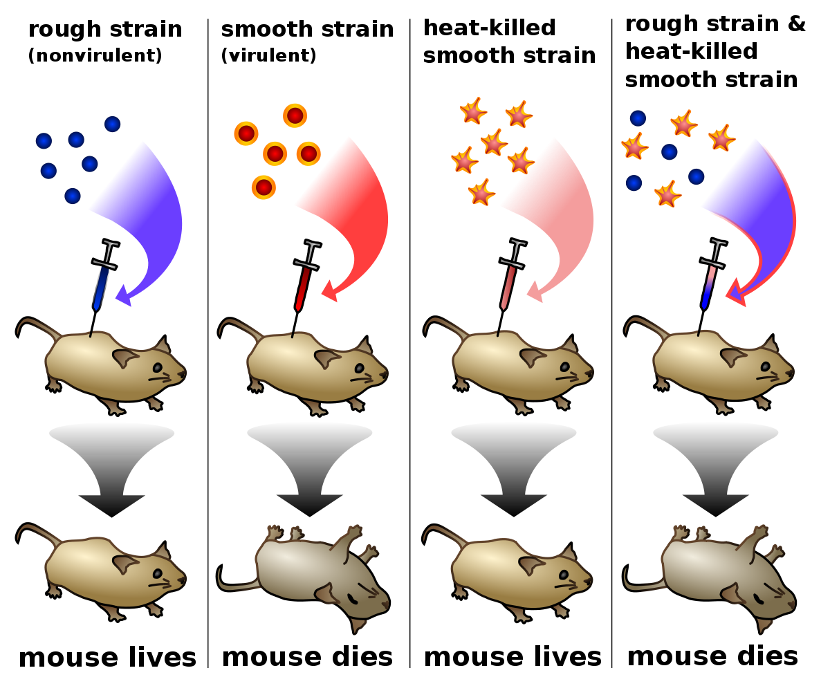
In 1928, Fredrick Griffith was investigating two forms of the bacterium that causes pneumonia
- one form is surround by a coating made of carbohydrates
- S form — colonies look smooth
- second form does not have smooth coating
- R — rough form
Griffith injected the bacteria into mice — the S type killed the mice
- when the S bacteria were killed with heat before injection, it didn't kill the mice → only live S bacteria cases the mice to die
- he then injected mice with a combination of heat-killed S bacteria and live R bacteria → the mice died
- live S bacteria was found in the dead mice
- Griffith concluded that some material had been transferred from the heat-killed S bacteria to the live R bacteria
- he called this %%transformation%% - material transferred to alter another organism
- figure: Griffith's Experiments

}}Avery identifies DNA as the transforming principle}}
Oswald Avery worked to find what the transforming principle was that Griffith had discovered
- they combined living R bacteria with an extract made from S bacteria
- they then developed a process to purify their extract
- they then performed a series of tests to find out of the transforming principle was DNA or protein
- %%qualitative tests%% - standard chemical tests showed that no protein was present; the tests revealed that DNA was present
- %%chemical analysis%% - the proportions of elements in the extract closely matched those found in DNA; proteins contain almost no phosphorus
- %%enzyme tests%% - when enzymes were added, the extract still transformed the R bacteria to the S form → transformation only failed to occur when an enzyme was added that specifically destroys DNA
}}Hershey and Chase confirm that DNA is the genetic material}}
Alfred Hershey and Martha Chase studied viruses that infect bacteria and came up with conclusive evidence for DNA as the genetic material in 1952
%%bacteriophage%% - a type of virus that takes over a bacterium's genetic machinery and directs it to make more viruses
phages are relatively simple — are a little more than a DNA molecule surrounded by a protein coat
- by discovering if the DNA or protein of a phage enters a bacterium could answer the question
- protein contains sulfer but little phosphorus, while DNA contains phosphorus but no sulfer
%%experiment 1%% - bacteria were infected with phages that had radioactive sulfer atoms in their protein molecules
- they used a kitchen blender and a centrifuge to separate the bacteria
- they found no significant radioactivity
%%experiment 2%% - DNA had radioactive phosphorus → radioactivity was present inside the bacteria
conclusion: the phages' DNA entered the bacteria, but the protein had not
}}Structure of DNA}}
}}DNA is composed of four types of nucleotides}}
nucleotides - the small units (monomers) that make up DNA
each nucleotide has 3 parts
- a phosphate group (one phosphorus with four oxygens)
- a ring-shaped sugar called deoxyribose
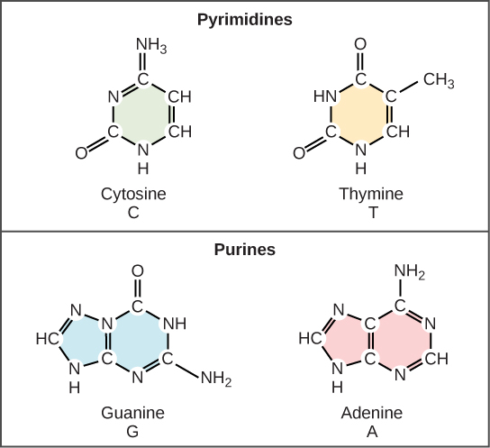
- a nitrogen-containing base (single or double ring built around nitrogen and carbon atoms)

Erwin Chargaff found that the same four bases are in the DNA of all organisms but the proportion of the four bases differs somewhat from organism to organism
- A = T and C = G relationships became known as Chargaff's rules
- aka %%base-pairing rules%%
}}Watson and Crick developed an accurate model of DNA's three-dimensional structure}}
In 1950 James Watson, an American geneticist, and British physician Francis Crick worked together to understand the structure of DNA
- they hypothesized that DNA might be a helix
%%X-ray evidence:%% Rosalind Franklin and Maurice Wilkins were studying DNA using x-ray crystallography
- when DNA is bombarded with x-rays, the atoms diffract them in a pattern that can be captured on film
- Franklin's x-rays showed an X surrounded by a circle
- suggested that DNA is a helix consisting of two strands that are a regular, consistent width apart
%%the double helix:%% Watson and Crick made models of metal and wood to figure out the structure of DNA
- placed the sugar-phosphate backbones on the outside and the bases on the inside
- found that if they paired double-ringed nucleotides with single-ringed nucleotides, the bases fit
double helix - two strands of DNA wind around each other like a twisted ladder
}}Nucleotides always pair in the same way}}
The DNA nucleotides of a single strand are joined together by covalent bonds connecting the sugar of one nucleotide to the phosphate of the next
The DNA double helix is held together by hydrogen bonds between the bases in the middle
- each hydrogen bond individually is weak, but together maintain the structure
%%base pairing rule%%: thymine always pairs with adenine, cytosine always pairs with guanine
- due to sizes of the bases and ability to form hydrogen bonds
- A & T form 2 hydrogen bonds with each other
- C & G form 3 hydrogen bonds with each other
}}DNA Replication}}
}}Replication copies the genetic information}}
replication - the process by which DNA is copied during the cell cycle
- assures that every cell has a complete set of identical genetic information
- %%a single DNA strand can serve as a template, or pattern, for a new complementary strand%%
- from free nucleotides available within the nucleus
- completed molecules are identical to the original molecule
- DNA can be accurately replicated over and over
}}Proteins carry out the process of replication}}
Enzymes and other proteins replicate the DNA
- DNA does nothing more than store information
- helicases - enzymes that start the process by unzipping the double helix to separate the strands of DNA
- other proteins hold the strands apart while they serve as templates
- nucleotides that float free can then pair up with nucleotides of existing DNA strands
%%DNA polymerases%% - a group of enzymes that bond the new nucleotides together
- results in in two complete molecules of DNA, each identical to the original strand
- also carry out a proofreading step that quickly removes nucleotides that have base-paired incorrectly during replication
- DNA polymerases and DNA ligase are also involved in repairing DNA damaged by harmful radiation or toxic chemicals in the environment
%%Replication Process:%% (in eukaryotes and is similar to prokaryotes)
- helicase enzymes begin to unzip the double helix at numerous places along the chromosome — called %%origins of replication%%
- origins of replication - short stretches of DNA that have a specific sequence of nucleotides
- hydrogen bonds connecting base pairs are broken
- original molecule separates
- bases on each strand are exposed
- the process of unzipping DNA proceeds in two directions at the same time
- the DNA molecule of a eukaryotic chromosome has many origins where replication can start simultaneously, shortening the total time needed for replication
- the sugar-phosphate backbones run in opposite directions and are said to be "%%antiparallel%%"
- one by one, free nucleotides pair with the bases exposed as the template strands unzip between what is known as %%replication forks%%
- DNA polymerases bond the nucleotides together and form new strands complementary to each template
- on one template, DNA replication occurs in a smooth, continuous way in one direction — called the leading strand
- on the other template, replication occurs in a discontinuous, piece-by-piece way in the opposite direction — lagging strand
- these pieces are called %%Okazaki fragments%%
- %%DNA ligase%% - an enzyme which then links the pieces together into a single DNA strand
- two identical molecules of DNA result, each with one strand from the original molecule and one new strand
- DNA replication is called semiconservative because one old strand is conserved, and one new strand is made
DNA replication varies in prokaryotes and eukaryotes — prokaryotes have a single, circular piece of DNA (plasmid) in their cytoplasm
- replication begins when regulatory proteins bind to a certain point on the plasmid and proceeds in two directions until the whole plasmid is copied
- the two copies are then put into different cells during cell division
Eukaryotes have about 1000 times more DNA than prokaryotes — chromosomes are linear and tightly packed with proteins in the nucleus
- replication may begin at dozens are even hundreds of places at the same time and proceed in both directions until the whole chromosome is copied
- remain close to each other (sister chromatids) until they are separated during mitosis or meiosis (anaphase/anaphase II)