


2.1.1 Cell structure
2.1.1 Cell structure (g-k)
Cell structure - Notes
2.1.1g: the ultrastructure of eukaryotic cells and the functions of the different cellular components
Define the terms “eukaryotic cell” and “ultrastructure”. (F)
Draw diagrams of a typical plant and animal cell, labelling the structures of the cell and annotating with their function.
Draw a table outlining the structure and function of the nucleus, nucleolus, nuclear envelope, rough and smooth endoplasmic reticulum (ER), Golgi apparatus, ribosomes, mitochondria, lysosomes, chloroplasts, the plasma membrane, centrioles, cell wall, flagella and cilia. (F)
State 3 similarities and 3 differences between a typical plant and animal cell.
Draw and label a diagram of a mitochondrion.
Draw and label a diagram of a chloroplast.
Draw a diagram showing the relative sizes of different cellular components. (S+C)
2.1.1h: photomicrographs of cellular components in a range of eukaryotic cells.
Identify organelles from images produced by light microscopy, TEM and SEM. (F)
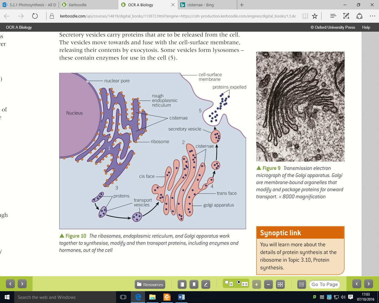
2.1.1i: the interrelationship between the organelles involved in the production and secretion of proteins.
Draw a flow chart that shows how different organelles and molecules are involved in the process of protein production and trafficking in a cell. (F)
2.1.1j: the importance of the cytoskeleton
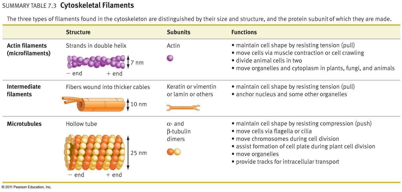
Outline the structure of the 3 components of the cytoskeleton. (F)
Describe the functions of the cytoskeleton in a cell. (F)
Describe the importance of the cytoskeleton in movements of chromosomes, cilia, flagella and vesicles.
Describe the importance of the cytoskeleton in the shape and behaviour of neutrophils.
2.1.1k: the similarities and differences in the structure and ultrastructure of prokaryotic and eukaryotic cells.
Define the term “prokaryotic cell”. (F)
List examples of both prokaryotic and eukaryotic cells.
Draw a diagram of a prokaryotic cell, label the structures and annotate with their function.
Draw a table outlining the structure and function of the cell wall, ribosomes, bacterial flagellum, plasma membrane, plasmid, bacterial chromosome, cytoskeleton, pili and slime capsule in prokaryotic cells. (F)
Draw table comparing eukaryotic and prokaryotic cells. (F)
Describe the endosymbiotic theory. (S+C)
Cell structure
There are two main types of cell. Eukaryotic cells (cells with a nucleus and membrane-bound organelles) and Prokaryotic cells (cells without a nucleus or membrane-bound organelles)
The ultrastructure of cells describes the structures that cells are made of (e.g. ribosomes, cytoskeleton, mitochondria, cell wall etc.)
The kingdoms of life with eukaryotic cells are plantae, animalia, fungi and protoctista. The kingdom of life with prokaryotic cells is the prokaryotae (this includes the bacteria, archaea and cyanobacteria)
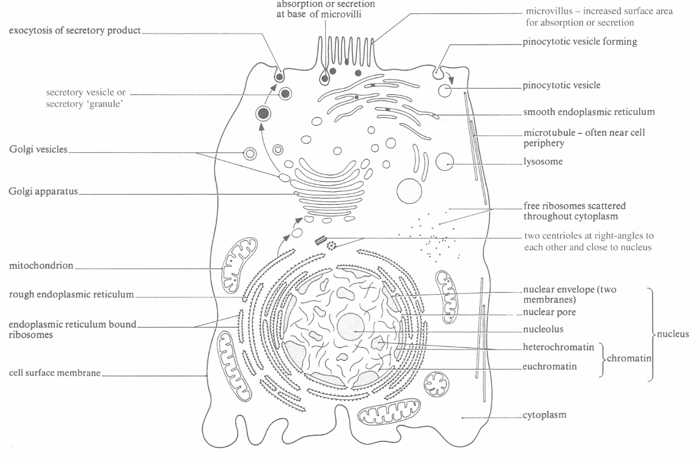
An animal cell
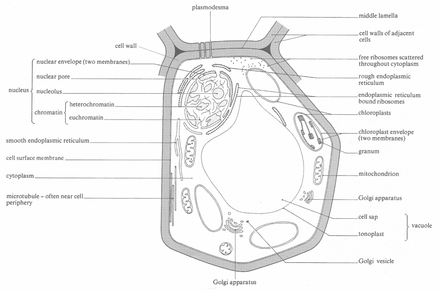
A plant cell
Plant and animal organelles
Organelle = a subcellular structure (often membrane-bound) with a specialised function
Structure (and in plants and/or animals) | Description of structure | Description of function |
Cell wall (Plants only) | A layer of cellulose outside the plasma membrane. It may have holes in it that join adjacent cells called plasmodesmata. An associated thinning of the cell wall at these locations is called a pit. [In Fungi the cell wall is made of a molecule called chitin] | Allows the cell to become turgid by preventing the cell from bursting. It determines the shape of the cell when it’s turgid. It is freely permeable. Plasmodesmata allow the movement of substances between cells without passing across the plasma membrane. |
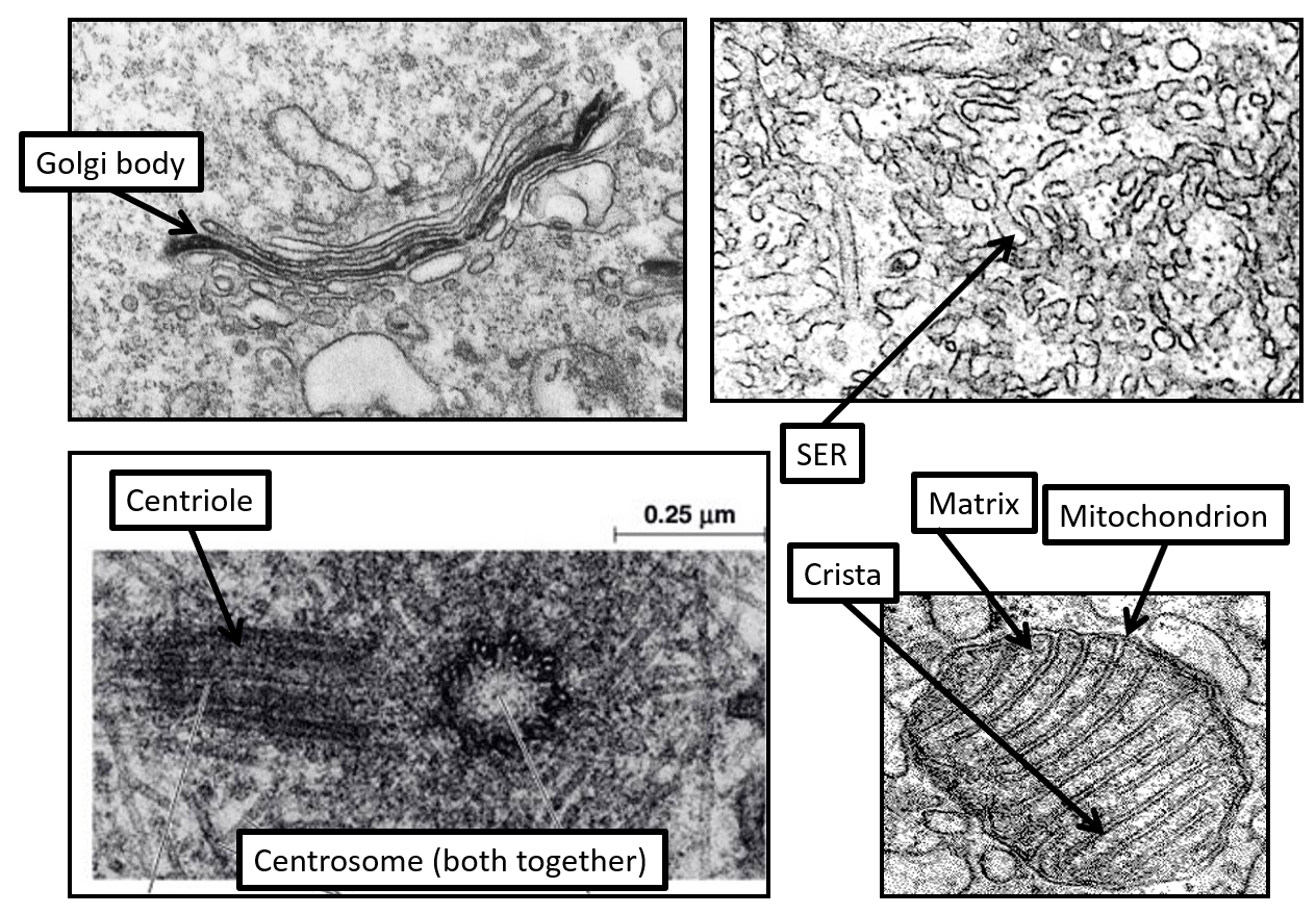
Centrioles / Centrosome (animals and only some plants – best to say absent in plants) | The centrosome consists two centrioles perpendicular to each other. Each centriole is a tube made of microtubules. | Involved in the assembly and organisation of microtubules – particularly the spindle fibres during cell division |
Chloroplasts (Plants only) | Double membrane layer around the outside with an internal membrane system suspended in a gel called the stroma as well. (Full details of structure follow this table) | The site of photosynthesis |
Cytoplasm / cytosol (Plants and Animals) | Technically termed the cytosol this is the gel within the plasma membrane. | The site of any reaction that the cell performs that doesn’t occur within an organelle. Organelles are suspended in the cytosol and molecules can pass through the cytosol from one organelle to another. |
Cytoskeleton (Plants and animals) | A network of 3 types of protein fibres (microtubules, microfilaments and intermediate fibres) (more details of structure come later in this pack) | Stabilises cell shape Allows organelles to be moved about the cell Allows some cells to move Coordinates the movement of chromosomes in cell division. |
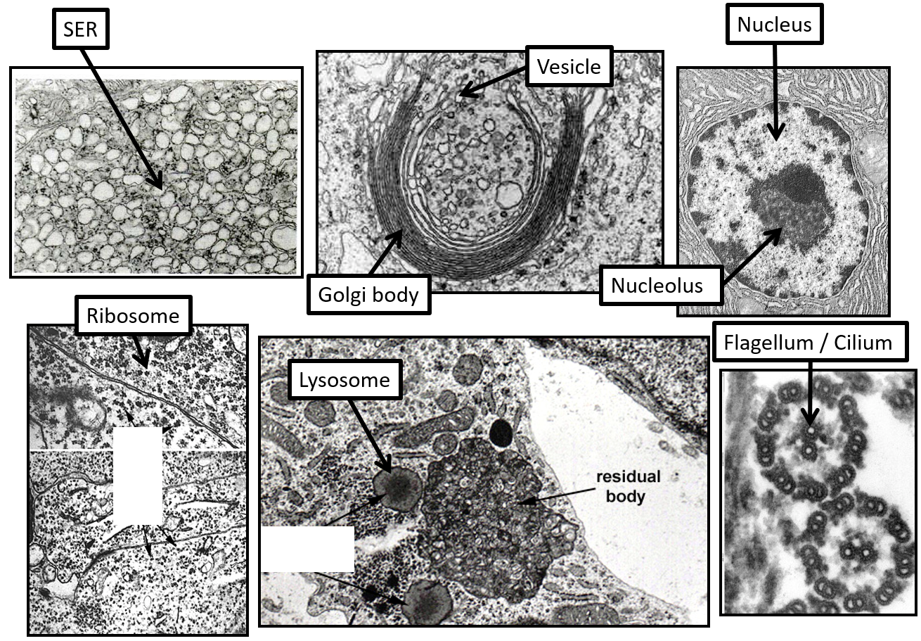
Flagella and cilia (Animals only, except for the occasional plant cells that have flagella) | Thin finger-like projections of the plasma membrane containing cytoplasm and a 9+2 arrangement of microtubules. Flagella are longer than cilia. | They move in a whip-like way to either move cells or move substances over the surface of cells. Cilia can be used to move cells or move substances such as mucus out of the lungs. Flagella move cells (e.g. sperm) |
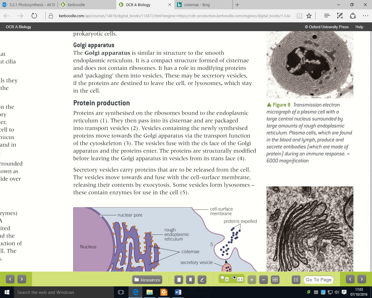
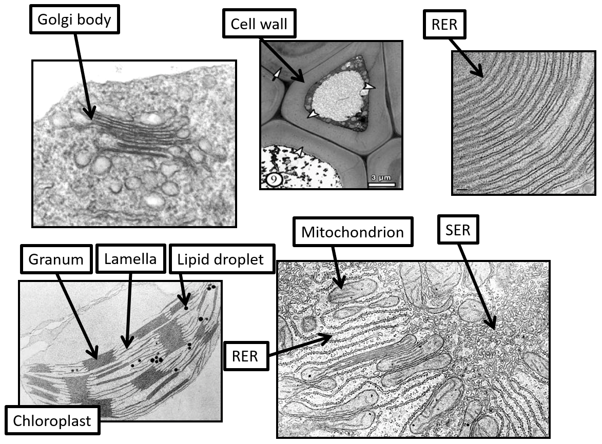
Golgi apparatus / body (Plants and animals) | A collection of flattened sacs called cisternae made of membrane with an inner space called the lumen. The cis face of the Golgi apparatus faces the RER, the trans face is the other side. | Modifies proteins and packages them in vesicles for transport around the cell or secretion out of it. |
Large permanent vacuole (Plants only) | A large membrane-bound sac. The membrane surrounding it is called the tonoplast and the contents are called cell sap. | Stores water, ions, waste products and a range of other molecules for a variety of functions. When it swells due to water entering it causes cells to be turgid. |
Lysosomes (Animals, in plants organelles with this function exist but tend to be called vacuoles) | A membrane-bound sac that contains hydrolytic enzymes. | Used for breaking down faulty organelles or engulfed bacteria. Also used in autolysis (programmed cell death). |
Microvilli (Animals only) | Finger-like projections of the plasma membrane. | Increase the surface area of the cell for absorption. |
Mitochondria (Plants and animals) | Double membrane layer around the outside of a gel called the matrix. The inner membrane is heavily folded. (Full details of structure follow this table) | Site of aerobic respiration to generate ATP for cell functions. |
Nuclear envelope (Plants and animals) | A double membrane layer containing holes called nuclear pores surrounding the nucleus. The membrane is continuous with the endoplasmic reticulum membrane. | Allows the entry and exit of molecules from or into the nucleus. The nuclear pores control the passage of very large molecules into or out of the nucleus. |
Nucleolus (Plants and animals) | A dark staining region of the nucleus that is not membrane-bound. It contains a high density of nucleic acids. | The site of ribosome production |
Nucleus (Plants and animals) | Contains DNA as linear chromosomes, surrounded by the nuclear envelope. | Coordinates the functions of the cell. |
Plasma / cell surface membrane (Plants and animals) | A double layer of phospholipids with proteins and cholesterol embedded within it. It surrounds the cell contents. (more details will be in the membranes topic) | Allows control of what enters and leaves the cell. |
Ribosomes (Plants and animals) | Made of protein and RNA. Not membrane-bound. Can be free in the cytoplasm or bound to RER. 80S | Site of protein synthesis. |
Amyloplast (Plants only) | A membrane-bound sac containing starch | Acts as a starch store for plants. |
Rough endoplasmic reticulum (RER) (Plants and animals) | A network of tubes or sacs called cisternae surrounded by membrane. The membrane has ribosomes attached to its external surface. The space inside is called the lumen or cisternal space | Proteins fold into their shape within the RER and are transported within the RER before being packaged into transport vesicles. |
Smooth endoplasmic reticulum (SER) (Plants and animals) | A network of tubes or sacs called cisternae surrounded by membrane. The membrane does not have ribosomes attached to its external surface. The space inside is called the lumen or cisternal space. | Site of lipid and carbohydrate synthesis. |
Vesicle (transport or secretory) (Plants and animals) | Small spheres enclosed by a membrane. They contain a variety of molecule types depending on their function. | Transport molecules from one organelle to another (e.g. RER to Golgi) or out of the cell (i.e. secreted). They are also used to embed molecules in the plasma membrane. |
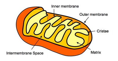
The details of mitochondrial structure
Two membranes surround the matrix
The inner membrane is heavily folded and contains ATP synthase (the site of ATP synthesis)
The folds are called cristae
Contain 70S ribosomes and circular DNA
The details of chloroplast structure
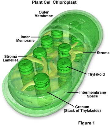
Two membranes surround the stroma
A third membrane system (made of thylakoid membrane) within the stroma has flattened sacs called thylakoids, arranged in stacks called grana. Extended thylakoids called lamellae join grana together.
The thylakoid membrane contains chlorophyll
Contain 70S ribosomes and circular DNA
Contain starch granules/grains and lipid droplets.
Protein production and transport (protein trafficking)
 The cytoskeleton
An example:
A neutrophil is a type of white blood cell that can engulf and destroy pathogens (a phagocyte) it crawls around using the cytoskeleton, manipulates its shape to surround and engulf bacteria (endocytosis) and then directs lysosomes to where the engulf bacterium is to digest the bacterium. All this requires use of the cytoskeleton.
Images of plant and animal organelles
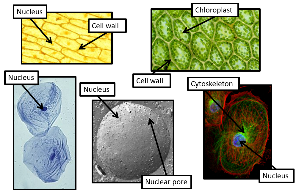
Light Microscopy
Light Microscopy
Laser Scanning Confocal Microscopy
Scanning Electron Microscopy
Light Microscopy
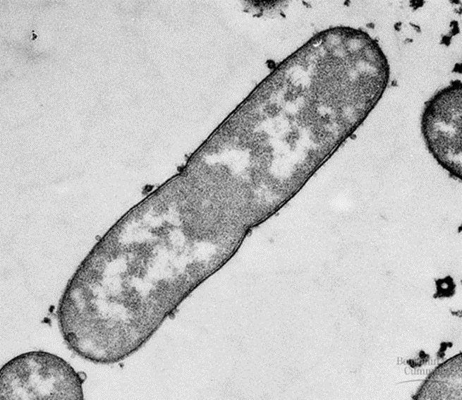
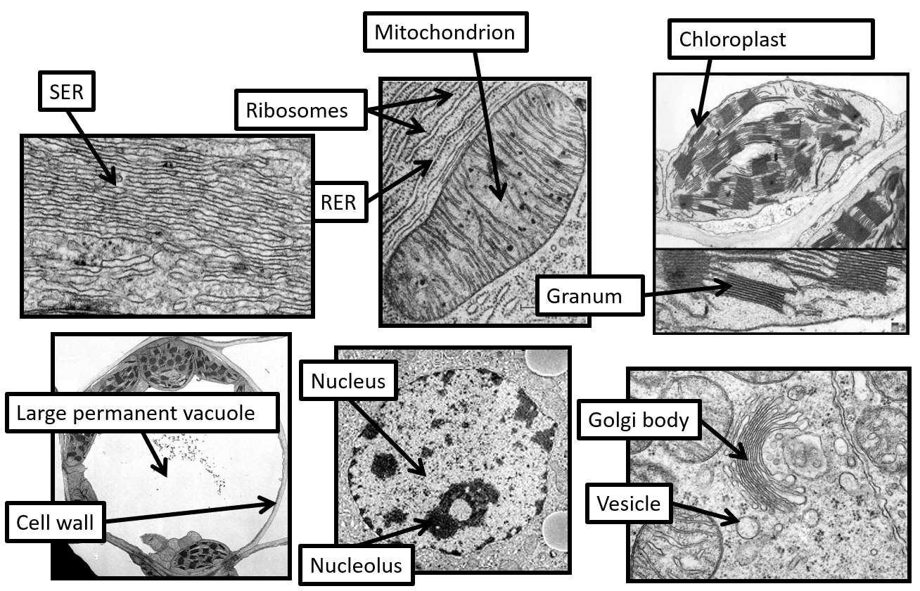 All the images below (and on the next couple of pages) are from Transmission Electron Microscopy
A prokaryotic cell
Prokaryotic cells have no membrane-bound organelles within their cytoplasm and so their structure is much simpler than eukaryotic cells.
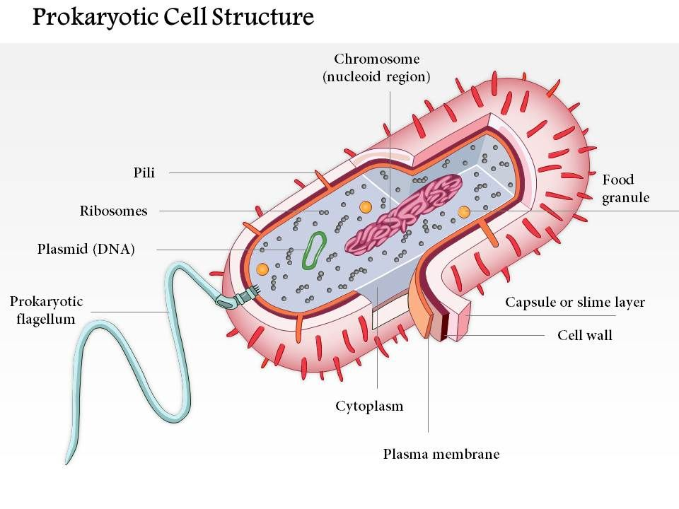
The chromosome is a loop of DNA and so often referred to as circular DNA. The DNA can be referred to as ‘naked’ as it is not bound to proteins called histones (which eukaryotic DNA is). The DNA controls the functioning of the cell.
Prokaryotes can also have small loops of DNA called plasmids. These contain genes for particular characteristics (e.g. antibiotic resistance) and can be copied and given to other bacteria.
Prokaryotes have 70S ribosomes for protein synthesis
Prokaryotes have a plasma membrane, a cell wall made of a peptidoglycan called murein (to prevent bursting by osmosis) and a slime capsule made of polysaccharides (to protect from dehydration and from host immune systems).
Prokaryotes can have protein hairs called pili on their surface for attachment to surfaces.
Prokaryotes may have a flagellum for swimming. It is a spinning protein fibre and very different from the eukaryotic flagellum.
Prokaryotes have a cytoskeleton but it is less complex than the eukaryotic one.
Prokaryotes don’t use mitosis or meiosis for reproduction, they use a process called binary fission.
Sometimes an infolding of the plasma membrane is seen (called a mesosome). It could be for increasing the surface area for reactions involved in photosynthesis or respiration but there is uncertainty about whether it is a real structure or an artefact of the preparation process for electron microscopy.
Comparing prokaryotic and eukaryotic cells
Feature | Prokaryotic cell | Eukaryotic cell |
Types of organisms | All unicellular Bacteria / Archaea Cyanobacteria (photosynthetic) | Plants, Animals, Fungi, Protoctists (uni- or multicellular) |
Cell size | Usually 0.5-5µm diameter | Usually 20-40µm diameter |
Nucleus | Absent | Present |
Location of genetic material | Free in the cytoplasm | In a nucleus |
Form DNA is in | Circular DNA | Linear chromosomes |
DNA’s association with other molecules | Limited / so “naked” DNA | Associated with proteins called histones |
Extra chromosomal DNA | Circular DNA called plasmids | Only present in mitochondria and chloroplasts. No plasmids in the cytoplasm |
Ribosomes | Present – smaller 70S (18nm diameter) | Present – larger 80S (22nm diameter) |
Organelles | No membrane-bound organelles | Mitochondria / Chloroplasts / Endoplasmic reticulum / Golgi apparatus / vesicles etc. |
Cell wall | Present (made from peptidoglycan / murein) | Sometimes present (Cellulose in plants, Chitin in Fungi) |
Slime capsule | Layer around the cell, outside the cell wall | Absent |
Flagellum | Not 9+2 – different structure to those in eukaryotes | 9+2 arrangement of microtubules |
Cell surface membrane | Present | Present |
Reproduction | Binary fission | Asexual or Sexual |
Cytoskeleton | Present | Present, more complex |
Pili | Present | Absent |