Circulation
Important definitions:
Platelets: Small fragments of cells which clot the blood after injury
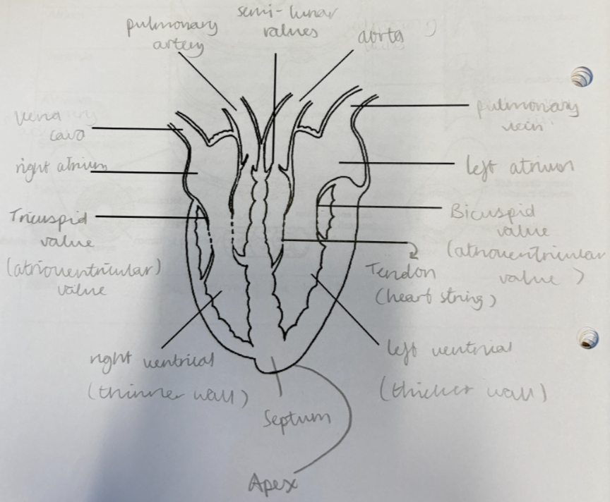
Aorta: The main artery that pumps blood to the body from the left side of the heart
Ventricles: Receive blood from the atria-large chambers at the bottom of the heart
Vena Cava: The main vein that brings deoxygenated blood back to the heart
Pulmonary artery: Carries deoxygenated blood from heart to lungs to pick up oxygen and give up carbon dioxide
Pulmonary vein: Carries oxygenated blood from lungs to heart
AV valve: Prevent backflow of blood from the ventricles back into the atria and pump it into the pulmonary artery (right) and the aorta (left)
Tricuspid: Valve between the right atrium and the right ventricle
Bicuspid: Valve between the left atrium and the left ventricle
Cardiac: Term relating to the heart
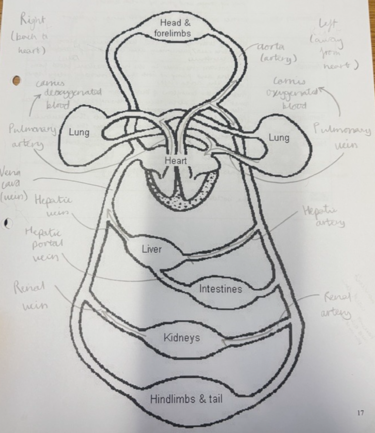
Renal: Term relating to the kidneys
Hepatic: Term relating to the liver
Hepatic portal vein: Carries nutrient rich blood from the intestines to the liver
Septum: Separates the right and left side of the heart
Apex: The bottom of the heart
Septum: Wall of the heart that separates the left and right side
Coronary arteries: Tiny vessels which supply the cardiac cells with blood
Semi-lunar valves: Found at the base of the arteries and ensure blood leaves the heart and doesn’t return to the ventricles
Blood:
Blood is a special type of tissue consisting of millions of cells in a fluid and has 2 main roles:
Transport substances around the body such as oxygen and glucose
defence against disease
COMPOSITION OF BLOOD
Plasma
Red blood cells
White blood cells
Platelets
PLASMA
This is the liquid part of the blood and consists of 90% water. It is naturally a pale yellow colour (straw coloured)
The remaining 10% consists of a mixture of substances that are either dissolved or suspended in it.
Function
Plasma transports soluble substances around the body, e.g. minerals, vitamins, digested food products such as glucose, chemical waste products such as urea and carbon dioxide
It also carries chemical messengers such as hormones and helps redistribute heat energy around the body.
Red Blood Cells:
Produced in red bone marrow, lifespan of ~4 months, destroyed by liver/spleen.
Most abundant blood cell.
Structure & Adaptations:
Biconcave shape → Increases surface area for oxygen uptake and flexibility to pass through capillaries.
No nucleus → More space for haemoglobin.
Contain haemoglobin → A red iron-containing protein that binds with oxygen to form oxyhaemoglobin for transport.
Haemoglobin & Iron:
Haemoglobin needs iron to function.
Iron-rich foods (e.g., red meat, dark green vegetables) are essential for healthy red blood cell production.
Carbon Monoxide Poisoning:
Haemoglobin binds more easily to carbon monoxide (CO) than oxygen.
CO is found in car exhaust fumes and cigarette smoke.
Inhalation of CO reduces oxygen transport, leading to oxygen starvation in tissues, which can cause serious harm or death.
Platelets:
Platelets are small fragments of cells with no nucleus, produced in the bone marrow.
There are approximately 400,000 platelets per cm³ of blood.
Their main function is blood clotting, which helps:
Prevent excessive blood loss when a blood vessel is cut.
Stop pathogens from entering the bloodstream through wounds.
Blood Clotting Process:
When a blood vessel is damaged, the normally smooth vessel walls become rough.
Platelets stick to the rough edges and release a chemical signal.
This chemical triggers the conversion of the soluble plasma protein fibrinogen into insoluble fibrin.
Fibrin forms a network of fibers, creating a mesh across the wound.
Red blood cells and platelets get trapped in the mesh, forming a blood clot that seals the wound.
Over time, the clot hardens into a scab, protecting the area while new skin forms underneath.
Diseases:
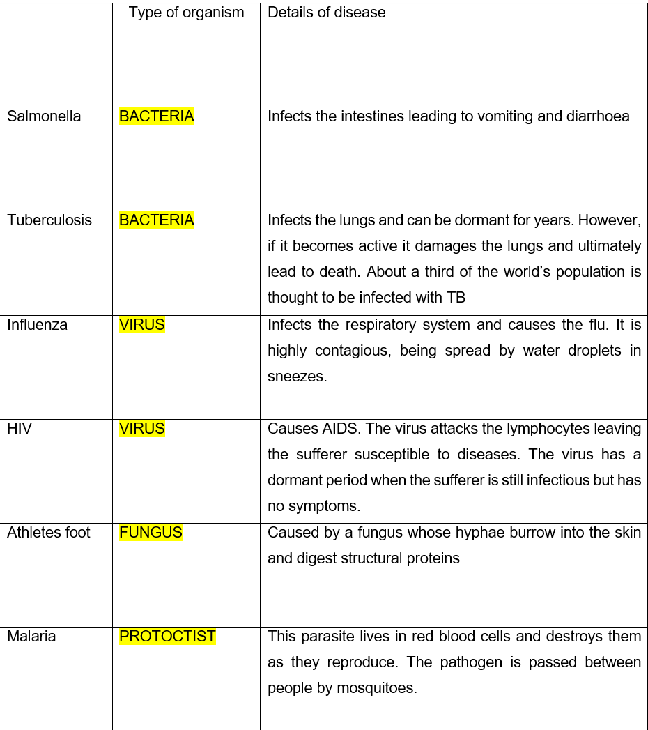
Microorganisms such as bacteria and viruses that cause diseases are known as pathogens
White Blood Cells:
Larger than red blood cells, have a nucleus, and are fewer in number (~8,000 per cm³ of blood).
Function: Protect the body by defending against pathogens (disease-causing microorganisms).
Pathogens can harm the body by damaging tissues or releasing toxins.
Difference between red and white blood cells:
Red blood cells are smaller, more numerous, and lack a nucleus, while white blood cells are larger, fewer, and have a nucleus. Red blood cells transport oxygen, whereas white blood cells defend against infections.
Body's Defence System
First line of defence:
Skin acts as a physical barrier against pathogens.
Mucus in airways traps microbes; stomach acid (HCl) kills ingested pathogens.
If these barriers fail, white blood cells take over.
There are two types of white blood cells:
Phagocytes – Engulf and digest pathogens.
Lymphocytes – Produce antibodies to target specific pathogens and create memory cells for future immunity.
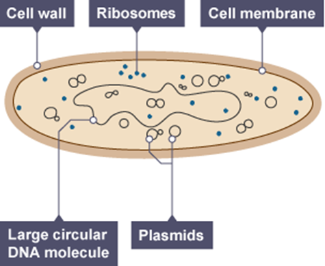
Phagocytes:
They are made in the red bone marrow and only live a few days.
They are larger than red blood cells and are an irregular shape. They have a multi-lobed nucleus and granular cytoplasm.
They destroy foreign substances such as bacteria by engulfing and digesting them by a process called phagocytosis.

Lymphocytes
Lymphocytes are produced in the lymphatic system and can live for several years. They have a spherical shape, a large nucleus, and a small amount of non-granular cytoplasm.
Function:
Lymphocytes produce proteins called antibodies that target microorganisms entering the bloodstream.
Antibodies are produced in response to proteins found on the surface of bacteria and viruses. A protein that triggers an immune response is called an antigen (found on the outside of pathogens).
Each lymphocyte produces antibodies that target only one specific antigen. When this antigen enters the body, the lymphocyte produces large quantities of the correct antibody.
Antibodies destroy pathogens in various ways:
They can cause bacteria or viruses to clump together.
They promote phagocytosis.
They prevent the microorganism from invading host cells.
Some bacteria or viruses release toxins, and antibodies that target these are called anti-toxins.
Immunity and the Immune System
After a person has had a disease like measles, the body is usually protected from getting it again. The person is said to be immune to the disease because:
When the virus enters the body, lymphocytes produce antibodies to defeat it, and the person recovers.
Some lymphocytes become memory cells that can recognize the virus if it re-infects the person.
Memory cells produce antibodies faster and in larger quantities during a re-infection, known as the secondary immune response.
This allows the body to defeat the virus before the person becomes ill, making them immune.
Vaccination
Vaccination takes advantage of this immune process.
A person is injected with a weakened or dead form of the pathogen.
This does not cause the disease, but the body recognizes the pathogen as foreign and makes antibodies.
Memory cells form to recognize the real pathogen, and if infected later, they produce antibodies more quickly and in larger amounts.
The pathogen is destroyed before illness occurs, making the person immune.
Bacterial infections can be treated with antibiotics, which kill the pathogen.
Other diseases can be caused by fungi (like athlete's foot) or protoctists (like malaria).
Pathogens:
An organism that can cause a disease is called a pathogen. The most common types are bacteria and viruses.
Circulation
The circulatory system is the transport system of the human body. It consists of blood, a fluid tissue that circulates around the body in blood vessels. The heart acts as a pump to keep the blood moving. Blood is made up of plasma, a straw-colored fluid containing blood cells.
Blood Vessels
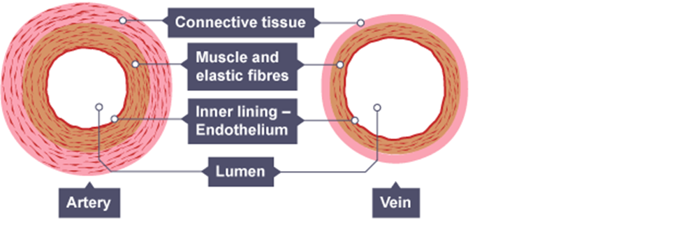
Arteries
Arteries carry blood away from the heart to all regions of the body.
They have a small lumen and thick elastic walls to withstand the high pressure from the heart pumping blood.
Near the tissues, arteries branch into smaller vessels called arterioles.
Veins
Capillaries reunite to form venules, which then join to form veins.
Veins carry blood back to the heart from the tissues.
They have a large lumen and thinner walls than arteries since they don’t need to withstand as much pressure.
Most veins are located between muscles, and when the muscles contract, they help move blood by squashing the veins.
To ensure blood flows in the correct direction, veins have one-way semi-lunar valves, which is important when moving blood against gravity.
Capillaries
Capillaries are tiny vessels found between arterioles and venules, thinner than a human hair. Their walls are just one cell thick, allowing for a short distance between blood and cells, which speeds up diffusion.
Capillaries form a network in tissues, ensuring every cell is less than 0.1mm away.
Oxygen, glucose, and other useful products diffuse from the blood into cells.
Waste products like carbon dioxide and urea diffuse from cells into the blood for excretion.
The Heart
The heart is divided into two halves by a vertical partition:
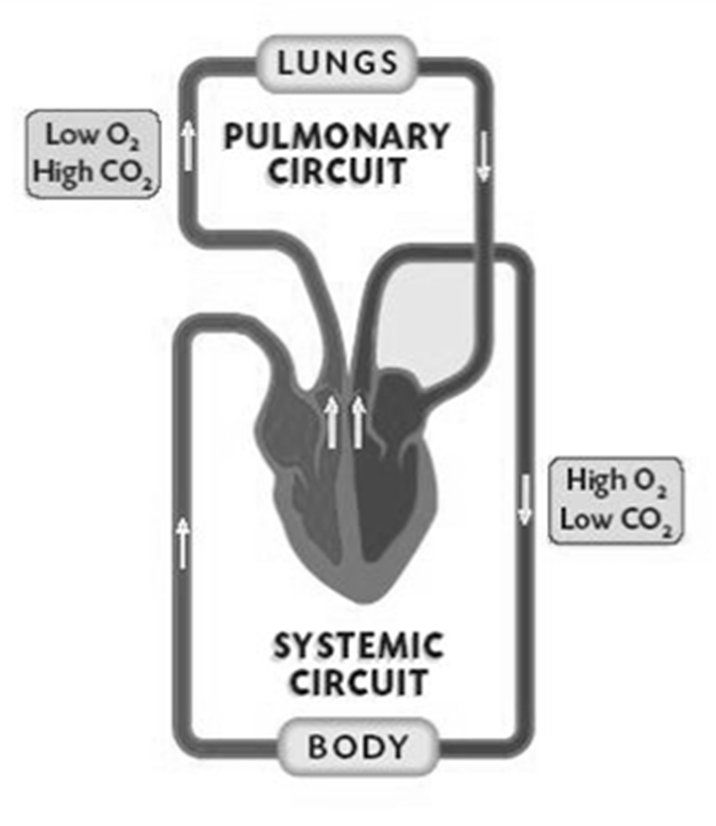
Right side of the heart
Deoxygenated blood arrives from the body and is pumped to the lungs to gain oxygen and remove CO2.
Left side of the heart
Oxygenated blood arrives from the lungs and is pumped to the rest of the body.
The human body has a double circulatory system, with one circuit serving the lungs and the other serving the body. Blood passes through the heart twice for every complete circuit.
Structure of the circulatory system:
Structure of the heart:
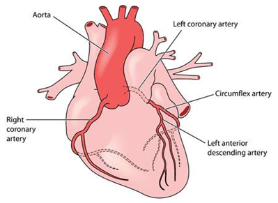
The walls of the heart are made from cardiac muscle that can contract without any stimulus from the brain
It obtains the food and oxygen it requires from the coronary blood vessels which run through
Many heart problems are due to these being blocked
The Heart
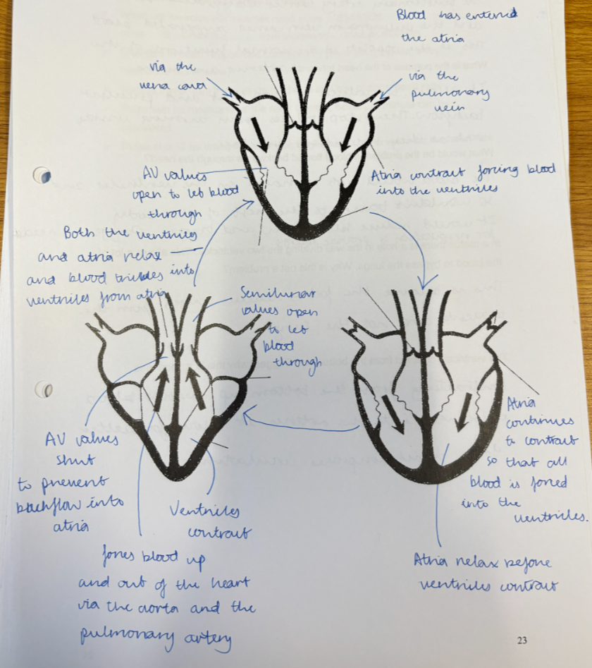
The heart has four chambers: two atria and two ventricles.
Blood enters the atria via the vena cava and pulmonary vein. Atria have thin walls as they only pump blood to the ventricles.
Ventricles have thicker walls and contract to pump blood to the lungs (pulmonary artery) or body (aorta).
Valves prevent backflow:
Tricuspid valve (right atrium to right ventricle)
Bicuspid valve (left atrium to left ventricle)
Semi-lunar valves at the start of major blood vessels leaving the ventricles.
Action of the Heart
Both sides of the heart contract together:
The atria contract, pushing blood through the tricuspid and bicuspid valves into the ventricles.
The ventricles contract, forcing blood out:
Right ventricle → pulmonary artery → lungs
Left ventricle → aorta → body
Cuspid and semi-lunar valves prevent backflow, while tendons keep valves in place.
The cardiac cycle:
Heart Rate and Exercise
The heart beats around 70 times per minute at rest.
During exercise, muscles need more oxygen for increased respiration in mitochondria, requiring a higher heart rate.
Heart rate is controlled by the brain (medulla) and hormones (adrenaline).
Nerves from the brain can adjust heart rate.
Pulse can be measured at the neck or wrist and calculated in beats per minute.
For accuracy, pulse should be timed for 15-30 seconds.
Cormmss:

Coronary Heart Disease
Coronary arteries are narrow and supply oxygen to the heart.
Fatty deposits like cholesterol can block them, narrowing the arteries further.
This restricts oxygen supply to the heart muscle, leading to a heart attack.
A heart attack occurs because the lack of oxygen prevents ATP production, causing heart cells to die.
Risk Factors for Heart Disease
Diet high in animal fats (cholesterol).
Smoking – nicotine causes cholesterol buildup in arteries.
Lack of exercise – increases heart rate and slows recovery to resting rate.
Adrenaline
A hormone produced by the adrenal glands above the kidneys.
Released in response to stress or threats.
Prepares the body for action by triggering specific physiological changes.
Effects of Adrenaline
Heart – Increases heart rate.
Muscles – Increases respiration for more energy.
Brain – Supplies more oxygen for alertness.
Liver – Converts glycogen to glucose for energy.
Eyes – Pupils dilate for better vision.
A common side effect is the feeling of "butterflies" when nervous, caused by reduced blood flow to the digestive system as more blood is directed to muscles, liver, and brain.
Artificial Pacemaker
Used to treat irregular heartbeats.
Inserted under the skin with an electrode connected to the heart via a vein.
Sends electrical impulses to regulate heart contractions.