Tissues etc notes
TISSUE TYPES, INJURY AND REPAIR NOTES
- In the tissue level of organisation groups of similarly specialised cells that work together to perform a similar function
- Histology is the study of tissues. A biopsy is the removal of living tissue for microscopic examination
- Four major types of tissue
- Connective tissue – transports; protects, supports and binds tissues and organs; stores energy as fat (adipose); provides immunity. Cells are sparse in an extracellular matrix containing a ground substance and protein fibres (collagen – for tensile strength, elastic for stretch, and reticular for frameworks).
- Muscular tissue - produces movement via contraction and generates body heat. Cells closely arranged (have intercellular junctions).
- Epithelial tissue - covers body surfaces, lines hollow organs, body cavities and ducts; forms glands. Cells are closely arranged (have intercellular junctions) into sheets or glands
- Nervous tissue - detects changes in the body & responds by generating nerve impulses. Cells are closely associated with each other (via synapses) to form networks.
- In some tissue types, adjacent cells are connected using a variety of intercellular junctions
- Intercellular junctions include:
- Tight Junctions (occluding junctions) are found where a leakproof seal is needed between cells. They keep materials from leaking out of organs like the stomach and bladder.
- Adherens Junctions (anchoring junctions) make an adhesion belt that keeps tissues from separating as they stretch (epithelial, muscular) and/or contract (muscular).
- Desmosomes (anchoring junctions) act as “spot welds”. They hook into the cytoplasm of adjacent cells
- Hemidesmosomes (anchoring junctions) are half-welds that join cells to the basement membrane.
- Both desmosomes and hemidesmosomes are widely distributed in tissues especially those subjected to severe mechanical stress eg cardiac muscle, other muscle, and the epidermis
- Gap Junctions are pores (connexons) that allow small substances like ions to pass between cells. Allow rapid communication between cells. Especially important in cardiac muscle.
- Germ layers that each tissue type develops from are:
- Connective - Mesoderm
- Muscular - Mesoderm
- Epithelial – Ectoderm, Endoderm, Mesoderm
- Nervous - Ectoderm
- Epithelial tissue
- Epithelia are classified according to cell shape (squamous – flat, thin tile-like, cuboidal, and columnar) and number of layers
- Simple Epithelia are composed of a single thin layer of cells and are concerned primarily with movement/transport of substances from one body compartment to another.
- Simple squamous epithelium – allows rapid diffusion; found lining the lungs and capillaries
- Simple cuboidal epithelium – cubes make tubes; found in ducts and tubules
- Simple columnar epithelium – allows absorption and secretion as well as providing some protection; lines the digestive tract and often contain goblet cells and may be ciliated
- Pseudostratified columnar epithelium - appears to have several layers but all cells are attached to the basement membrane; may contain cilia and goblet cells and are found in the upper respiratory tract (ciliated) and vas deferens (non-ciliated)
- Stratified Epithelia - contains 2 or more layers of cells and are thicker and stronger than simple epithelia and acts as a protective covering.
- Stratified squamous epithelium (widespread) - Keratinized – found in superficial layers of the skin. Non-keratinized – found lining wet surfaces which are exposed to the exterior e.g. mucosae mouth, esophagus & vagina.
- Stratified cuboidal and columnar epithelia (rare) - Locations include the pharynx, sweat glands and part of the male urethra
- Stratified transitional epithelium – stretches, found in the bladder
- Endocrine glands - Ductless, secretions diffuse directly into blood vessels. All secretions are hormones.
- Exocrine glands - Secretions empty through ducts to the epithelial surface. Secretions include mucus, sweat, oil, saliva, tears, digestive enzymes etc.
- Muscular tissue
- Skeletal Muscle = Under voluntary control. Contracts to pull on bones or skin. Produces gross body movements or facial expressions.
- Characteristics of skeletal muscle cells are: Long, cylindrical cells called fibres. Striated (due to arrangement of the contractile proteins into sarcomeres). Multinucleate (more than one nucleus).
- Cardiac Muscle = Under involuntary control. Found only in the myocardium of the heart wall. Function is to pump blood.
- Characteristics of cardiac muscle cells are branched and attached to other cardiac muscle cells at intercalated disks. Striated (due to arrangement of the contractile proteins). One nucleus per cell.
- Smooth Muscle = Under involuntary muscle. Found in walls of hollow organs such as stomach and other digestive organs, uterus, and blood vessels.
- Characteristics of smooth muscle: cells are spindle-shaped, no visible striations, one nucleus per cell.
- Connective tissue
- The most abundant and widely distributed tissue type in the body
- Usually highly vascular and supplied with many nerves - Exceptions are cartilage, ligaments and tendons
- Contain sparse cells separated by a non-living extracellular matrix (composed of a ground substance and fibres).
- Ground substance - mostly water along with adhesion proteins and polysaccharide molecules.
- Protein Fibres - 3 types: Collagen (white – tensile strength); Elastic (yellow – stretch); Reticular (form fine meshwork)
- Loose connective tissue = Areolar; adipose, reticular
- Areolar Connective Tissue is the most widely distributed in the body. Used to attach skin and underlying tissues, and as a packing between glands, muscles, and nerves
- Adipose tissue is located in the subcutaneous layer deep to the skin and around organs and joints. Functions to insulate, cushion (protection) and store energy.
- Reticular connective tissue is a network of interlacing reticular fibers and cells. It forms a scaffolding (stroma) used by cells of lymphoid tissues such as the spleen and lymph nodes
- Dense connective tissue = Regular; irregular; elastic
- Contains very few cells (e.g. fibroblasts – produce collagen)
- Contains numerous, thick, dense fibres; fibres are packed a lot closer than in loose connective tissue
- Dense Regular Connective Tissue is found in tendons and ligaments. Has bundles of collagen fibres arranged in parallel patterns; provides strength along one axis
- Dense Irregular Connective Tissue is found in dermis, submucosa of the digestive tract, the lymph nodes and spleen, among other places as well as in tendons and ligaments. Has collagen fibres that are randomly arranged; provides strength in many different directions
- Elastic Connective Tissue allows stretching of certain tissues like the elastic arteries (the aorta) and in the lungs and is also found in a few ligaments e.g. those between adjacent vertebrae. Has freely branching elastic fibres; allow stretching
- Cartilage = Hyaline; fibrocartilage; elastic
- Hyaline cartilage is the most abundant type of cartilage. Forms most of the foetal skeleton prior to birth. It covers the ends of long bones and parts of the ribs, nose, trachea, bronchi, and larynx.
- Fibrocartilage is a very strong, tough cartilage; absorbs shock. Forms intervertebral discs and found in the knee joints (menisci)
- Elastic cartilage makes up the malleable part of the external ear and nose and the epiglottis. Provides strength and elasticity.
- Bone (osseous) tissue = Compact; spongy
- Compact bone makes up the external layer of all bones. Consists of a solid matrix of calcium and phosphate salts arrange in osteons; osteons are not found in spongy bone
- Spongy bone is found in the ends of long bones and within most short bones. Consists of bone arranged in an irregular network of trabeculae; has lots of small spaces within it often filled with red bone marrow
- Liquid connective tissue = Blood; lymph
- Blood is found in the blood vessels and heart. Is composed of blood cells in a liquid matrix (plasma) containing dissolved fibres (fibrinogen)
- Lymph is found in lymphatic vessels. Is similar to blood plasma, contains lymphocytes.
TISSUE DAMAGE, INJURY & REPAIR
- Causes include trauma, disease (homeostatic imbalance) or simple wear and tear
- In medicine physical trauma is an injury to living tissue caused by an extrinsic/external agent
- Two main types of physical trauma are:
- Blunt force trauma—when an object or force strikes the body, often causing haematoma and/or broken bones
- Penetrating trauma—when an object pierces the skin or body, usually creating an open wound eg a needle or knife
- Strains and sprains = wear and tear
There are 2 types of tissue repair:
- Regeneration - Replacement of destroyed tissue by the same kind of cells (parenchymal cells – the cells that make up the tissue – do this). No scarring
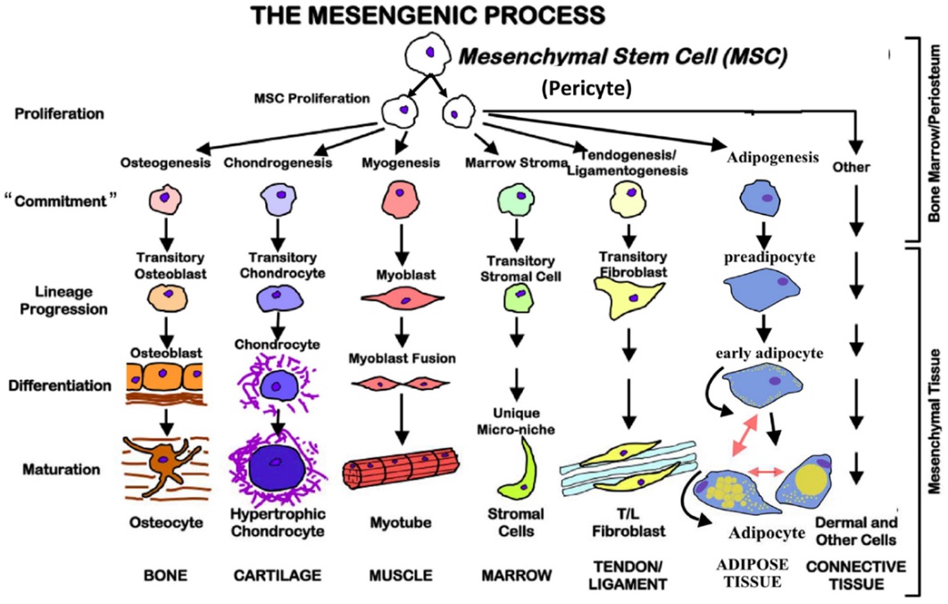
- Mesenchymal cells (in bone marrow and the periosteum) are multipotent adult stem cells. They can differentiate into many types of tissue cells needed for regeneration to repair damaged tissue
- Fibrosis - Replacement of destroyed tissue by dense (fibrous) connective tissue. Forms scar tissue
- Carried out by fibroblasts.
- When an injury (or infection) occurs fibroblasts:
- Stop making collagen, change shape and travel to the area of injury
- Release inflammatory products, destroying damaged tissue for phagocytosis
- Then start making collagen and repair the area of damage with dense (fibrous) connective tissue which forms scar tissue
- When finished, they migrate back to where they came from and return to their normal shape and function
- Whether regeneration or fibrosis occurs depends on: Type of tissue damaged and severity of the injury
- Tissues that regenerate easily:
- Epithelial tissue (skin and mucous membranes)
- Loose connective tissues, bone and blood
- In these tissues the parenchymal cells divide to replace the damaged tissue with new tissue of the same type
- Tissues that regenerate poorly and have limited capacity for tissue repair:
- Nervous, muscle, dense connective tissue, and cartilage
- In these tissues fibrosis occurs, and the damaged tissue is replaced largely with scar tissue reducing functionality
Connective Tissue Repair
- Fibroblasts are the most common cells in connective tissue, some are able to transform into any of the other types of connective tissue cells for regeneration, others are responsible for fibrosis (forms scar tissue)
- Mesenchymal cells can differentiate into many types of connective tissue cells needed for regeneration
- Haematopoietic stem cells (make blood cells) are only found in red bone marrow
- Repair of dense connective (eg tendons and ligaments) and cartilage tissues in adults is limited by:
- their intrinsic hypocellularity (ie a lack of cells)
- a dense extracellular matrix that limits cellular migration to, and local proliferation at, an injury site
- Sprain = The stretch or tear of ligaments. Affects reinforcement of joints and range of motion
- Grade 1 - the ligament is stretched but not torn.
- Grade 2 - the ligament is partially torn. Can be inflammation and bruising
- Grade 3 – the ligament is completely torn or ruptured. There is inflammation and brusing
- In severe cases, joints can become unstable
- bones can move out of alignment
- joint may extend beyond its normal range of motion.
- Severe sprains sometimes require surgery to repair torn ligaments
- Bone has a high capacity for repair. Bone Fractures = break in a bone
- Types of bone fractures
- Closed (simple) fracture—break that does not penetrate the skin
- Open (compound) fracture—broken bone penetrates through the skin
- Bone fractures are treated by reduction and immobilization
- Repair of Bone Fractures
- Haematoma (blood-filled swelling) is formed
- The break is splinted by fibrocartilage to form a callus – Phagocytes remove cellular debris and fibroblasts deposit collagen to form the callus
- Fibrocartilage callus is replaced by a bony callus of spongy bone
- Bony callus is remodeled to form a permanent patch – the spongy bone is replaced by compact bone
Muscle Tissue Repair
1. Skeletal muscle
- Skeletal muscle cells/fibres cannot divide to repair damage but can lay down new protein and enlarge (hypertrophy)
- Skeletal muscle contains stem cells, called satellite cells found underneath the basal lamina
- These mononucleated quiescent cells which divide (slowly) when muscle is damaged. After dividing, these cells fuse with existing muscle fibres, to regenerate and repair the damaged fibre. Capable of repairing limited damage.
- Remaining damaged tissue is replaced by scar tissue.
2. Cardiac muscle is lacking in stem cells for tissue regeneration
- Muscle tissue has a relatively poor capacity for the repair of dead or damaged cells. Damaged tissue is replaced by scar tissue.
3. Smooth muscle regenerates from stem cells called pericytes which are found in some blood vessels
- capable of slow and limited repair
- regenerates and repairs much more readily than skeletal and cardiac muscle tissue
- Myofibrosis - Replacement of muscle tissue by connective tissues (scar tissue). This tissue can not contract therefore scar tissue reduces the functionality of muscle
- Strain = Stretching or tearing of skeletal or cardiac muscle fibres
- the muscle has been stretched beyond its limits or it has been forced to contract too strongly
- Classified depending on the severity of muscle fibre damage:
- Grade I – mild: only a few muscle fibers are stretched or torn. Muscle is intact and has normal strength
- Grade II – moderate: with a greater number of injured fibers. There is inflammation, loss of strength and may be bruising (due to blood vessel damage)
- Grade III - tears the muscle all the way through. Complete loss of muscle function. There is inflammation and bruising. May require surgery.
Nervous Tissue Repair
- Nerve cells are amitotic therefore do not divide and can not replace damaged cells
- The PNS has some capacity for repair and regeneration
- axons are able to regrow as long as the cell body is intact, and they have contact with Schwann cells
- The CNS is largely incapable of self-repair and regeneration; damaged CNS tissue undergoes gliosis
- formation of scar tissue composed of glial cells (neuroglia)
- recently it has been discovered that certain areas of the adult brain possess neural stem cells, although to what extent they are capable of repairing damage to neurons or neuroglia is still uncertain
Epithelial Tissue Repair
- Epithelial covering and linings are often under constant heavy wear and tear and therefore must be highly regenerative
- This occurs either by division and differentiation of stem cells (eg in the epidermis), or division of parenchymal cells (eg endothelial cells in the blood vessels)
- Glandular tissue
- Many exocrine glands have a continuous loss of cells which have to be constantly replaced by new ones (regeneration) eg the liver; sebaceous glands (replace cells that disintegrate during secretion)
- Stem cells have been identified in some endocrine glands eg pituitary, adrenal, pancreas
Open Wound Healing (skin repair)
- There are two kinds of wound-healing:
- Epidermal wound healing occurs following superficial wounds that affect only the epidermis. Usually return to normal function
- Deep (open) wound healing occurs when an injury extends to the dermis and subcutaneous layer. Usually results in loss of some function and development of scar tissue
- Events in Wound Healing
- Inflammation and Haemostasis - injured blood vessels bleed, and haemostasis occurs, inflammatory chemicals are released and uninjured capillaries become very permeable, clotting proteins migrate into area. A clot walls off the injured area. The clot surface dries and forms a scab.
- Organisation and blood supply restored - growth of new capillaries, the blood clot is replaced with granulation tissue, epithelium begins to regenerate, fibroblasts produce collagen fibres to bridge the gap, debris is phagocytized
- Regeneration and fibrosis - Regeneration of surface epithelium, scab detaches, fibrous tissue matures (epithelium thickens and begins to resemble adjacent tissue). Results in a fully regenerated epithelium with underlying scar tissue
Burns
- Tissue damage caused by excessive heat, electricity, radioactivity, or corrosive chemicals that denature (break down) the proteins in the skin cells
- Immediate threat = Dehydration and electrolyte imbalance, leading to renal shutdown and circulatory shock
- Burns are graded according to their severity
- A first-degree burn involves only the epidermis
- A second-degree burn destroys the epidermis and part of the dermis
- A third-degree burn is a full-thickness burn (epidermis, dermis, and subcutaneous layer)
- Burns are critical if:
- >25% of the body has second-degree burns
- >10% of the body has third-degree burns
- Third degree burns on the face, hands, feet, or perineum
- When the burn area >70%, more than half the victims die
- The rule of nines is used to quickly estimate the surface area affected by a burn
- Several factors influence skin healing after burn injuries, e.g., the causes, the degree and size of burn, and the patient’s general condition and type of graft or materials for covering burn wounds.
- Depending on burn severity, the healing process may result in different consequences:
- Superficial burns recover within two weeks and cause minimal scarring.
- The re-epithelization of partial thickness burns is ensured by keratinocyte migration from skin dermal appendages within a few hours of the injury.
- In deeper burns, the healing starts around the edges, but not at the centre.
- Improving burn wound care and healing is a specialist field
Haemostasis (repairing blood vessels)
- Haemostasis is the stoppage of bleeding to prevent blood loss.
- Occurs in 3 stages:
- Vascular spasm
- Vasoconstriction causes blood vessel to spasm and decreases blood loss
- Platelet plug formation
- Collagen fibres from the connective tissue are exposed by a break in a blood vessel
- Platelets become “sticky” and cling to fibres
- Anchored platelets release chemicals to attract more platelets (positive feedback)
- Platelets pile up to form a platelet plug
- Blood clotting/coagulation
- A series of reactions in which blood is transformed from a liquid to a gel
- Injured tissues release chemicals and calcium ions which trigger a clotting cascade
- Prothrombin activator converts prothrombin to thrombin (an enzyme) → Thrombin joins fibrinogen proteins into insoluble fibrin → Fibrin forms a meshwork (the basis for a clot) which traps RBCs to form the clot). Note: Clots are broken down by fibrinolysis
- Blood usually clots within 3 to 6 minutes
- The clot remains as the endothelium regenerates
- The clot is broken down after tissue repair by fibrinolysis (not part of haemostasis)
- Undesirable Clotting
- Thrombus: A clot in an unbroken blood vessel which may block circulation, leading to tissue death. Can be deadly in areas like the heart
- Embolus: A thrombus that breaks away and floats freely in the bloodstream and can later clog vessels in critical areas such as the heart, lungs and brain
- Bleeding Disorders
- Thrombocytopenia: Platelet deficiency. Even normal movements can cause bleeding from small blood vessels that require platelets for clotting
- Haemophilia: Hereditary bleeding disorder. Symptoms include prolonged bleeding, especially into joint cavities. Normal clotting factors are missing
- Haematoma
- A collection of coagulated blood outside a blood vessel but within the body (c.f. haemorrhage = active, ongoing bleeding)
- Can be seen under the skin or nails as bruises (aka contusions)
- Can also happen deep inside the body where they may not be visible- may sometimes form a mass or lump that can be felt
- During the healing process, oxygen-rich blood to loses oxygen (red → purple/blue), then the RBC degrade and haemoglobin breaks down biliverdin (green) → bilirubin (yellow)→ haemosiderin (brown)
- Phagocytosis clears the breakdown products from the area