13.11 structure of skeletal muscle
compare similarities/differences between typical animal cell to muscle fibre
similarities
endoplasmic/sarcoplasmic reticulum
contain mitochondria
enclosed in plasma membrane
differences
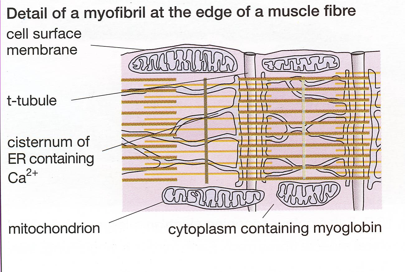
more mitochondria in muscle fibres (site of aerobic respiration - produces ATP for muscle contraction)
mitochondria at edge in muscle fibre
sarcoplasmic membrane has t-tubules
muscle → muscle fibre → myofibrils → sarcomere → actin ad myosin
skeletal muscles
stripey due to protein filaments
contain two protein called ACTIN + MYOSIN
each muscle made of muscle cells called muscle fibres
muscle fibres same as most cells containing cytoplasm, mitochondria + endoplasmic reticulum
very long, sometimes called acellular as don’t seem to have distinct cell membranes + may have many nuclei (multinucleate).
structure of skeletal muscle fibres
skeletal muscles comprise of numerous bundles of long, cylindrical muscle fibres.
individual cells are fused to form muscle fibres during development to avoid weakness at junctions between cells + increase overall muscle strength.

Key components of muscle fibre include:
Sarcolemma - cell-surface membrane of muscle fibre.
Sarcoplasm - cytoplasm of muscle fibre. interwoven w special sarcoplasmic reticulum (modified ER) + infoldings of sarcolemma (T tubules)
Transverse tubules (T tubules) - extensions of sarcolemma that transmit electrical signals, ensures entire muscle receives impulse to contract simultaneously. formed when membrane folds into cell
Sarcoplasmic reticulum - specialised endoplasmic reticulum that’s responsible for storing + releasing calcium ions.
Myofibrils - subcellular structures designed for contraction, made up of sarcomeres. longitudinally running fibres - gives it striations
Multiple nuclei - Skeletal muscle fibres have many nuclei because several cells merge to form one muscle fibre.
Mitochondria - many for lots of ATP energy release for muscle contraction.
the structure of myofibrils
Myofibrils: core units of muscle fibres, contain organised bundles of protein filaments.
filaments slide past each other to enable muscle contraction
the structure of a sarcomere
myofibrils made up of repeating units called sarcomeres
main filaments in sarcomere:
myosin filaments - Thick filaments composed of long rod-shapes with bulbous heads that project to the side.
actin filaments - Thin filaments composed of two strands twisted around each other.
under microscope, myofibrils exhibit distinctive banding pattern - corresponds to arrangement of myosin and actin within each sarcomere
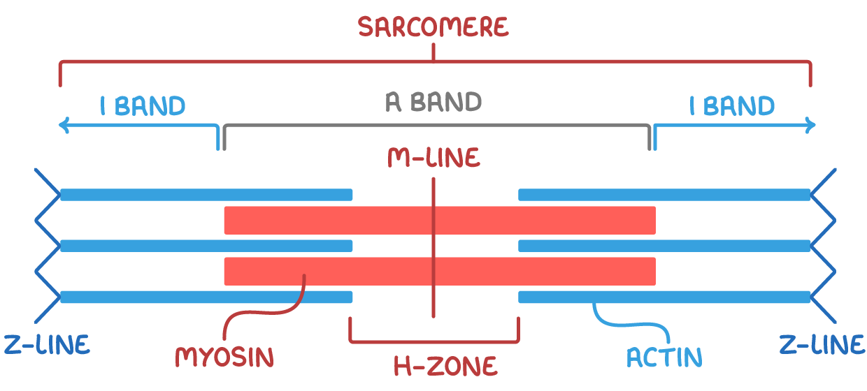
Key sections of sarcomere:
A band (dark band) - Area with both myosin and overlapping actin filaments (darker in colour).
I band (light band) - area contains only light actin filaments (lighter in colour). represents length of thicker Myosin strands.
Z-lines - joins sarcomeres end to end (central line within I band). where actin fibres cross link
M-line - central line of sarcomere. protein that holds myosin together (like the Z line for actin.)
H-zone - area wi only myosin filaments (central lighter region in A band).
sliding of actin + myosin filaments within sarcomeres is fundamental to process of muscle contraction. During muscle contraction, sarcomeres shorten, causing change in pattern of light + dark bands.