Somatic Sensory System I Touch
Identify and describe different types of sensory receptors in the skin
Introduction
Receptors are distributed throughout body instead of being in a small specialized region
Can think of touch as a group of at least four senses rather than single one
Touch
Temperature
Pain
Body position
Skin Layers
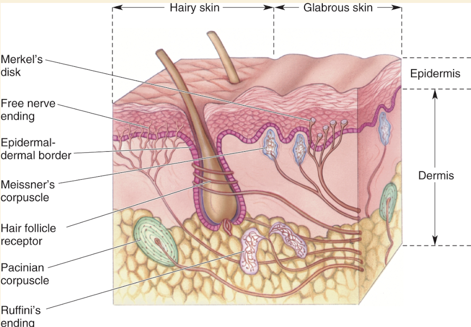
Hairy skin = skin with hair
Glabrous skin = skin with no hair
Mechanoreceptor of Skin
Make up most of sensory receptors in somatic sensory system & sensitive to physical distortion (bending/stretching)
Mechanoreceptors monitor:
Skin contact
Pressure in the heart
Blood vessels
Stretching of digestive organs
Urinary bladder
Force against the teeth
@ heart of receptors are unmyelinated axon branches sensitive to:
Stretching
Bending
Pressure
Vibration
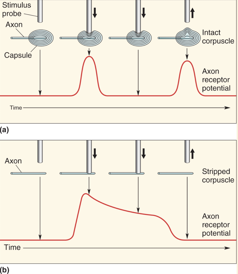
Pacinian corpuscle
Each human hand has 2500 (highest amount in fingers)
Lies deep in dermis
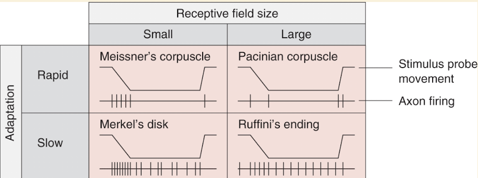
Large receptive fields
Stimulus probe pressed against skin → respond quickly at first then stop firing even though stimulus continues
Aka rapidly adapting
Most sensitive to vibrations around 200-300 Hz
Has football shaped capsule w/ 20-70 layers of connective tissue w/ axon in the middle
Capsule compressed → energy transfer to nerve terminal → membrane deforms → mechanosensitive channels open → current flowing generates receptor potential (depolarizing) → AP
Capsule layers have fluid between them, if stimulus maintains to where axon terminal is no longer deformed, receptor potential dissipates
Pressure release → events reverse themselves
Layered campsule makes pacinian corpuscle sensitive to vibrating, high-frequency stimuli & unresponsive to steady pressure
Has some of largest & fastest axons that originate in the skin
Ruffini’s endings
Both hairy & glabrous skin
Slightly smaller than pacinian corpuscles
Large receptive fields
Stimulus probe pressed against skin → generates more sustained response during long stimulus
Slowly adapting
Meissner’s corpuscles
one-tenth size of pacinian corpuscles
Located in ridges of glabrous skin
Small receptive field
Stimulus probe pressed against skin → respond quickly at first then stop firing even though stimulus continues
Aka rapidly adapting
Most sensitive to vibrations around 50 Hz
Merkel’s disks
Consist of a nerve terminal & flattened, non-neural epithelial cell
Small receptive field
Stimulus probe pressed against skin → generates more sustained response during long stimulus
Slowly adapting
Merkel cells make synapses onto nerve terminals
Both cell & axon terminal mechanically sensitive
Channel Piezo2 opens in response to pressure → depolarization → synaptic release of NTM → nearby nerve ending excites
The nerve ending is also mechanically sensitive bc of second unknown ion channel in its membrane
Krause end bulbs
Lie in border regions of dry skin & mucous membrane
Nerve terminals look like knotted balls of string
Hair follicles
Innervated by free nerve endings
Bending of hair → deformation of follicle → stretches, bends, or flattens nearby nerve endings → increase/decrease AP frequency
Mechanoreceptors vary in stimulus frequencies, pressures, & receptive field sizes
Allow us to differentiate feelings of skin
Selectivity depends on structure of special ending
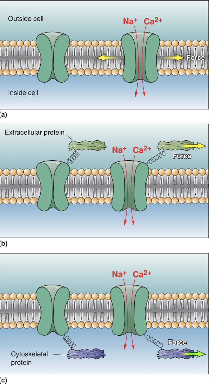
Mechanosensitive Ion Channels
Membranes of unmyelinated axon terminals have mechanosensitive ion channels
Converts mechanical force into change of ionic current
Force applied to channels alters gaiting
Can enhance or decrease channel opening
Force can be applied by:
Membrane is stretched or bent
Connections between channels & extra cellular proteins
Intracellular cytoskeletal components
Mechanical stimuli may trigger release of second messengers that regulate ion channels
Describe how a sensory event is encoded in action potentials in sensory fibers
Primary Afferent Axons
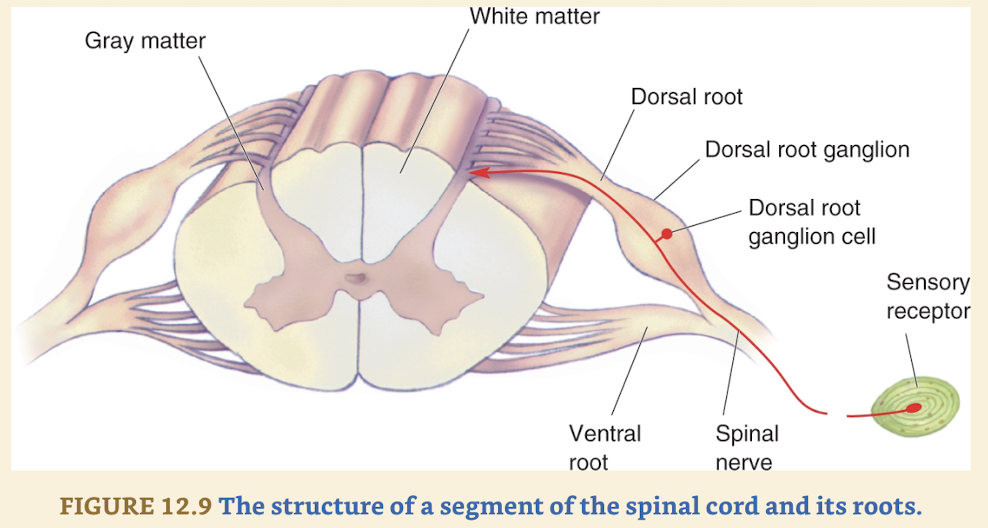
Axons brining info from somatic sensory system to:
Spinal cord
Enter thru dorsal roots
Brain stem
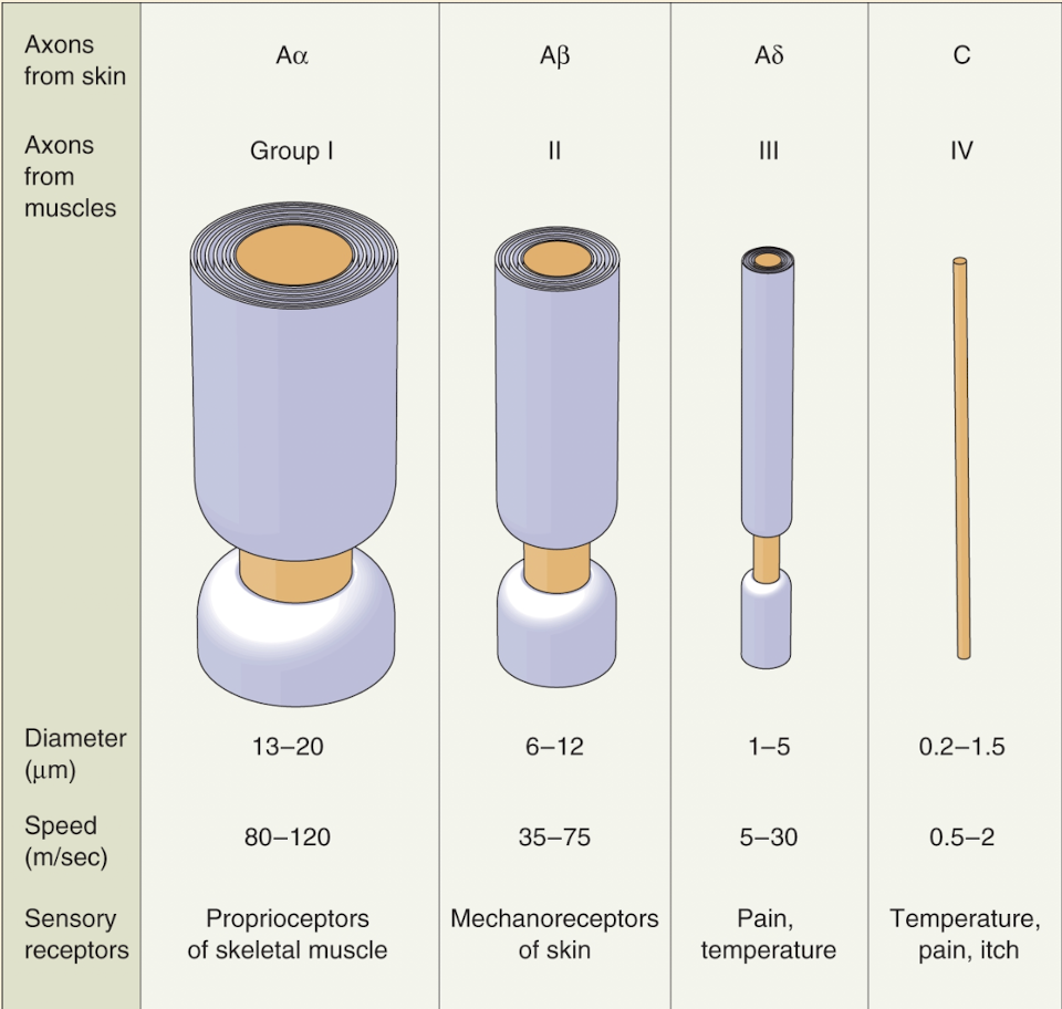
Have varying diameters
Size correlates to type of sensory receptor attached to
In order of decreasing size, axons from skin sensory receptors are called:
Aa, AB, Ao, and C
Axons of similar size by innervating muscles & tendons are called groups:
I, II, III, IV
Group C (or IV) are unmyelinated, rest are myelinated
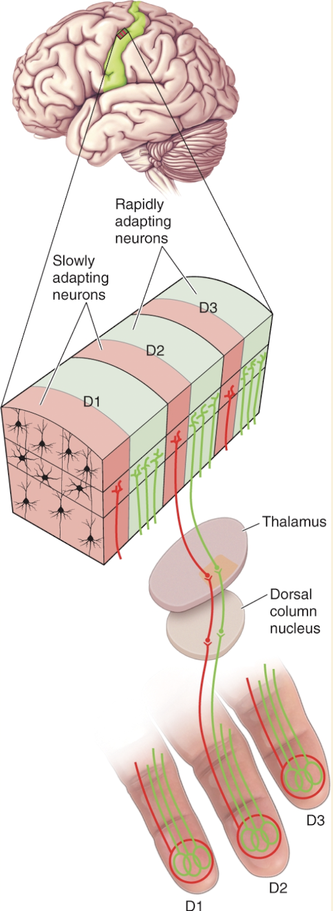
Distinguish somatic sensory receptors based on their receptive field size
Compare and contrast rapidly-adapting and slowly-adapting receptor firing patterns
Relate the structure of the pacinian corpuscle to adaptation in the receptor
Discuss why our ability to discriminate a somatic sensory stimulus across different regions of the body varies
Two Point Discrimination
Measure of spatial resolution is two point discrimination test
Use paperclip w/ two ends and move them until it feels like the same point
Two point discrimination varies twentyfold across body
Finger tips have highest resolution
Higher density of mechanoreceptors
Receptors have small receptor fields
More brain tissue devoted to sensory info
May be special neural mechanisms devoted to high resolution discriminations
Describe the somatosensory pathways to the brain
Sensory Organization of the Spinal Cord
Neurons that receive sensory input from primary afferents are called second-order sensory neurons
Most lie within dorsal horns
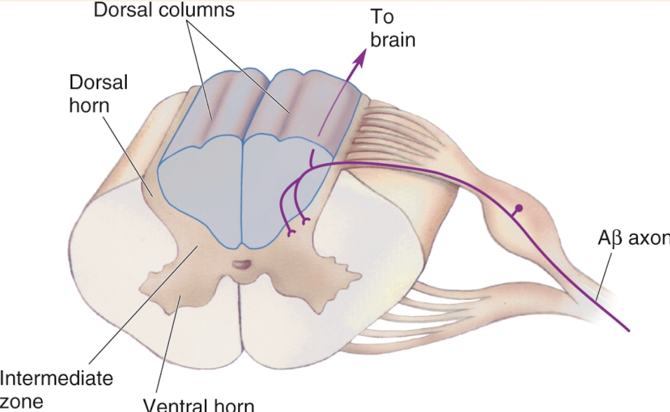
Large myelinated AB axons enter thru dorsal horn & branch → branch synapses in deep part of dorsal horn on second-order sensory neurons
These connections can initiate or modify variety of rapid & unconscious reflexes
Other branch of AB primary afferent axon ascends straight to brain
Responsible for perception & making complex judgements about stimuli touching skin
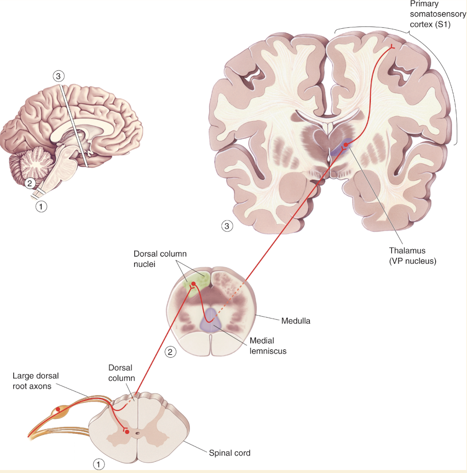
The Dorsal Column-Medial Lemniscal Pathway
Pathway that serves touch only
Ascending branch of large sensory axons enters ipsilateral dorsal column, white matter tract medial to dorsal horn
Dorsal columns carry info about tactile sensation & limb position toward brain
Composed of primary sensory axons & second order axons from neurons in spinal gray matter
Axons of dorsal column terminate in dorsal column nuclei
At this point in pathway info is still represented ipsilaterally
However axons from cells of dorsal column nuclei arch toward ventral & medial medulla & decussate
From that point on somatic sensory system of one side of brain is concerned with sensations from other side of body
Axons of dorsal column nuclei ascend within conspicuous white matter tract caled medial meniscus
ML rises thru medulla, pons, & midbrain
ML axons synapse upon neurons of VP nucleus of thalamus
Thalamic neurons of VP nucleus project to specific regions of primary somatosensory cortex (S1)
Relay nuclei: sensory info is transferred unchanged thru nuclei in brain stem & thalamus on its way to cortex (NOT TRUE)
Alot of transformation takes place instead
Info is altered every time it passes through set of synapses in the brain
Trigeminal Touch Pathway
Somatic sensation of face is supplied mostly by the large trigeminal nerves that enter brain thru pons
There are twin trigeminal nerves (one on each side) & each break up into three peripheral nerves that innervate:
Face
Mouth areas
Other two-thirds of the tongue
Dura matter covering brain
Additional sensations from skin around ears, nasal areas, & parynx is provided by:
Facial (VII)
Glossopharyngeal (IX)
Vagus (X)
The large-diameter sensory axons of the trigeminal nerve carry tactile information from skin mechanoreceptors → synapse onto second-order neurons in the ipsilateral trigeminal nucleus, which is analogous to a dorsal column nucleus → axons from the trigeminal nucleus decussate and project into the medial part of the VP nucleus of the thalamus → information is relayed to the somatosensory cortex
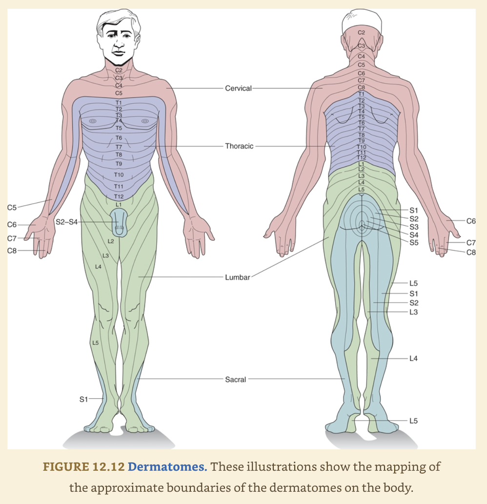
Define dermatome
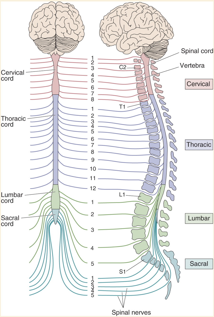
Segmental Organization of the Spinal Cord
Each spinal nerve consisting of dorsal root & ventral root axons passes thru notch between vertebrae of spinal column
30 spinal segments are divided into 4 groups named after where nerves originate
Cervical (C): 1-8
Thoracic (T): 1-12
Lumbar (L) : 1-5
sacral (S): 1-5
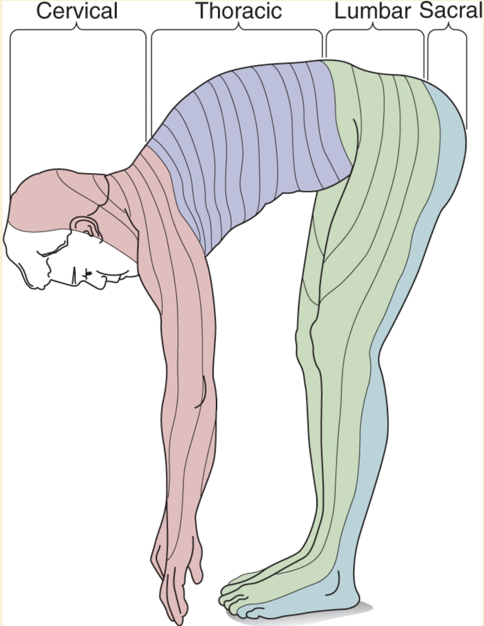
Dermatome: area of skin innervated by right & left dorsal roots of single spinal segment
1:1 correspondence between dermatomes & spinal segments
Dermatomes delineate set of bands on body surface when mapped
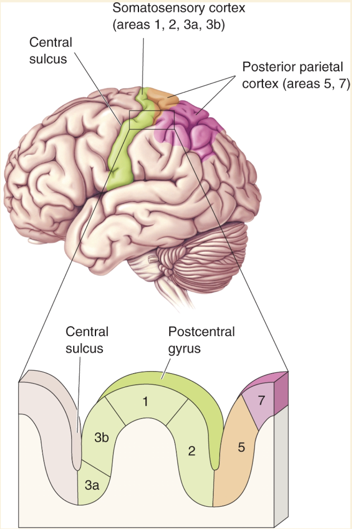
Somatosensory Cortex
Cerebral cortex processes the most complex levels of somatosensory processing
Most located in parietal lobe
Brodmann’s area 3b (now primary somatosensory cortex S1) is on post central gyrus
Recieves input from VP nucleus of thalamus
Neurons are very responsive to somatosensory stimuli
Lesions here impair somatic sensation
When electrically stimulated evokes somatic sensory experiences
Other cortical areas that also process somatic sensory information
3a
1
2
All above on post central gyrus
5
7
All above on posterior parietal cortex
The somatic sensory cortex (S1) has a layered structure, similar to other neocortical areas
Thalamic inputs to S1 primarily terminate in layer IV
Layer IV neurons send projections to neurons in the other cortical layers
S1 neurons with similar inputs and response properties are organized vertically into columns that span all layers
Describe the organization of the primary somatosensory cortex
Discuss an example of how the organization of the somatosensory cortex is plastic
Cortical Somatotopic Map Plasticity (Reorganization of S1)
Researchers have long asked what happens to the somatotopic map in the primary somatosensory cortex (S1) when sensory input from a body part (e.g., a finger) is lost.
Questions included:
Does the cortical “finger area” go unused or atrophy?
Or is this region taken over by inputs from other sources?
These questions have important implications for recovery after peripheral nerve injury.
Merzenich’s Experiments on Cortical Plasticity
In the 1980s, Michael Merzenich and colleagues at the University of California, San Francisco conducted key experiments to test these possibilities.
Subjects: Adult owl monkeys.
Step 1 – Initial Mapping:
The regions of S1 responsive to stimulation of the hand were carefully mapped using microelectrodes.
Step 2 – Finger Amputation:
Digit 3 (third finger) was surgically removed.
Step 3 – Remapping after Several Months:
When S1 was mapped again, the cortical area that had represented the amputated digit was now activated by stimulation of adjacent digits (2 and 4).
→ This showed a major rearrangement of cortical circuitry underlying somatotopy.
Figure 12.23 Summary
(a, b): The normal somatotopic map of the monkey’s hand shows each finger mapped onto a distinct region of S1.
(c): After digit 3 removal, the representations of digits 2 and 4 expand into the area previously devoted to digit 3.
(d): When digits 2 and 3 are selectively stimulated (increased activity), their cortical representations expand as well.
Conclusions from the Experiments
Cortical maps are dynamic — they change depending on sensory experience and input activity.
Decreased input (e.g., amputation) causes neighboring representations to expand into the unused area.
Increased input or activity (e.g., frequent use or stimulation) causes that region’s cortical representation to grow.
This demonstrates map plasticity, a fundamental property of cortical organization.
Broader Evidence of Plasticity
Similar types of map plasticity have been demonstrated in other cortical areas:
Visual cortex
Auditory cortex
Motor cortex
This shows that experience-dependent reorganization is a widespread phenomenon in the brain.
Evidence in Humans
Amputees often experience “phantom limb sensations” — perceiving feelings from a missing limb when other body parts are touched.
These sensations typically arise from stimulation of skin regions whose cortical representations border that of the missing limb.
Example: Touching the face can evoke sensations in a phantom arm.
Functional brain imaging shows that cortical areas once devoted to the missing limb are now activated by stimulation of adjacent regions (like the face).
This demonstrates that S1 cortex is not left inactive but reorganized after limb loss.
However, this plasticity can also create confusion — sensory input from one region is now misinterpreted as coming from the missing limb.
Adaptive vs. Maladaptive Plasticity
While cortical plasticity may be adaptive (ensuring cortex doesn’t go unused), it can also produce mismatches between stimulation and perception, as seen in phantom limb phenomena.
Therefore, not all cortical reorganization results in beneficial outcomes.
Experience-Driven Plasticity in Musicians
String instrument players (e.g., violinists) provide another striking example of cortical map plasticity.
Their left hand fingers (used for fingering strings) receive continuous, fine tactile stimulation.
The right hand, used mainly for bowing, receives less individual finger input.
Functional imaging shows a greatly enlarged cortical representation for the left-hand fingers in these musicians.
This is considered an enhanced version of the normal remapping process that occurs in all people as their sensory experiences change over time.
Compare and contrast: retinotopy and somatotopy
Cortical Somatotopy
Stimulation of S1 surface can cause somatic sensations localized to specific part of the body
Moving stimulator around S1 will cause sensation to move across body
Receptive fields of many S1 neurons produce map of the body on the cortex
Somatotopy: mapping of the body’s surface sensations onto a structure in the brain
Somatotopic maps generated by electrical stimulation & neuronal recording methods are similar
The map is sometimes called a homunculus
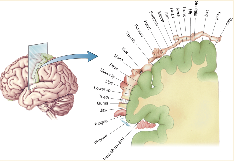
The relative size of cortex devoted to each body part is correlated with the density of sensory input received from that part
Size on the map is also related to the importance of the sensory input from that part of the body