Cells: Organization and Communication (February 11, 2025)
Cell are the Building Blocks of Life
Cells are highly organized and dynamic
The human body contains trillions of cells.
Cells differ in both shape and size.
The study of cells is called cytology.
All Cells Have Similar Characteristics
Current cell theory holds:
All living things are composed of cells.
All cells arise from preexisting cells through cell division.
Cells contain hereditary material, which they pass to daughter cells during cell division.
The chemical composition of all cells is quite similar.
The metabolic processes associated with life occur within cells.
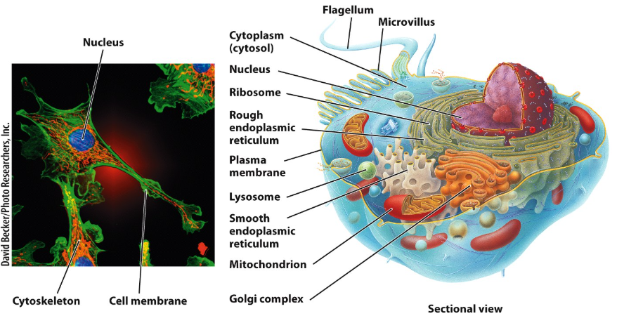
The Cell is a Highly Organized Structure That Has Three Basic Parts
A barrier called the plasma membrane (or cell membrane)
Plant cells and bacteria have a cell wall next to their plasma membrane.
An area where the cell’s genetic material is stored.
A nucleus is animal and plant cells.
A nucleoid in bacterial cells.
A fluid called cytosol.
Found between plasma membrane and nucleus.
Filled with organelles, each with a function vital to the life of the cell.
Early Life Forms were Prokaryotic
Millions of years ago, prokaryotic cells adapted to the extreme environments of early Earth.
Today, they survive as bacteria and Archaebacteria
Prokaryotic cells have no internal membrane-bound organelles.
Although they have genetic material, they have no nucleus
Their genetic material is confined to the nucleoid region in their cytoplasm
They contain ribosomes
These permit prokaryotic cells to produce the proteins they require for life.
Eukaryotic Cells Have a Nucleus and Membrane-Bound Organelles
Eukaryotic cells are believed to have evolved from prokaryotic ancestors.
Endosymbiosis theory proposes that some early prokaryotic cells engulfed (absorbed) other smaller, energy-producing prokaryotic cells.
The engulfed prokaryotic cells survived inside their “host cells.”
Both co-evolved symbiotically.
Plant, animal, and fungal cells are eukaryotic.
Plant Cells
Plant cells have most of the same organelles as animal cells.
Plant cells also have additional organelles that are not found in animal cells.
The cell wall lies next to the plasma membrane to provide structural stability to the cell.
The central vacuole maintains cell pressure (turgor)
The central vacuole consists of water and nutrients.
Chloroplasts produce sugars through the process of photosynthesis.
The sugars contain energy which is used by the plants as nutrients.
The sugars (with their energy) also are passed on to the organisms that eat the plants.
Many believe that chloroplasts originated as bacteria that were “adopted” (engulfed) by the primitive ancestors of plant cells through endosymbiosis.
The Cell Membrane Separates the Cell From the Extracellular Fluid
The cell membrane is composed of two layers of phospholipids, which are interspersed with proteins, lipids, cholesterol, and sugars.
Phospholipids are arranged in a double layer, or bilayer.
The hydrophilic, water-loving, polar heads of the phospholipid molecules are oriented toward the aqueous environment both inside and outside the cell.
The hydrophobic, water-fearing, non-polar lipid portions of the phospholipid molecules are sandwiches in the center of the bilayer.
Proteins and lipids associated with the cell membrane have sugars attached to their external surface.
They are called glycoproteins and glycolipids.
Cell Membrane Structure
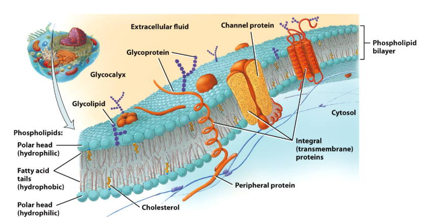
The Glycocalyx
The glycoproteins and glycolipids form a layer on the cell membrane called the glycocalyx.
The glycocalyx is unique and defines cells as belonging to a specific organism.
Both blood type and tissue type are defined by the specific structures on the glycocalyx.
Each person’s white blood cells carry a group of identifying proteins called the human leukocyte antigens (HLAs).
These serve as markers indicating that our cells belong to us.
HLA is used to match tissues before organ transplants.
HLA is inherited — close tissue matches often occur within immediate family.
The Cell Membrane Has a Fluid Mosaic Structure
The basic structure of the plasma membrane is a continuously swirling fluid with consistency similar to olive oil.
Cholesterol molecules separate phospholipid fatty avid tails, which contributes to the fluid nature of the plasma membrane.
The membrane proteins also are in constant motion, floating within the phospholipid bilayer.
Membrane fluidity enables membranes to de-form and change their overall shape during cell locomotion.
Movement Across the Membrane
The membrane is a semipermeable barrier
It provides a means for
Nutrients to enter the cell
Waste products to exist the cell.
Movement across the membrane occurs in two ways
Passive transport
Active transport
Passive Transport
Passive transport does not require the cell to expend energy to move molecules across membranes down their concentration gradients.
Includes diffusion, facilitated diffusion, and filtration.
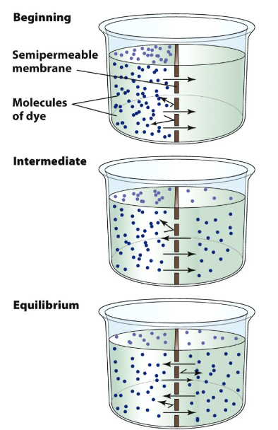
Diffusion is the movement of a substance towards the area of its lower concentration,
Diffusion of water across a semipermeable membrane is termed osmosis.
Water moves in a direction across the cell membrane that equalizes solute concentrations on each side of the membrane.
Locations with higher solute concentrations have lower water concentrations.
“Pull” water towards them.
Water moves down its concentration gradient.
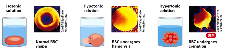
Osmosis
Usually, the extracellular fluid is isotonic to the cells.
Water flows equally into and out of the cell.
A hypotonic solution may cause a cell to burst.
Has water with a lower concentration of solutes than the cytosol.
A hypertonic solution may cause a cell to shrivel up.
Has water with a higher concentration of solutes than the cytosol.
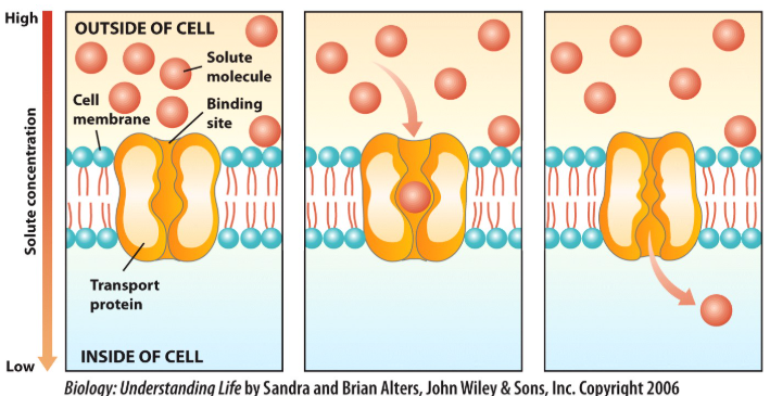
Facilitated Diffusion
The cell does not require to expend energy to transport molecules.
Uses integral membrane proteins (transporter proteins) to move molecules across the cell membrane.
Molecule to be transported binds to transporter protein on one side of bilayer.
Protein “passes” (transports) molecule to other side of membrane.
Solutes are transported across the membrane down their concentration gradients.
From area of high solute concentration to area of low solute concentration.
This is the main method by which glucose is moved into cells.
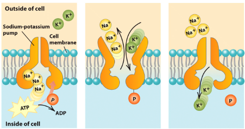
Active Transport
Cell must expend energy to transport molecules or ions across cell membrane.
Using energy derived from the hydrolysis (breakdown) of ATP into ADP
Molecules or ions are transported across the membrane against their concentration gradients.
From area of low solute concentration to area of high solute concentration.
Integral membrane proteins (transporter proteins, or “pumps”) move molecules or ions across the cell membrane.
Molecule or ion to be transported binds to transporter protein on one side of bilayer (on the side where there is a lower concentration of the material).
Using energy transporter protein “pumps” molecule or ion to other side of membrane (to side where there is a higher concentration of the material)
Active Transporter Pumps Often Have Reciprocal Functions
Pumping one molecule or ion into the cell while simultaneously removing a second from the cell.
The sodium/potassium ATPase pumps two K+ into the cell while pumping three Na+ out of the cell.
Active Transport Concentrates Molecules an Ions Inside and Outside of Cells
Active transport accounts for
Establishing potassium and sodium ion concentrations inside and outside of the cell membranes of nerve cells in preparation for nerve impulse transmission.
The almost complete uptake of digested nutrients from the small intestine blood by the kidneys.
The collecting of iodine in thyroid gland cells.
Endocytosis and Exocytosis
Active transport also can move molecules into or out of the cell, in bulk.
In endocytosis
Extracellular molecules and particles are taken into the cell via vesicle formation.
In exocytosis
Secretory products or waste products are removed form the cell.

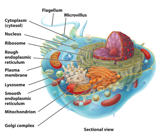
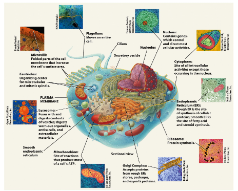
The Components of a Cell are Called Organelles
Each organelle plays a role in regulating the life and death of cells.
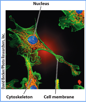
Cytoskeleton
The cytosol is a highly organized “chemical soup” complete with a support structure called the cytoskeleton.
The cytoskeleton provides
Shape and structural support for the cell.
A scaffold for suspending and moving organelles within the cell.
The cytoskeleton continuously changes shape.
Forming and breaking down and reforming.
Giving cells a plasticity, or fluidity, that allows them to change shape or move organelles quickly.
The Cytoskeleton is Composed of Three Types of Filamentous Proteins
All three types of cytoskeleton contribute to cell shape — but each also has its own specific function.
Microfilaments
Long filaments constructed of actin protein subunits.
Responsible for cellular locomotion and muscle contractions.
Establish the basic shape and strength of the cell.
Intermediate filaments
Strong cables of protein subunits.
Protein type depends on type of intermediate filament.
Stronger than microfilaments - protect cell from mechanical stresses
Microtubules
Long tiny tubules made of tubulin protein subunits
Are instrumental in chromosome movement during cell division.
Also used as tracks for organelle movement.
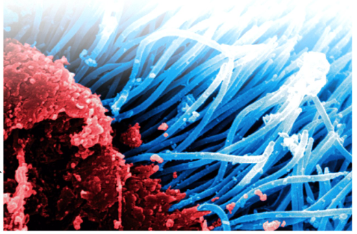
Flagella and Cilia
Flagella are single, long, whip-like structures that propel the cell forward
The only human cell that moves by flagellum is the sperm.
Cilia are shorter extensions that look like hairs or eyelashes.
Cilia line the upper respiratory tract, moving mucus upward and sweeping out debris and pathogens.
Cilia also line the fallopian tubes, moving the egg from the ovary to the uterus.
The Endoplasmic Reticulum
Endoplasmic reticulum or ER (literally “within fluid network”)
The membranes of the ER are directly connected to the double membrane surrounding the cell nucleus.
Humans Have Two Types of Endoplasmic Reticulum
Rough endoplasmic reticulum (RER)
Processing and sorting area for proteins synthesized by the ribosomes that stud its outer membrane.
Ribosomes are small nonmembrane-bound organelles composed of protein and ribosomal RNA that function as protein factories.
Synthesize proteins that may be incorporated into other organelles or into the plasma membrane.
Smooth endoplasmic reticulum (SER)
Responsible for the synthesis of fatty acid and steroid hormones.
SER has no attached ribosomes.
Both SER and RER produce vesicles filled with product ready for the next step in processing.
Vesicles usually move substances from to the cell membrane for exocytosis.
Or to the Golgi complex for further packaging.
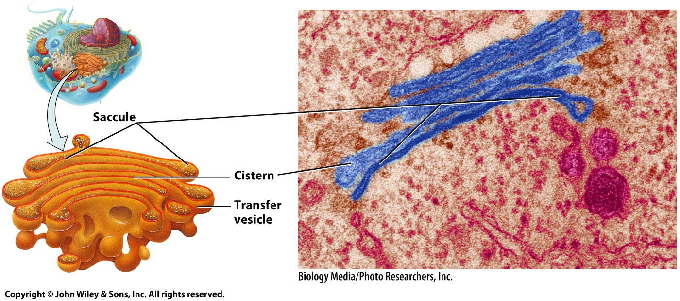
The Golgi Complex is Involved with Processing Proteins and Fatty Acids
The Golgi complex, or Golgi apparatus, is found near the end of the SER that is farthest from the nucleus.
Resembles a stack of pancakes called saccules.
Saccules are slightly curved, with concave and convex faces.
Concave portions face the ER; convex portions face plasma membrane
Vesicles are found at the edges of these saccules.
Vesicles that leave the Golgi complex migrate all over the cell.
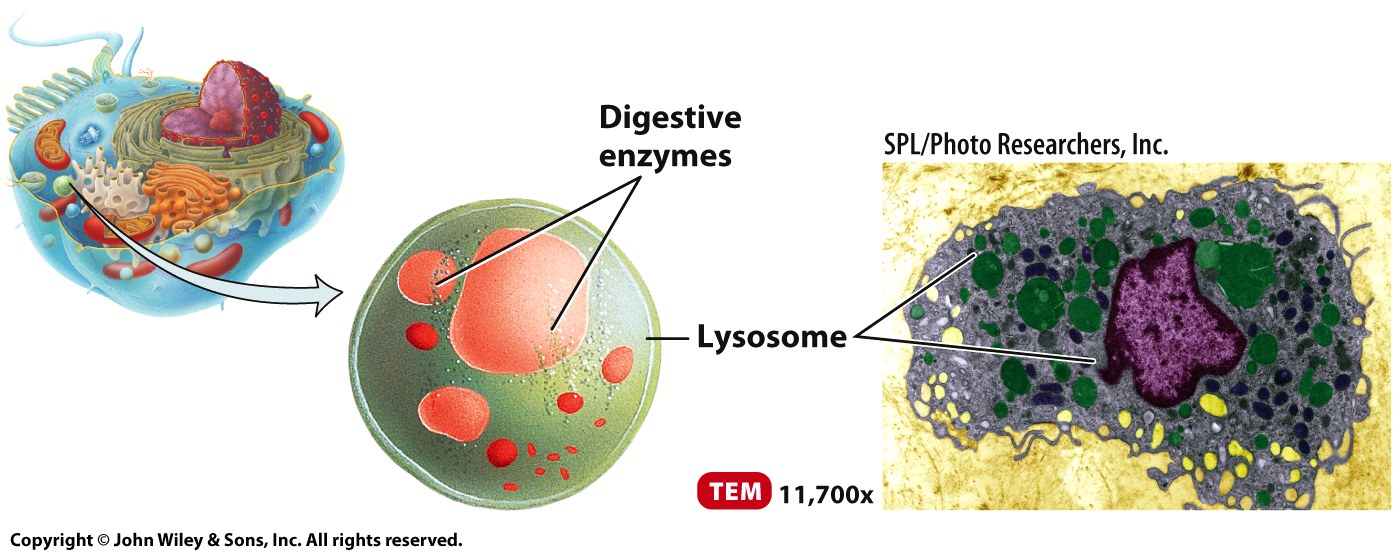
Lysosomes are Chemical Packages that Contain Hydrolytic Enzymes
Produced by the Golgi complex.
When a lysosome (lyse means to break open or break apart) fuses with an endocytic vesicle, it pours its contents into the vesicle.
The hydrolytic enzymes break down the vesicle’s contents.
Phagocytosed bacteria are routinely destroyed in the body by lysosomal activity.
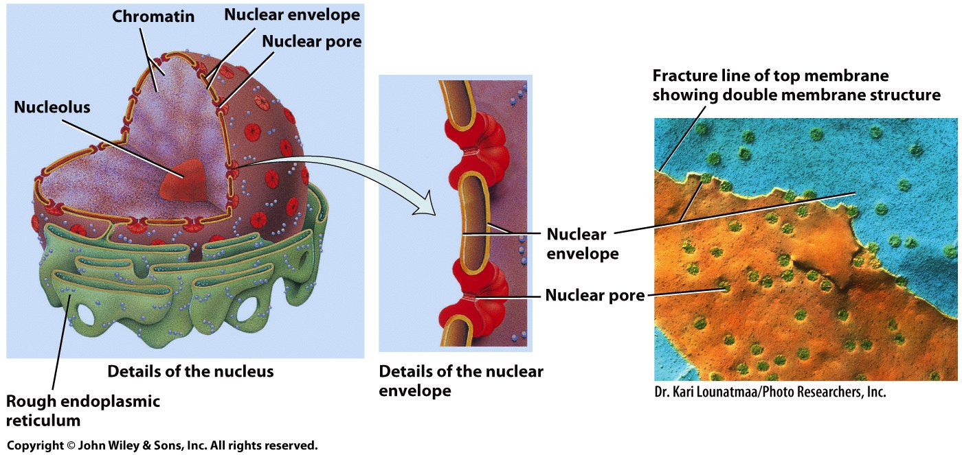
The Nucleus Contains a Cell’s Genetic Library
Usually the largest organelle in a eukaryotic cell.
It is covered by two phospholipid bilayers, called the nuclear envelope.
The envelope is punctuated by nuclear pores, which also molecules to enter and exit the nucleus.
The small size of the nuclear pores prevents the genetic material (DNA) from leaving the nucleus.
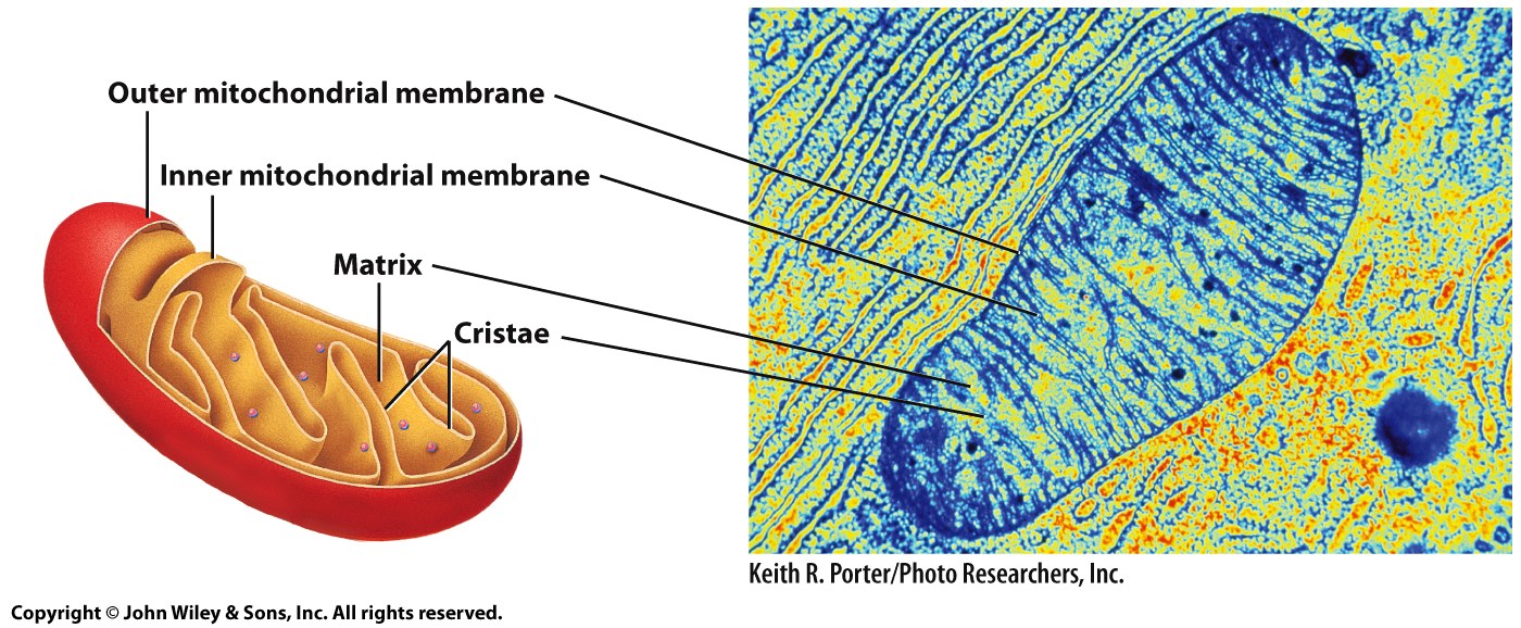
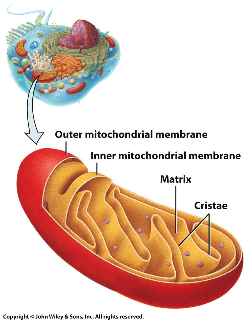
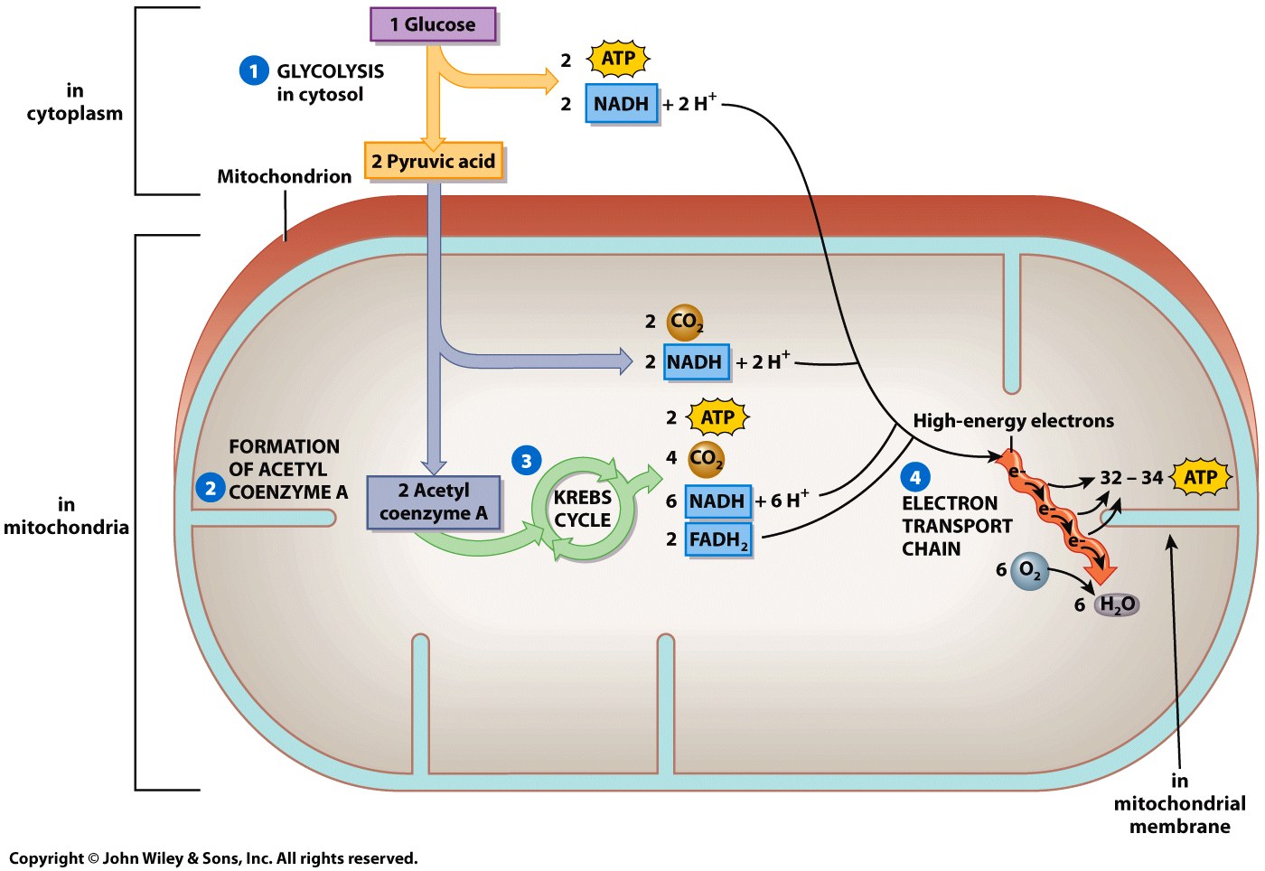
Mitochondria Covert Nutrients into Usable Energy in the Form of ATP
Have a smooth outer membrane and a folded inner membrane (cristae)
Mitochondria require oxygen, and produce carbon dioxide in their endless production of ATP.
Cellular respiration
Cellular Respiration Occurs in Four Steps

Cell Communication
Cell communicate with one another to function as a tissue
Tissues send signals throughout the organ for the organ to function properly.
Organs in a particular system communicate to carry out the system’s process.
The signals sent from cell to cell include information
The timing of cell divisions, the health of adjacent cells, and the status of the external environment.
Cells communicate with one another via chemical messengers or through physical contact.
Cell signaling occurs via three routes
Each route differs in the speed and distance of the signal transmission.
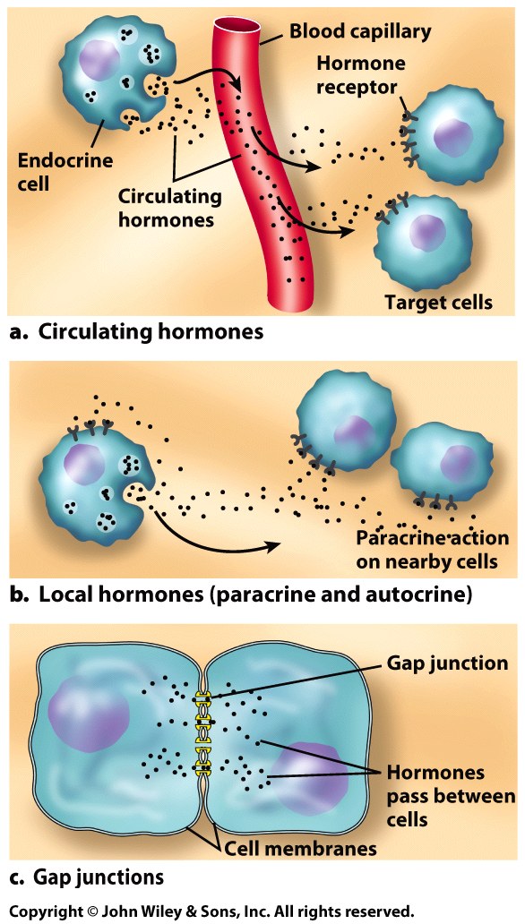
Cell Signaling
Circulating hormones can be released into the bloodstream, potentially reaching every cell.
Much hormonal communication is long distance, carrying information to distant cells that will alter their functioning.
Target cells are often remote from the secreting cells.
Paracrine (local hormones) can be released to affect only cells in the vicinity.
Mostly used when quick responses are required
Cells that are infected with a virus may secrete the paracrine interferon.
Interferon alerts the surrounding cells, warning them of the viral invasion, helping them to fight it.
Neurons use paracrine to stimulate nearby nerve, muscle, or glandular cells by releasing short-lived chemicals called neurotransmitters.
Neurons must respond instantly to information; therefore they secrete neurotransmitters directly into the space between cells.
Cells Can Interact with other Cells Directly Through Cell-to-Cell Junctions
Cell-to-cell junctions occur in tissues like your skin and heart muscle tissue, where cells are in direct contact with one another.
For example, skin cells are knit tightly together, so any change in one cell will be immediately passed to the next.
Gape junctions are used for instantaneous communication.
Occur across very short distances and are extremely specific.
Immediate and short-lived.
Important between heart cells.