S & P Intro to Vision
Visible Light
electromagnetic spectrum
light as wave or particle
nanometer = 10^-9 of a meter
The Eye - transduces light to neutral firing
Pupil - where light enters the eye
Cornea - first site of focusing on surface of eye (80%)
Lens - Flexible focuser in the eye (20%)
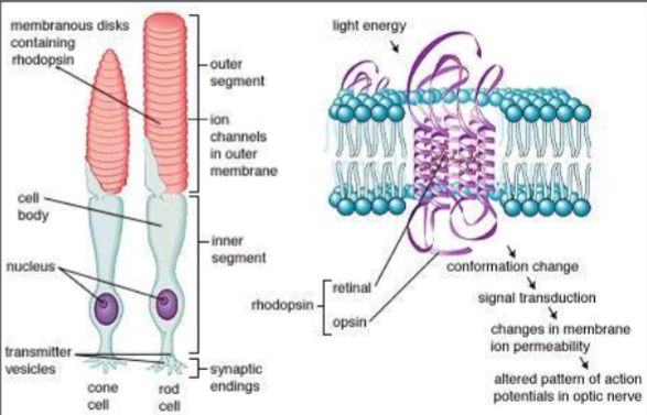
Retina - contains rods and cones, the transducers of light
Optic nerve - carry the electrical signal from the eye to the brain
Sclera - outer white part of your eye
Lens Accommodation
The accommodates for close vision by tightening the ciliary muscles, allowing the pliable crystalline lens to become more rounded
Ciliary muscles relaxed, fibers taut, lens at minimum strength for distant vision
Ciliary muscles contracted, fibers slack, lens rounds to greater strength for close vision
Light rays from distant objects are nearly parallel and don’t need as much refraction to bring them to a focus
Light rays from close objects diverge and require more refraction for focusing
Focusing Problems
Emmetropia - normal vision
Hyperopia - farsighted
Myopia - nearsighted
Astigmatism - distortion
Presbyopia - old vision
Corrective Lenses
Lenses focus light at the back of the eye
Concave - nearsighted
Convex - farsighted
Transduction of Light
Light isomerizes (changes from one shape to another) Rhodopsin
Leads to action potential
A rod will respond to 1 photon
7 photons needed to see
Eye Structure and Retinal Array
the retina lines the back of the eye and consists of rod and cone photoreceptors as four types of neurons:
second-order bipolar
horizontal cells
third-order retinal ganglion cells (RGCs)
amacrine cells
muller glial cells fill the spaces between the neurons
the pigment epithelium, critical for photoreceptor function, underlies the retina
photoreceptors and RGCs are most susceptible to blinding retinal disease
progress in combating photoreceptor degeneration has been made, but there are few strategies to address RGC loss
Distribution of Rods and Cones
Fovea - only cones (-50,000)
Periphery - rods and cones (6 million cones, 120 million rods)
Macular degeneration - destroys fovea
Retinitis pigmentosa - destroys periphery
Blind Spot
site where optic nerve exits the eye
no receptors there
brain fills in so you don’t see it
Dark Adaptation Curve
Scotopic - night vision
Photopic - day vision
More on Dark Adaptation
Measure cone adaptation - use light placed on the fovea - no rods
Measure rod adaptation - need a rod monochromat - no functioning cones
can measure the isolated response of the rods
can’t use normal vision observer as there are cones in the periphery
Process of adaptation
once light is out, both rods and cones increase sensitivity
3-5 minutes cones finish adapting
around 25-30 minutes, rods finish adapting
rod-cone break is where rods start to determine the curve of adaptation
Differences in Adaptation - Rods and Cones
Why do rods take longer to adapt than cones?
Visual pigment regeneration
when light hits the retina, rhodopsin isomerizes
this causes the visual pigment to bleach
as some pigments are bleaching, some are regenerating
this process is faster in cones than rods
Detached retina - separates retina from pigment epithelium (contains regenerating enzymes)
caused by blow to the head
prevents process of pigment regeneration
person becomes blind in area of detachment
Spectral Sensitivity of Rods and Cones
Threshold is the inverse of sensitivity
high sensitivity means low threshold
low sensitivity means high threshold
higher sensitivity means you need less light to see it
lower sensitivity means you need more light to see it
Purkinje Shift - at dusk
shift from red to blue
shift from cones to rods as light decreases
rods are more sensitive to blue wavelengths
hues change as the spectral sensitivities of the two receptors are different
Absorption Spectrum
how much light is absorbed
peak is most absorbed wavelength
spectral sensitivity is related to absorption spectrum
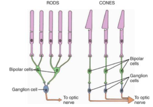
Neural Convergence
Order of processing:
linear processing is Receptors → bipolar cells → ganglion cells → “brain”
lateral processing by horizontal cells and amacrine cells
Horizontal cells - processing between receptors and bipolar cells
Amacrine cells - processing between ganglion cells
Neural convergence - one neuron receives signals from many cells
Rods vs Cones
Rods - more convergence, better in low light because of convergence and greater sensitivity
Cones - little to no convergence, better resolution due to little convergence, not good in low light
Get lateral inhibition with rods
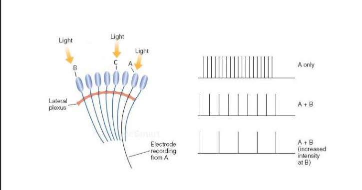
Lateral Inhibition - related to convergence
Limulus (horseshoe crab) - huge eyes or ommatidia
easy to study lateral inhibition
light excites Cell A
add light to B, cell A fires even less
add more light at B, cell fires even less
cell A is being inhibited by cell B activity
Simultaneous Contrast
examples of lateral inhibition at work

Direct or Indirect Perception?
We think that we perceive directly
We perceive through our senses and it’s all in our head - more indirect
Seems very real to us
But our senses distort the world (lateral inhibition is just one example)